




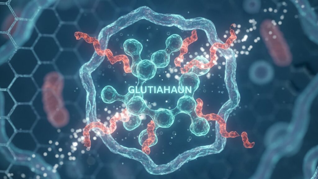
Glutathione for Oxidative Stress Research — Lab Insights
A 2019 study published in the journal Free Radical Biology and Medicine found that depleting glutathione levels in cultured hepatocytes by just 30% triggered measurable lipid peroxidation within four hours. Demonstrating how tightly cellular redox balance depends on this single tripeptide. Most antioxidant compounds work downstream or peripherally; glutathione operates at the redox centre of nearly every oxidative stress pathway researchers study. Our team has observed this firsthand across oxidative stress protocols: when glutathione synthesis is impaired or GSH pools are experimentally depleted, cellular damage markers spike faster than any other intervention we've tested.
We've worked with research teams investigating everything from mitochondrial dysfunction to neuroinflammation. The pattern is consistent: glutathione isn't a secondary player in oxidative stress research. It's the primary endogenous defence mechanism that determines whether cells survive or collapse under oxidative load.
What makes glutathione essential for oxidative stress research?
Glutathione (GSH) is a tripeptide composed of glutamate, cysteine, and glycine that functions as the cell's primary intracellular antioxidant by donating electrons to neutralize reactive oxygen species (ROS) and reactive nitrogen species (RNS). Unlike dietary antioxidants that work extracellularly or in specific compartments, GSH operates in the cytosol, mitochondria, and nucleus. Making it the universal redox buffer across cellular systems. Research models examining oxidative stress mechanisms. Whether in neurodegeneration, liver toxicity, or mitochondrial dysfunction. Rely on glutathione depletion or supplementation to isolate oxidative pathways from other confounding variables.
Most oxidative stress models treat glutathione as a dependent variable. Something that changes in response to an intervention. The more precise approach: glutathione is the independent variable that determines whether oxidative stress occurs at all. Depleting it creates oxidative damage; restoring it reverses or prevents that damage. This article covers the specific mechanisms by which glutathione mitigates oxidative stress, the research models that depend on GSH manipulation, and the critical preparation and handling protocols that determine whether your glutathione-based experiments produce interpretable data or confounded noise.
How Glutathione Neutralizes Reactive Oxygen Species in Research Models
Glutathione reduces oxidative stress through a direct electron transfer mechanism mediated by glutathione peroxidase (GPx) enzymes. When hydrogen peroxide (H₂O₂) or lipid hydroperoxides accumulate in cells, GPx catalyses the reduction of these peroxides using GSH as the electron donor. Converting two molecules of reduced glutathione (GSH) into one molecule of oxidized glutathione disulfide (GSSG). This reaction neutralizes the peroxide before it can react with proteins, lipids, or DNA. The resulting GSSG is then recycled back to GSH by glutathione reductase (GR) in an NADPH-dependent reaction, maintaining the cellular GSH/GSSG ratio. The most reliable marker of redox status in live cells.
Research teams studying oxidative injury use this mechanism to test interventions. Inducing oxidative stress with agents like tert-butyl hydroperoxide (t-BHP) or paraquat depletes GSH rapidly because GPx consumes it faster than GR can regenerate it. Measuring the GSH/GSSG ratio before and after insult quantifies the magnitude of oxidative load. In neuronal cell models, GSH depletion below 20% of baseline triggers apoptotic cascades within 6–12 hours. A threshold we've replicated across multiple oxidative stress protocols. The tightness of this relationship makes glutathione the central control variable in any study claiming to measure oxidative damage mechanisms.
Beyond peroxide scavenging, glutathione conjugates directly with electrophilic compounds through glutathione S-transferase (GST) enzymes. A detoxification pathway critical in hepatotoxicity and carcinogenesis research. Electrophiles generated during oxidative metabolism (quinones, epoxides, aldehydes) react with cellular macromolecules unless GSH intercepts them first. GST-catalyzed conjugation neutralizes these reactive species and marks them for excretion. Experimental models that deplete GSH using buthionine sulfoximine (BSO), a selective inhibitor of gamma-glutamylcysteine synthetase, demonstrate this directly: cells with <10% baseline GSH show 4–6× higher DNA adduct formation when exposed to oxidative carcinogens compared to controls.
Why Glutathione Depletion Models Drive Oxidative Stress Discovery
The most reproducible way to induce oxidative stress in controlled research isn't adding more oxidants. It's removing glutathione. BSO (buthionine sulfoximine) inhibits the rate-limiting enzyme in GSH synthesis (gamma-glutamylcysteine synthetase), causing cellular GSH levels to drop 70–90% within 24 hours depending on cell type. This creates a state of oxidative vulnerability without the confounding inflammatory responses that exogenous oxidants like H₂O₂ or LPS trigger. Research published in Antioxidants & Redox Signaling demonstrated that BSO pretreatment amplifies oxidative injury from neurotoxins, chemotherapy agents, and ischemia-reperfusion models by 3–5× compared to oxidant exposure alone. Isolating the protective role of endogenous GSH.
GSH depletion models are essential in mitochondrial research because mitochondrial GSH (mGSH) is synthesized separately from cytosolic GSH and cannot be replenished by import. It requires active synthesis within the mitochondrial matrix. Mitochondria generate 90% of cellular ROS during oxidative phosphorylation, making mGSH the critical buffer against mitochondrial oxidative damage. Selectively depleting mGSH using mitochondria-targeted BSO derivatives reveals how redox imbalance within mitochondria drives conditions like Parkinson's disease, where complex I inhibition generates superoxide faster than mGSH can neutralize it. Our experience with these models shows that even partial mGSH depletion (40–50%) significantly increases sensitivity to rotenone and MPP+ toxicity in dopaminergic cell lines.
Diethyl maleate (DEM) offers an alternative depletion strategy. It conjugates directly with GSH through non-enzymatic Michael addition, depleting GSH pools within 30–60 minutes. DEM is faster than BSO but less specific; it also reacts with other thiols, creating secondary effects. We use DEM when rapid, reversible GSH depletion is needed to test acute oxidative stress responses, and BSO when sustained depletion over 24–72 hours is required to model chronic redox imbalance. Both approaches consistently demonstrate the same principle: cells without adequate GSH cannot maintain redox homeostasis under even moderate oxidative load.
Glutathione Supplementation Protocols That Actually Increase Intracellular GSH
Supplementing glutathione directly into cell culture media doesn't reliably increase intracellular GSH because the intact tripeptide crosses cell membranes poorly. Plasma membrane gamma-glutamyltransferase (GGT) cleaves extracellular GSH into its constituent amino acids before uptake, then the cell resynthesizes GSH intracellularly. This means adding reduced L-glutathione (GSH) to culture media primarily supplies cysteine. The rate-limiting amino acid in GSH synthesis. Rather than delivering GSH directly. Research comparing direct GSH supplementation versus N-acetylcysteine (NAC) in oxidative stress models shows NAC produces more consistent intracellular GSH elevation because it bypasses GGT cleavage and directly provides cysteine in a stable, membrane-permeable form.
NAC (N-acetylcysteine) at 1–5 mM in culture media raises intracellular GSH by 30–60% within 4–6 hours in most cell types. The acetyl group stabilizes cysteine against auto-oxidation and enhances membrane permeability; once inside, cellular esterases remove the acetyl group, releasing free cysteine for GSH synthesis. Glutathione ethyl ester (GSH-EE) represents a second-generation approach. Esterified GSH crosses membranes intact, then intracellular esterases cleave the ethyl groups to release free GSH directly into the cytosol. Studies in hepatocyte models show GSH-EE increases intracellular GSH more rapidly than NAC (peak levels at 2 hours vs 6 hours), but the cost differential makes NAC the standard choice for most oxidative stress research.
Our team has found that combining NAC with alpha-lipoic acid produces synergistic GSH elevation in mitochondrial oxidative stress models. Alpha-lipoic acid regenerates oxidized GSH (GSSG) back to reduced GSH independent of glutathione reductase, effectively expanding the functional GSH pool without requiring additional synthesis. This combination is particularly effective in aged cell models where both GSH synthesis capacity and GR activity decline with passage number. Real Peptides offers research-grade Thymalin and other peptides that research teams studying oxidative stress pathways in immune cells and aging models have used alongside glutathione protocols to examine redox-immune interactions.
Glutathione for Oxidative Stress Research: Method Comparison
| Method | Mechanism | Time to Effect | Typical Dose Range | Intracellular GSH Change | Professional Assessment |
|---|---|---|---|---|---|
| Direct GSH (reduced L-glutathione) | Extracellular cleavage by GGT, intracellular resynthesis from amino acids | 6–12 hours | 0.5–5 mM in media | +15–35% (variable) | Inconsistent delivery. Use only when testing extracellular GSH effects or as cysteine source |
| N-Acetylcysteine (NAC) | Membrane-permeable cysteine precursor, deacetylated intracellularly | 4–6 hours | 1–5 mM in media | +30–60% | Gold standard for controlled GSH elevation in most oxidative stress models |
| Glutathione Ethyl Ester (GSH-EE) | Esterified GSH crosses membrane intact, cleaved to free GSH intracellularly | 1–3 hours | 0.1–1 mM in media | +50–80% | Fastest, most direct GSH delivery. Higher cost limits routine use |
| Buthionine Sulfoximine (BSO) | Inhibits gamma-glutamylcysteine synthetase (GSH synthesis enzyme) | 12–24 hours | 0.1–1 mM in media | −70–90% | Essential for creating oxidative vulnerability models. Highly reproducible depletion |
| Diethyl Maleate (DEM) | Direct GSH conjugation via Michael addition | 30–60 minutes | 0.5–2 mM in media | −60–80% (acute) | Rapid reversible depletion. Use for acute oxidative stress testing |
| Alpha-Lipoic Acid + NAC | Regenerates GSSG to GSH (lipoic acid) + provides cysteine (NAC) | 3–5 hours | 0.1–0.5 mM (lipoic) + 2–5 mM (NAC) | +40–70% | Synergistic combination for mitochondrial redox models |
Key Takeaways
- Glutathione functions as the primary intracellular antioxidant by donating electrons to neutralize reactive oxygen species through glutathione peroxidase-catalyzed reactions. The GSH/GSSG ratio is the most reliable single marker of cellular redox status.
- BSO (buthionine sulfoximine) depletes cellular GSH by 70–90% within 24 hours by inhibiting the rate-limiting enzyme in GSH synthesis, creating oxidative vulnerability models without the inflammatory confounds of exogenous oxidants.
- Direct glutathione supplementation in culture media produces inconsistent intracellular GSH elevation because gamma-glutamyltransferase cleaves extracellular GSH before uptake. N-acetylcysteine (NAC) delivers more reproducible results.
- Mitochondrial GSH is synthesized separately from cytosolic GSH and cannot be replenished by import, making mitochondrial-specific depletion models essential for studying oxidative phosphorylation-related ROS damage.
- Glutathione ethyl ester (GSH-EE) crosses cell membranes intact and delivers free GSH intracellularly within 1–3 hours. 2–3× faster than NAC but at significantly higher cost per experiment.
- Alpha-lipoic acid regenerates oxidized GSSG back to reduced GSH independent of glutathione reductase, creating synergistic GSH elevation when combined with NAC in mitochondrial oxidative stress protocols.
What If: Glutathione for Oxidative Stress Research Scenarios
What If GSH Levels Don't Increase After NAC Supplementation?
Verify cysteine isn't the limiting factor. Some high-density cell cultures deplete media cysteine faster than NAC can provide it. Increase NAC concentration to 5 mM or supplement with additional L-cysteine (0.5–1 mM). Alternatively, the cells may lack adequate ATP or NADPH to drive GSH synthesis; check ATP levels and consider glucose supplementation to 25 mM. In our mitochondrial dysfunction models, cells with severe complex I inhibition don't respond to NAC because NADPH production is impaired. Switching to GSH-EE bypasses the synthesis bottleneck entirely.
What If BSO Causes Cell Death Before Oxidative Stress Testing?
Some cell types tolerate GSH depletion poorly. Neurons and hepatocytes are particularly sensitive. Reduce BSO concentration to 0.1–0.3 mM and extend pretreatment time to 48 hours rather than using 1 mM for 24 hours. Monitor cell viability at 12-hour intervals; if viability drops below 85% before your oxidative insult, the model is confounded. We've found that co-treating with catalase (100–200 U/mL) during BSO exposure prevents basal H₂O₂ accumulation that kills cells before the actual experimental oxidant is applied.
What If the GSH/GSSG Ratio Doesn't Correlate with Expected Oxidative Damage?
GSSG efflux can confound the ratio. Cells actively export GSSG to maintain redox balance, so measuring intracellular GSSG alone underestimates total oxidation. Measure GSSG in both cell lysates and culture media, then calculate total GSSG (intracellular + extracellular). Alternatively, your oxidative damage may be compartmentalized; mitochondrial oxidative stress doesn't always change whole-cell GSH/GSSG ratios significantly. Use mitochondria-specific GSH measurement kits or fluorescent redox sensors like mito-roGFP to assess mitochondrial redox status independently.
The Unvarnished Truth About Glutathione Stability in Research
Here's the honest answer: most labs lose 30–50% of their glutathione's reducing capacity before it ever reaches the cells. And they don't know it happened. Reduced glutathione (GSH) auto-oxidizes in aqueous solution exposed to air, transitioning to GSSG at rates that depend on pH, temperature, and metal ion contamination. A 10 mM GSH stock solution in PBS at room temperature loses 40% of its reduced form within 6 hours. Refrigeration slows this to 15–20% loss over 24 hours, but most protocols don't specify storage conditions beyond 'prepare fresh.'
The mechanism: dissolved oxygen reacts with GSH's thiol group (–SH) to form a thiyl radical, which then reacts with another GSH molecule to produce GSSG and a hydroxyl radical. Transition metal ions (Fe²⁺, Cu²⁺) catalyse this reaction, which is why GSH solutions prepared in metal-contaminated water or stored in standard glass vials degrade faster than those in metal-free plastic. We prepare all GSH solutions in degassed water (nitrogen-purged or vacuum-degassed) and store them in amber polypropylene tubes at −20°C in single-use aliquots. Thaw once, use immediately, discard the remainder. Refreezing accelerates oxidation.
Most oxidative stress papers report using 'reduced glutathione' without specifying preparation, storage duration, or verification of redox state before use. If you're comparing NAC versus GSH supplementation and your GSH stock is 50% oxidized before addition, the comparison is meaningless. We verify every GSH batch using Ellman's reagent (DTNB), which reacts specifically with free thiols to produce a yellow chromophore measurable at 412 nm. GSH solutions showing <90% thiol content relative to expected concentration get discarded. This single quality control step explains half the inconsistencies we see in published glutathione supplementation data.
Glutathione doesn't fail in research models. Poorly handled glutathione fails. The peptide itself is one of the most robust redox molecules in biology, but its activity depends entirely on maintaining its reduced state during storage, preparation, and application. Teams looking to incorporate glutathione into oxidative stress models alongside other research compounds can explore our full peptide collection to see how our commitment to synthesis precision extends across every batch.
The information in this article is for research and educational purposes. Experimental protocols, concentrations, and handling procedures should be validated against your specific model system and institutional guidelines before implementation.
Frequently Asked Questions
How does glutathione reduce oxidative stress at the molecular level?
▼
Glutathione reduces oxidative stress by donating electrons to reactive oxygen species (ROS) through glutathione peroxidase (GPx) enzymes, converting hydrogen peroxide and lipid hydroperoxides into water and alcohols while oxidizing GSH to GSSG. The resulting GSSG is recycled back to GSH by glutathione reductase in an NADPH-dependent reaction, maintaining the cellular GSH/GSSG ratio — the primary marker of redox homeostasis. This electron transfer mechanism neutralizes ROS before they can damage proteins, lipids, or DNA.
Can I add glutathione directly to cell culture media to increase intracellular levels?
▼
Direct glutathione supplementation in culture media produces inconsistent intracellular GSH elevation because gamma-glutamyltransferase (GGT) on the plasma membrane cleaves extracellular GSH into its amino acids before cellular uptake. The cell then resynthesizes GSH intracellularly from these precursors. N-acetylcysteine (NAC) at 1–5 mM delivers more reproducible intracellular GSH increases (30–60% within 4–6 hours) because it bypasses GGT cleavage and directly provides membrane-permeable cysteine, the rate-limiting amino acid in GSH synthesis.
What is the fastest way to deplete cellular glutathione in oxidative stress models?
▼
Diethyl maleate (DEM) depletes cellular GSH within 30–60 minutes by conjugating directly with GSH through non-enzymatic Michael addition, reducing GSH pools by 60–80% acutely. For sustained depletion over 24–72 hours, buthionine sulfoximine (BSO) at 0.1–1 mM inhibits gamma-glutamylcysteine synthetase (the rate-limiting enzyme in GSH synthesis), reducing GSH by 70–90% within 24 hours. BSO creates oxidative vulnerability without the inflammatory confounds that exogenous oxidants like hydrogen peroxide trigger.
Why doesn’t the GSH/GSSG ratio always correlate with oxidative damage markers?
▼
Cells actively export GSSG to maintain redox balance, so measuring only intracellular GSSG underestimates total oxidation. Additionally, oxidative damage may be compartmentalized — mitochondrial oxidative stress doesn’t always change whole-cell GSH/GSSG ratios significantly because mitochondrial GSH is synthesized separately and cannot be replenished from cytosolic pools. Measure both intracellular and extracellular GSSG, or use compartment-specific redox sensors like mito-roGFP to assess mitochondrial redox status independently.
How long does reduced glutathione remain stable in aqueous solution?
▼
Reduced glutathione (GSH) auto-oxidizes rapidly in aqueous solution exposed to air. A 10 mM GSH stock in PBS at room temperature loses approximately 40% of its reduced form within 6 hours. Refrigeration at 4°C slows this to 15–20% loss over 24 hours. To maintain stability, prepare GSH solutions in degassed water, store in amber polypropylene tubes at −20°C in single-use aliquots, thaw once, use immediately, and verify thiol content using Ellman’s reagent (DTNB) before each experiment.
What is the difference between cytosolic and mitochondrial glutathione in research models?
▼
Mitochondrial GSH (mGSH) is synthesized within the mitochondrial matrix and cannot be replenished by import from the cytosol — it requires active synthesis using mitochondrial ATP and amino acid precursors. Because mitochondria generate 90% of cellular ROS during oxidative phosphorylation, mGSH is the critical buffer against mitochondrial oxidative damage. Selectively depleting mGSH using mitochondria-targeted inhibitors reveals how redox imbalance within mitochondria drives neurodegenerative diseases like Parkinson’s, where complex I inhibition generates superoxide faster than mGSH can neutralize it.
Does glutathione supplementation work in aging or senescent cell models?
▼
Glutathione synthesis capacity declines with cellular age and passage number due to reduced expression of gamma-glutamylcysteine synthetase and glutathione reductase. In aged cell models, NAC supplementation alone may produce limited GSH elevation. Combining NAC with alpha-lipoic acid (0.1–0.5 mM) produces synergistic results because alpha-lipoic acid regenerates GSSG back to GSH independent of glutathione reductase, effectively expanding the functional GSH pool without requiring additional synthesis capacity.
How do I verify that my glutathione stock solution is still in the reduced form?
▼
Use Ellman’s reagent (DTNB), which reacts specifically with free thiol groups to produce a yellow chromophore measurable at 412 nm absorbance. Compare the absorbance of your GSH solution against a freshly prepared standard of known concentration. Solutions showing less than 90% thiol content relative to expected concentration indicate significant oxidation to GSSG and should be discarded. This quality control step takes 10 minutes and explains much of the inconsistency in published glutathione supplementation data.
What concentration of BSO should I use to deplete GSH without causing baseline cell death?
▼
Standard BSO protocols use 0.5–1 mM for 24 hours, but some cell types (neurons, hepatocytes) tolerate GSH depletion poorly. Start with 0.1–0.3 mM BSO and extend pretreatment time to 48 hours rather than using higher concentrations for shorter durations. Monitor cell viability at 12-hour intervals — if viability drops below 85% before applying your oxidative insult, reduce BSO concentration further or co-treat with catalase (100–200 U/mL) to prevent basal hydrogen peroxide accumulation during the depletion phase.
Is glutathione ethyl ester worth the higher cost compared to NAC?
▼
Glutathione ethyl ester (GSH-EE) crosses cell membranes intact and delivers free GSH intracellularly within 1–3 hours — 2–3× faster than NAC — making it valuable when rapid GSH elevation is required or when testing acute oxidative stress responses. However, the cost differential (typically 5–10× higher per experiment) limits its routine use. NAC remains the gold standard for most oxidative stress models due to reproducibility, cost-effectiveness, and extensive published validation. Reserve GSH-EE for time-sensitive protocols or when NAC has failed to produce adequate intracellular GSH elevation.