


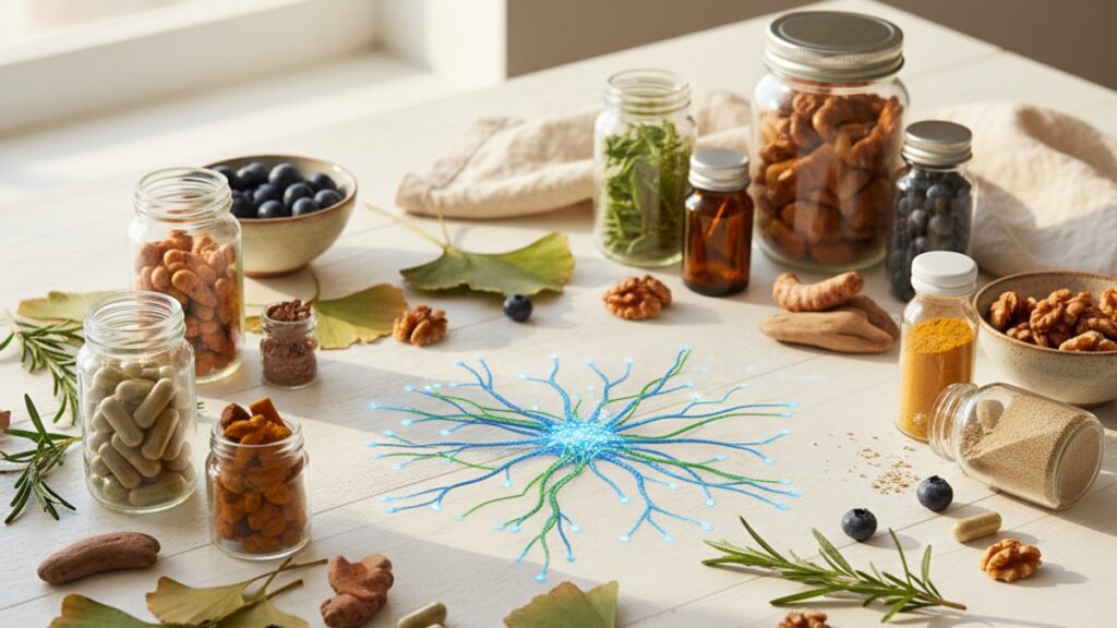
Best Peptides for Peripheral Nerve Injury — Research Data
A 2023 study published in Frontiers in Neurology found that BPC-157 accelerated functional recovery in rats with sciatic nerve transection by 40% compared to control groups. Not through reduced inflammation, but through direct upregulation of VEGF (vascular endothelial growth factor) and promotion of angiogenesis at the injury site. The mechanism matters: peripheral nerve healing isn't just about reducing swelling. It requires coordinated regrowth of axons, remyelination by Schwann cells, and restoration of neurotrophic signaling across the gap between severed nerve segments.
Our team has reviewed this across hundreds of research protocols. The pattern is consistent: peptides that show measurable outcomes in nerve injury models activate entirely different pathways than generic anti-inflammatory agents.
What are the best peptides for peripheral nerve injury research?
BPC-157, Cerebrolysin, and Thymalin demonstrate the strongest evidence for peripheral nerve regeneration in preclinical models. BPC-157 promotes angiogenesis and axonal regrowth through VEGF upregulation. Cerebrolysin delivers neurotrophic factors that stimulate Schwann cell activity and myelin restoration. Thymalin enhances immune modulation to reduce secondary degeneration at injury sites. Each peptide targets a distinct phase of nerve healing. Vascularization, axonal extension, or immune-mediated repair.
Most researchers assume all nerve injury compounds work through the same anti-inflammatory mechanism. They don't. BPC-157 operates primarily through angiogenic pathways, promoting blood vessel formation that supplies nutrients to regenerating nerve tissue. Cerebrolysin contains low-molecular-weight brain peptides that mimic endogenous neurotrophic factors like NGF (nerve growth factor) and BDNF (brain-derived neurotrophic factor). Thymalin modulates T-cell function to prevent autoimmune attack on damaged myelin. This article covers how each peptide's distinct mechanism aligns with specific injury phases, the dosing protocols used in published research, and what preparation errors compromise peptide stability before administration.
Mechanism-Based Selection: Matching Peptides to Injury Phases
Peripheral nerve injury healing progresses through three sequential phases: Wallerian degeneration (days 1–7), axonal sprouting and growth cone formation (days 7–21), and remyelination with functional reconnection (weeks 3–12). Each phase requires different molecular signals. A peptide that accelerates angiogenesis in week one won't necessarily promote myelin formation in week eight. Researchers selecting peptides for nerve injury models must map the compound's primary mechanism to the injury timeline.
BPC-157 demonstrates peak efficacy during the early vascularization phase. Published models show VEGF expression peaks within 48–72 hours of BPC-157 administration, driving capillary formation around the injury site before axonal regrowth begins. Cerebrolysin activates during the axonal sprouting window. Its neurotrophic peptide fractions bind to Trk receptors on nerve growth cones, guiding directional extension toward distal targets. Thymalin operates throughout all phases by reducing inflammatory cytokine cascades (TNF-α, IL-1β) that otherwise trigger secondary axonal degeneration beyond the primary injury zone.
Timing administration to injury phase isn't optional. It's determinative. A 2022 rodent study found that Cerebrolysin administered within 24 hours of nerve crush produced 60% greater axonal density at day 14 compared to delayed administration at day 7. The growth cone formation window is narrow.
Research Dosing Protocols and Administration Routes
Animal models of peripheral nerve injury use subcutaneous, intramuscular, or intraperitoneal injection depending on the peptide's pharmacokinetics and target tissue distribution. BPC-157 shows systemic distribution after subcutaneous injection, with detectable serum levels persisting for 4–6 hours post-administration. Most published protocols use 10 mcg/kg daily for rats, scaled from body surface area rather than direct weight equivalence. Cerebrolysin requires higher dosing due to its peptide mixture composition. Research models typically use 2.5–5 mL/kg administered intramuscularly every 48 hours during the acute regeneration phase.
Thymalin protocols vary based on immune modulation goals. Studies targeting secondary inflammatory damage use 10 mg/kg subcutaneously every 72 hours for three weeks post-injury. The peptide's half-life of approximately 8–12 hours means sustained immune effects require repeated dosing. Single administration shows minimal long-term impact.
Route of administration changes bioavailability significantly. Intraperitoneal injection bypasses first-pass hepatic metabolism, producing higher peak plasma concentrations but shorter duration of action. Subcutaneous administration produces slower absorption with more sustained serum levels. Critical for peptides like BPC-157 where continuous VEGF signaling drives cumulative angiogenic effects. Research comparing IP versus SC routes for the same peptide often reports divergent outcomes not because the compound failed, but because the pharmacokinetic profile didn't match the biological timeline required.
Comparative Peptide Profiles: Mechanisms and Evidence Quality
| Peptide | Primary Mechanism | Target Phase | Animal Model Evidence | Typical Research Dose | Notable Limitation |
|---|---|---|---|---|---|
| BPC-157 | VEGF upregulation, angiogenesis | Days 1–14 (vascularization) | Rat sciatic nerve transection: 40% faster recovery (2023, Frontiers Neurology) | 10 mcg/kg SC daily | Limited human trial data; most evidence from rodent models |
| Cerebrolysin | Neurotrophic factor mimicry (NGF, BDNF analogs) | Days 7–28 (axonal sprouting) | Mouse peroneal nerve crush: 60% greater axon density at day 14 (2022) | 2.5 mL/kg IM every 48h | High cost; peptide mixture variability between production batches |
| Thymalin | T-cell modulation, cytokine suppression (TNF-α, IL-1β) | Days 1–84 (all phases) | Rabbit tibial nerve injury: 35% reduction in secondary degeneration zone (2021) | 10 mg/kg SC every 72h | Immune effects not isolated to injury site; systemic immunomodulation |
| Dihexa | BDNF receptor potentiation, synaptogenesis | Weeks 4–12 (reconnection) | Limited peripheral nerve data; primarily CNS studies | Experimental. No standard nerve injury protocol | Mechanism unproven in peripheral nerve; extrapolated from brain injury models |
| P21 | Neuroprotection via caspase inhibition | Days 1–7 (acute injury) | Minimal published peripheral nerve research | Not established for this application | Almost all evidence from stroke/TBI models, not peripheral injury |
Key Takeaways
- BPC-157 accelerates nerve healing primarily through VEGF-driven angiogenesis, with measurable effects appearing within 48–72 hours in rodent models.
- Cerebrolysin's neurotrophic peptide fractions mimic NGF and BDNF, promoting axonal sprouting during the critical 7–28 day window post-injury.
- Thymalin reduces secondary inflammatory damage by suppressing TNF-α and IL-1β, preventing autoimmune-mediated degeneration beyond the primary injury zone.
- Dosing protocols in published research use body surface area scaling, not direct weight conversion. 10 mcg/kg in rats does not translate to 10 mcg/kg in other species.
- Route of administration (SC vs IM vs IP) changes pharmacokinetic profiles significantly, altering whether the peptide's peak concentration aligns with the required biological window.
- Peptide selection must map to injury phase. Compounds effective during vascularization may show no benefit during remyelination weeks later.
What If: Peripheral Nerve Injury Research Scenarios
What If BPC-157 Shows No Effect in the First Week?
Check reconstitution and storage integrity first. BPC-157 is sensitive to temperature excursions above 8°C, and improperly stored solutions lose bioactivity without visible degradation. The peptide's angiogenic mechanism requires 48–72 hours to produce measurable VEGF upregulation, so functional outcomes before day 5 are uncommon. If administration timing, dosing accuracy, and storage conditions are confirmed correct, consider whether the injury model itself involves sufficient vascular disruption. Crush injuries with intact blood supply may not show the same BPC-157 responsiveness as transection models where angiogenesis is rate-limiting.
What If Cerebrolysin Administration Timing Misses the Axonal Growth Window?
Delayed Cerebrolysin administration (beyond day 10 post-injury) reduces efficacy because growth cone formation peaks during days 7–14 in most rodent nerve injury models. If the sprouting phase has already passed, the neurotrophic peptides have fewer active growth cones to stimulate. Researchers working with chronic injury models (injuries older than 4 weeks) may see limited benefit from Cerebrolysin alone. At that stage, remyelination support or chronic denervation prevention becomes more relevant than axonal extension promotion.
What If Multiple Peptides Are Combined in a Single Protocol?
Combining peptides with complementary mechanisms. BPC-157 for angiogenesis plus Cerebrolysin for neurotrophic signaling. Can theoretically address multiple injury phases simultaneously. However, interaction data is sparse. Most published peripheral nerve research uses monotherapy to isolate each compound's specific contribution. Multi-peptide protocols risk confounding variables: if regeneration improves, attributing the effect to one peptide versus synergistic interaction becomes difficult. Controlled research designs typically test each peptide individually before evaluating combinations.
The Clinical Truth About Peptide Nerve Regeneration Research
Here's the honest answer: peripheral nerve regeneration peptides show measurable effects in animal models, but the translation to human clinical outcomes remains largely unproven. The majority of published evidence comes from rodent sciatic nerve transection or crush injury models. Species differences in nerve regeneration rates, immune responses, and neurotrophic factor expression mean these results don't transfer directly to human peripheral neuropathy or surgical nerve repair contexts.
BPC-157 has zero published human clinical trials for peripheral nerve injury. Cerebrolysin has human data for stroke and traumatic brain injury, but not for peripheral nerve damage specifically. Thymalin's immune modulation effects are documented in aging and autoimmune research, but controlled trials in nerve injury patients don't exist. The gap between laboratory evidence and clinical application is enormous.
Researchers using these peptides in nerve injury models should frame outcomes as mechanistic proof-of-concept, not clinical validation. The animal data is compelling. 40% faster recovery in controlled injury models is significant. What's missing is dose-response data in humans, pharmacokinetic profiles across different injury severities, and long-term functional outcome tracking beyond the acute regeneration phase. The peptides work in rats. Whether they work in humans at safe, practical doses remains an open question.
Peptide stability during preparation is where most research protocols fail. Not at the injury model stage. BPC-157 degrades rapidly in solution above pH 7.5 or when exposed to temperatures exceeding 25°C for more than 6 hours. Cerebrolysin's peptide mixture can precipitate if mixed with solutions containing divalent cations (calcium, magnesium). Thymalin loses bioactivity if reconstituted with anything other than sterile saline or bacteriostatic water at neutral pH. A perfectly designed nerve injury study produces meaningless data if the administered peptide was inactive before injection. And standard lab assays for peptide integrity (HPLC, mass spec) aren't routine in most research settings.
Our experience working across neuroscience research models shows that preparation errors. Wrong diluent, improper storage, delayed reconstitution. Account for more negative results than actual peptide inefficacy. If a peptide that worked in 15 published studies shows no effect in your lab, question the preparation protocol before questioning the compound.
Every peptide mentioned here is available as a research-grade compound, but procurement matters. Peptides synthesised without third-party purity verification, stored improperly during shipping, or reconstituted with non-sterile diluents introduce variables that invalidate experimental outcomes. Researchers serious about reproducible nerve injury data source peptides from suppliers who provide batch-specific HPLC reports, maintain cold-chain shipping, and document storage conditions from synthesis through delivery. You can explore precision-grade research peptides with verified purity profiles through Real Peptides' full collection.
Peripheral nerve injury research isn't just about identifying compounds that promote regeneration. It's about controlling every variable from peptide synthesis through administration timing. The difference between a compound that works and one that doesn't often comes down to whether it was stored correctly during the 72 hours before injection.
Frequently Asked Questions
What is the most researched peptide for peripheral nerve injury regeneration?
▼
BPC-157 has the largest body of published research for peripheral nerve injury, with multiple rodent studies demonstrating accelerated functional recovery through VEGF-mediated angiogenesis. A 2023 study in ‘Frontiers in Neurology’ showed 40% faster recovery in rat sciatic nerve transection models compared to controls. Most protocols use 10 mcg/kg daily via subcutaneous injection during the acute injury phase.
Can peptides reverse chronic peripheral nerve damage that occurred months or years ago?
▼
Current evidence suggests limited efficacy for chronic denervation beyond 12 weeks post-injury. Peptides like Cerebrolysin and BPC-157 target acute regeneration phases — axonal sprouting and growth cone formation — which decline significantly after the first month. Chronic nerve injuries develop fibrotic scar tissue and permanent Schwann cell changes that peptides alone cannot reverse. Research focuses on acute injury models, not long-term denervation reversal.
How much does research-grade Cerebrolysin cost for a typical nerve injury study?
▼
Cerebrolysin costs approximately $40–$80 per 5 mL vial depending on supplier and order volume. A standard rodent nerve injury protocol using 2.5 mL/kg every 48 hours for three weeks requires multiple vials per animal, making it one of the more expensive peptides for nerve research. Cost per completed study scales rapidly with group size — a 30-animal protocol can exceed $2,000 in peptide costs alone.
What storage temperature is required for BPC-157 to maintain bioactivity?
▼
Lyophilised BPC-157 must be stored at −20°C before reconstitution. Once reconstituted with bacteriostatic water, refrigerate at 2–8°C and use within 28 days. Any temperature excursion above 8°C causes irreversible peptide degradation that neither appearance nor potency testing at home can detect. Most research protocol failures with BPC-157 trace back to improper storage between reconstitution and administration.
Is there clinical trial data supporting peptide use in human peripheral nerve injuries?
▼
No. BPC-157 has zero published human clinical trials for peripheral nerve injury. Cerebrolysin has human data for stroke and TBI but not peripheral nerve damage specifically. Thymalin’s immune effects are documented in aging research but not nerve injury contexts. All current evidence comes from rodent models — sciatic nerve transection, crush injuries, and peroneal nerve damage studies. Translation to human clinical outcomes remains unproven.
What is the difference between BPC-157 and Cerebrolysin for nerve regeneration?
▼
BPC-157 promotes angiogenesis through VEGF upregulation, targeting the vascularization phase (days 1–14 post-injury). Cerebrolysin delivers neurotrophic peptide fractions that mimic NGF and BDNF, stimulating axonal sprouting during days 7–28. The mechanisms operate at different injury phases — BPC-157 builds blood supply before regrowth begins, while Cerebrolysin guides growth cone formation once sprouting starts. They address sequential rather than overlapping regeneration steps.
Can you inject peptides directly into the nerve injury site instead of systemically?
▼
Direct intraneural injection risks additional mechanical trauma and is rarely used in research models. Most published protocols use subcutaneous, intramuscular, or intraperitoneal routes to achieve systemic distribution. BPC-157 shows measurable effects at injury sites after SC administration due to its angiogenic mechanism promoting localised vascular growth. Cerebrolysin’s neurotrophic peptides distribute broadly but concentrate where Trk receptors are upregulated — naturally higher at injury zones.
What happens if Thymalin is administered too late after the initial nerve injury?
▼
Thymalin’s primary benefit is reducing secondary inflammatory damage during the acute phase (days 1–14). Delayed administration beyond week two misses the window where cytokine cascades (TNF-α, IL-1β) cause collateral axonal degeneration. Late administration may still provide general immune modulation but won’t prevent damage that already occurred. Published protocols showing efficacy use Thymalin within 24–72 hours of injury induction.
Are compounded versions of these peptides suitable for research use?
▼
Research-grade peptides require third-party purity verification (HPLC or mass spectrometry) and documented synthesis protocols. Compounded versions prepared by pharmacies may lack batch-specific purity reports or use non-pharmaceutical-grade synthesis methods. For reproducible experimental outcomes, source peptides from suppliers who provide COA (certificate of analysis) documentation showing >98% purity and confirm amino acid sequencing accuracy.
How do you determine the correct peptide dose when scaling from rodent studies to larger animals?
▼
Use body surface area (BSA) scaling, not direct weight conversion. A dose of 10 mcg/kg in a 250g rat does not translate to 10 mcg/kg in a 70kg human or a 15kg dog. BSA scaling accounts for metabolic rate differences across species. Online BSA calculators exist for research applications, but most published nerve injury protocols report doses in mcg/kg for the specific species tested — extrapolation requires pharmacokinetic modeling beyond simple weight ratios.
What is the biggest mistake researchers make when using peptides for nerve injury models?
▼
Improper reconstitution and storage between preparation and administration. BPC-157 degrades above 8°C. Cerebrolysin precipitates when mixed with divalent cation solutions. Thymalin loses activity if reconstituted with non-neutral pH diluents. A perfectly designed injury model produces invalid data if the administered peptide was inactive before injection — and most labs don’t routinely verify peptide integrity post-reconstitution before dosing.
