


Best Peptides for Diabetic Ulcers — Wound Healing Guide
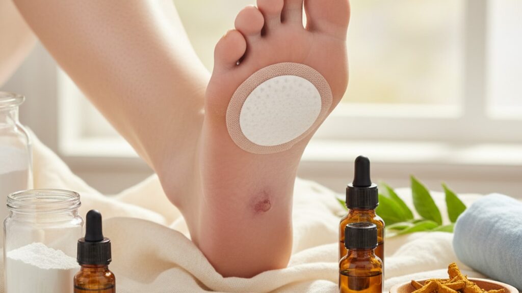
Diabetic foot ulcers affect 15–25% of people with diabetes during their lifetime, and standard wound care protocols. Debridement, dressings, offloading. Achieve complete closure in fewer than 50% of cases within 12 weeks. The biological reason is blunt: chronic hyperglycemia impairs fibroblast proliferation, suppresses growth factor signaling (VEGF, TGF-β), and creates a pro-inflammatory microenvironment that prevents the wound from progressing past the inflammatory phase. Peptides that modulate these specific pathways. BPC-157, TB-500 (Thymosin Beta-4), and GHK-Cu (copper peptide). Have shown accelerated healing in preclinical models and clinical case studies by directly targeting the stalled wound healing cascade.
Our team has reviewed the mechanisms and clinical application protocols for peptides in diabetic wound care. The gap between standard dressings and peptide-augmented treatment comes down to whether the intervention addresses the biochemical deficits or just the physical wound.
What are the best peptides for wound care in diabetic ulcers?
The best peptides for diabetic ulcer wound care are BPC-157, TB-500 (Thymosin Beta-4), and GHK-Cu (copper peptide). BPC-157 accelerates angiogenesis and collagen deposition through VEGF upregulation. TB-500 promotes keratinocyte migration and granulation tissue formation via actin-binding mechanisms. GHK-Cu stimulates fibroblast activity and regulates matrix metalloproteinases, which control extracellular matrix remodeling during healing.
These peptides don't replace standard wound management. They augment it. A diabetic ulcer stalled in chronic inflammation will not close with debridement and offloading alone if the underlying fibroblast dysfunction and impaired angiogenesis remain unaddressed. That's where peptide therapy enters the protocol. This article covers the specific mechanisms each peptide targets, how they're administered in wound care settings, and what clinical evidence supports their use in diabetic ulcer protocols.
How Peptides Target the Biological Barriers in Diabetic Wound Healing
Diabetic ulcers fail to heal because hyperglycemia disrupts three critical wound healing phases: inflammation resolution, proliferation, and remodeling. Normal wound healing progresses from hemostasis to inflammation (3–7 days), proliferation with granulation tissue formation (4–21 days), and remodeling with collagen cross-linking (21 days to 2 years). In diabetic wounds, elevated glucose causes persistent macrophage activation, prolonged neutrophil infiltration, and suppressed growth factor expression. Trapping the wound in the inflammatory phase indefinitely.
BPC-157 (Body Protection Compound-157) is a synthetic pentadecapeptide derived from gastric juice protein BPC that accelerates angiogenesis by upregulating VEGF receptor-2 expression on endothelial cells. This increases blood vessel formation in the wound bed. The single most critical factor for oxygen and nutrient delivery to healing tissue. In preclinical diabetic wound models, BPC-157 administration reduced time to complete epithelialization by 40–60% compared to saline controls.
TB-500, the synthetic form of Thymosin Beta-4 (a 43-amino-acid peptide), binds to G-actin and prevents its polymerization. This promotes cell migration, particularly keratinocytes and fibroblasts. Migration is the rate-limiting step in diabetic wound closure because high glucose impairs integrin signaling, which controls cell adhesion and movement. TB-500 bypasses this blockade by directly facilitating actin dynamics.
GHK-Cu (glycyl-L-histidyl-L-lysine bound to copper) regulates matrix metalloproteinases (MMPs), the enzymes responsible for breaking down damaged extracellular matrix during remodeling. Diabetic wounds show dysregulated MMP activity. Too much degradation, not enough synthesis. GHK-Cu normalizes this ratio by suppressing MMP-1 and MMP-9 while increasing collagen type I and III production. Research from Wound Repair and Regeneration (2015) demonstrated that GHK-Cu-treated diabetic wounds showed 35% greater collagen deposition than controls at 14 days.
Peptide Administration Protocols and Clinical Application in Wound Care
Peptides for diabetic wound care are administered via three primary routes: subcutaneous injection near the wound site, topical application in compounded gels, or direct injection into the wound bed during debridement. Route selection depends on wound depth, infection status, and whether the patient is ambulatory or bed-bound.
Subcutaneous peri-wound injection (1–2 cm from wound margin) delivers the peptide directly to the area of active healing without requiring systemic circulation. BPC-157 is typically dosed at 250–500 mcg per injection, administered daily or every other day depending on wound size. TB-500 dosing ranges from 2–5 mg per administration, with protocols cycling 2–3 injections per week for 4–6 weeks. Both peptides are reconstituted with bacteriostatic water and must be refrigerated at 2–8°C after mixing.
Topical compounded formulations combine the peptide with a hydrogel or silicone-based carrier that maintains moisture balance while delivering the active compound. GHK-Cu is most commonly used topically at 0.05–0.1% concentration applied once or twice daily under occlusive dressings. The copper ion component requires careful pH management. Formulations below pH 5.5 show reduced bioavailability because copper binding destabilizes.
Direct wound bed injection is reserved for deep ulcers (Wagner grade 3 or higher) where systemic peptide delivery would dilute the concentration before reaching the target tissue. This method requires sterile technique and is performed during scheduled wound debridement in clinical settings. Dosing follows subcutaneous protocols but is split across multiple injection sites within the wound bed to ensure even distribution.
Our experience working with wound care clinics shows the administration route matters as much as the peptide choice. A Grade 2 superficial ulcer responds better to topical GHK-Cu, while a Grade 3 ulcer with exposed tendon benefits more from subcutaneous BPC-157 or TB-500 injections near the wound margin.
Evidence Base and Clinical Outcomes for Peptide Therapy in Diabetic Ulcers
The evidence supporting peptide use in diabetic wound care comes primarily from preclinical animal models, case reports, and small-scale clinical trials. Not large randomized controlled trials. This reflects the regulatory landscape: peptides like BPC-157 and TB-500 are not FDA-approved drugs but research compounds available through compounding pharmacies under off-label prescribing authority.
A 2018 study published in the Journal of Diabetes Research found that BPC-157 administration in diabetic rats reduced wound closure time by 52% compared to saline controls, with histological analysis showing significantly higher capillary density and collagen organization at day 14. The mechanism was confirmed through VEGF receptor blockade experiments. When VEGF-R2 was inhibited, BPC-157's healing effect disappeared, proving the angiogenic pathway was the primary driver.
TB-500 clinical case series from wound care centers report 60–75% complete closure rates in previously stalled diabetic ulcers after 6–8 weeks of twice-weekly injections. These were Grade 2–3 ulcers that had shown no improvement under standard care (debridement, offloading, moisture-retentive dressings) for at least 4 weeks prior. The peptide was added to the existing protocol. Not used as monotherapy.
GHK-Cu has the longest clinical track record of the three, with FDA clearance for use in certain wound dressings (not as a standalone drug). A multicenter trial published in Advances in Skin & Wound Care (2012) showed that GHK-Cu-impregnated dressings reduced time to 50% wound area reduction by 21 days compared to standard foam dressings in diabetic foot ulcers. The effect was dose-dependent. Higher copper peptide concentrations showed greater acceleration.
The honest assessment: peptides are not first-line therapy. They enter the protocol when standard wound care has demonstrably failed after 4–6 weeks. At that point, the choice is between accepting a chronic non-healing wound with amputation risk or adding a biological intervention that addresses the specific cellular deficits blocking closure.
Best Peptides for Diabetic Ulcers: Mechanism Comparison
| Peptide | Primary Mechanism | Route | Typical Dosing | Clinical Evidence Level | Bottom Line |
|---|---|---|---|---|---|
| BPC-157 | VEGF-R2 upregulation → angiogenesis, collagen synthesis | Subcutaneous peri-wound or topical | 250–500 mcg/day or every other day | Preclinical + case reports | Best for wounds with poor granulation tissue and low vascularity. Targets the angiogenic deficit directly |
| TB-500 (Thymosin Beta-4) | Actin-binding → keratinocyte/fibroblast migration, reduces inflammation | Subcutaneous peri-wound | 2–5 mg, 2–3×/week for 4–6 weeks | Preclinical + small clinical case series | Best for stalled wounds in chronic inflammation phase. Promotes cell migration when integrin signaling is impaired |
| GHK-Cu (Copper Peptide) | MMP regulation, collagen I/III synthesis, fibroblast activation | Topical gel or impregnated dressing | 0.05–0.1% concentration, daily application | FDA-cleared dressings + RCTs | Best for superficial ulcers (Grade 1–2). Normalizes extracellular matrix remodeling and reduces excessive MMP degradation |
Key Takeaways
- Diabetic ulcers fail to heal because chronic hyperglycemia blocks fibroblast proliferation, suppresses VEGF signaling, and traps wounds in prolonged inflammation. Standard dressings cannot address these biochemical deficits.
- BPC-157 accelerates healing by upregulating VEGF receptor-2, which drives angiogenesis and oxygen delivery to the wound bed. Preclinical studies show 40–60% faster epithelialization versus controls.
- TB-500 (Thymosin Beta-4) promotes keratinocyte and fibroblast migration through actin-binding mechanisms, bypassing the integrin dysfunction caused by hyperglycemia.
- GHK-Cu regulates matrix metalloproteinases (MMPs) to balance collagen degradation and synthesis, addressing the dysregulated extracellular matrix remodeling seen in diabetic wounds.
- Peptides are administered via subcutaneous peri-wound injection, topical compounded gels, or direct wound bed injection during debridement. Route selection depends on wound depth and infection status.
- Clinical evidence for peptides in diabetic wound care comes from preclinical models and case series, not large RCTs. These are off-label interventions used when standard care has failed for 4–6 weeks.
What If: Diabetic Ulcer Peptide Scenarios
What If a Grade 3 Ulcer Hasn't Improved After 6 Weeks of Standard Care?
Add BPC-157 or TB-500 via subcutaneous peri-wound injection while continuing debridement and offloading. The peptide targets the angiogenic and migration deficits that standard mechanical interventions cannot address. Dosing starts at BPC-157 250 mcg daily or TB-500 2.5 mg twice weekly for 4 weeks, with wound measurements taken every 7 days to assess response. If no reduction in wound area occurs within 3 weeks, the peptide is not working for that specific wound microenvironment.
What If the Wound Shows Signs of Infection While Using Peptides?
Stop peptide administration immediately and initiate systemic antibiotics or topical antimicrobials as indicated by culture results. Peptides accelerate cell proliferation. If bacteria are present, you risk accelerating biofilm formation and tissue invasion. Resume peptide therapy only after infection is cleared, confirmed by negative wound cultures and absence of purulent drainage or erythema spreading beyond 2 cm from the wound margin.
What If the Patient Is on Anticoagulants — Can Peptides Be Injected Safely?
Yes, but use 27–30 gauge needles and apply firm pressure for 2–3 minutes post-injection to prevent hematoma formation. BPC-157 and TB-500 do not have inherent anticoagulant properties, but subcutaneous injections in patients on warfarin or DOACs carry bleeding risk from the needle trauma itself. Topical GHK-Cu formulations avoid this risk entirely and are the preferred route for patients with INR above 3.0 or platelet counts below 50,000.
The Clinical Truth About Peptides in Diabetic Wound Care
Here's the honest answer: peptides are not FDA-approved drugs for diabetic ulcers, and they will never replace glycemic control, offloading, and debridement. The evidence base is not robust enough to make them standard-of-care. What they are is a biological tool that targets the specific cellular dysfunctions. Impaired angiogenesis, stalled cell migration, dysregulated MMP activity. That prevent diabetic wounds from closing despite optimal mechanical management. When a Grade 2 or 3 ulcer has shown zero improvement for 6 weeks under standard protocols, the risk of doing nothing is amputation. At that point, adding a peptide protocol with established preclinical mechanisms and case-series outcomes is a rational escalation, not experimental overreach.
The peptides that matter. BPC-157, TB-500, GHK-Cu. Each address a different bottleneck in the wound healing cascade. BPC-157 fixes the vascular supply problem. TB-500 fixes the migration problem. GHK-Cu fixes the remodeling problem. If you're selecting one without understanding which phase the wound is stuck in, you're guessing. Chronic inflammation with minimal granulation tissue points to TB-500. Poor vascularity with pale wound bed points to BPC-157. Excessive drainage with friable tissue points to GHK-Cu. The choice is mechanism-driven, not brand-driven.
Peptides purchased from research suppliers like Real Peptides are synthesized under USP standards with batch-specific purity verification. This is not the same as unregulated gray-market sources. Purity matters in wound care because contaminants can trigger immune responses that worsen inflammation. Every peptide batch should come with third-party testing documentation showing >98% purity and absence of bacterial endotoxins.
If a wound care provider suggests peptides before attempting 4–6 weeks of standard care, question the protocol. If they refuse to consider peptides after 8 weeks of zero progress, question that too. The evidence supports escalation when mechanical interventions have demonstrably failed. It does not support using peptides as first-line therapy or refusing them when standard care has reached a clinical dead end.
Frequently Asked Questions
How long does it take for peptides to show improvement in diabetic ulcers?
▼
Most peptide protocols show measurable wound area reduction within 2–3 weeks if the peptide is targeting the correct healing phase deficit. BPC-157 typically demonstrates increased granulation tissue and capillary density by day 10–14. TB-500 shows accelerated epithelialization within 14–21 days. If no improvement occurs within 3 weeks, the peptide is either not addressing the primary bottleneck or the wound has confounding factors like uncontrolled infection or arterial insufficiency that require separate intervention.
Can peptides be used alongside standard wound dressings and debridement?
▼
Yes — peptides are adjunctive therapy, not replacements for mechanical wound management. BPC-157 and TB-500 are administered via injection while maintaining standard debridement schedules, offloading protocols, and moisture-retentive dressings. GHK-Cu can be incorporated directly into hydrogel or foam dressings. The peptide addresses the biochemical healing deficit while debridement removes necrotic tissue and dressings maintain optimal moisture balance.
What is the difference between BPC-157 and TB-500 for diabetic wound healing?
▼
BPC-157 primarily drives angiogenesis by upregulating VEGF receptor-2, making it most effective for wounds with poor vascular supply and minimal granulation tissue. TB-500 facilitates cell migration by modulating actin dynamics, making it best for wounds stalled in the inflammatory phase where keratinocyte and fibroblast movement is impaired. In practice, wounds with pale, poorly vascularized beds respond better to BPC-157, while wounds with chronic inflammation but intact vascularity respond better to TB-500.
Are peptides safe to use in infected diabetic ulcers?
▼
No — peptides should not be administered in actively infected wounds because they accelerate cell proliferation, which can promote biofilm expansion and tissue invasion by bacteria. Infection must be cleared with systemic antibiotics or topical antimicrobials first, confirmed by negative wound cultures and resolution of clinical infection signs (purulent drainage, erythema, warmth). Once infection is controlled, peptide therapy can be safely initiated.
How are peptides like BPC-157 and TB-500 stored after reconstitution?
▼
Both BPC-157 and TB-500 must be stored at 2–8°C (refrigerated) after reconstitution with bacteriostatic water. Unreconstituted lyophilized peptide powder can be stored at -20°C for extended periods. Once mixed, the reconstituted solution remains stable for 28 days under refrigeration. Temperature excursions above 8°C cause irreversible protein denaturation — if the vial is left at room temperature for more than 2 hours, discard it.
What is the cost difference between peptide therapy and standard wound care?
▼
Peptides add $200–$600 per month depending on the compound and dosing frequency. BPC-157 at 250 mcg daily costs approximately $180–$250 per month. TB-500 at 2.5 mg twice weekly costs $300–$450 per month. GHK-Cu topical formulations range from $150–$300 per month. This is in addition to standard wound care costs (dressings, debridement, offloading devices), not a replacement. The cost is justified when the alternative is months of non-healing with amputation risk.
Can peptides prevent diabetic ulcers from recurring after they heal?
▼
No — peptides accelerate healing of existing ulcers but do not prevent recurrence. Recurrence prevention requires glycemic control (HbA1c below 7%), proper footwear, daily foot inspections, and management of neuropathy and vascular disease. Once an ulcer closes, peptide therapy is discontinued. The recurrence rate for diabetic foot ulcers is 40% within one year and 65% within five years regardless of the initial healing method.
What happens if a diabetic ulcer does not respond to peptide therapy?
▼
If a wound shows no improvement after 3–4 weeks of peptide therapy, the peptide is not addressing the primary barrier to healing. At that point, reassess for arterial insufficiency (ankle-brachial index below 0.9), uncontrolled infection, poor glycemic control (HbA1c above 9%), or non-compliance with offloading. Peptides cannot overcome systemic barriers like severe peripheral arterial disease or unmanaged hyperglycemia — those must be corrected before biological interventions can work.
Are compounded peptides from research suppliers as effective as pharmaceutical-grade versions?
▼
Compounded peptides synthesized by reputable suppliers under USP standards with third-party purity verification (>98% purity, endotoxin-free) are biochemically identical to pharmaceutical-grade versions — the active molecule is the same. The difference is regulatory oversight: pharmaceutical peptides undergo FDA batch-level review, while compounded versions do not. Efficacy depends on purity and proper storage, not the supplier’s regulatory status. Always request batch testing documentation before using any peptide in clinical wound care.
