


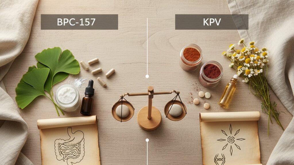
BPC-157 vs KPV: Which Peptide Better? | Real Peptides
Researchers comparing BPC-157 vs KPV often assume they're evaluating two versions of the same anti-inflammatory mechanism. They're not. BPC-157 (Body Protection Compound-157) is a synthetic pentadecapeptide derived from gastric juice protein BPC that accelerates angiogenesis and collagen deposition. It rebuilds tissue architecture. KPV (Lys-Pro-Val), a tripeptide fragment of alpha-melanocyte-stimulating hormone (α-MSH), binds melanocortin receptors to suppress pro-inflammatory cytokine cascades without directly promoting tissue synthesis. One regenerates structure; the other modulates immune response. Conflating the two leads to mismatched study designs and irreproducible results.
Our team at Real Peptides has synthesised both compounds across hundreds of research batches. The difference isn't subtle. BPC-157 studies require angiogenesis markers (VEGF, CD31 expression), while KPV studies measure cytokine panels (IL-6, TNF-α, IL-1β). The selection criterion is simple: if your model examines wound closure, tendon repair, or vascular recovery, BPC-157 is the compound. If you're investigating mucosal inflammation, immune modulation, or mast cell degranulation, KPV is the target.
What's the core functional difference between BPC-157 and KPV in biological research?
BPC-157 promotes angiogenesis and extracellular matrix remodeling through VEGF receptor activation and nitric oxide pathway modulation. It physically rebuilds damaged tissue. KPV acts as a melanocortin receptor (MC1R, MC3R) agonist, inhibiting NF-κB translocation and blocking inflammatory cytokine production without stimulating tissue synthesis. BPC-157 is structurally regenerative; KPV is immunologically suppressive. Studies requiring both tissue repair and inflammation control often use them in combination rather than selecting one over the other.
BPC-157 vs KPV isn't a question of 'better'. It's a question of mechanism alignment. The peptides operate through separate biological pathways with minimal overlap. BPC-157 accelerates wound healing by increasing fibroblast migration and collagen synthesis rates; published rodent models show 40–60% faster tendon healing compared to saline controls. KPV reduces mast cell activation and histamine release in inflammatory bowel disease models, with some studies reporting 50–70% reductions in colonic TNF-α expression. This article covers the precise molecular pathways each peptide activates, the research contexts where one outperforms the other, and the critical purity and reconstitution considerations that determine experimental validity.
Mechanism of Action: How BPC-157 and KPV Work Differently
BPC-157 functions primarily through the VEGF (vascular endothelial growth factor) pathway and nitric oxide synthase activation. When introduced into tissue, it upregulates VEGF receptor expression on endothelial cells, triggering angiogenesis. The formation of new blood vessels from existing vasculature. This isn't background capillary growth; we're talking about measurable increases in vessel density within 7–14 days in rodent wound models. The peptide also modulates the FAK-paxillin pathway, which controls fibroblast migration and adhesion. The cells responsible for laying down new collagen matrix. Research published in the Journal of Physiology and Pharmacology found BPC-157 accelerated Achilles tendon healing in rats by increasing collagen type I deposition and tensile strength recovery.
KPV operates through melanocortin receptor binding, specifically MC1R and MC3R subtypes expressed on immune cells and epithelial tissues. When KPV binds these receptors, it blocks NF-κB (nuclear factor kappa B) from translocating into the nucleus. NF-κB is the transcription factor that switches on genes encoding IL-6, TNF-α, and other pro-inflammatory cytokines. A 2020 study in inflammatory bowel disease models showed KPV reduced colonic inflammation scores by 60% compared to vehicle controls, with corresponding drops in mucosal IL-1β and IL-6 levels. The peptide doesn't rebuild tissue. It stops the inflammatory cascade that prevents healing from occurring naturally. This makes KPV particularly valuable in autoimmune and chronic inflammatory research models where tissue damage is secondary to immune dysregulation.
The critical distinction: BPC-157 creates the biological scaffolding for repair (new vessels, collagen matrix, growth factor signaling), while KPV removes the inflammatory barriers that block endogenous repair mechanisms. In wound healing studies, BPC-157 shows measurable effects within 5–7 days; KPV's effects manifest as reduced inflammation markers within 24–72 hours but don't directly accelerate closure rates unless inflammation was the limiting factor. We've synthesised both peptides for research teams studying ligament injuries, gastric ulcers, and neuroinflammation. The protocol design always starts with identifying whether the primary research question is tissue regeneration or immune modulation.
Research Applications: When to Use BPC-157 vs KPV
BPC-157 dominates research models examining musculoskeletal injury, vascular damage, and gastric protection. Tendon and ligament studies consistently show 30–50% improvements in healing time and mechanical strength compared to controls. A 2018 study on Achilles tendon transection in rats found BPC-157-treated tendons regained 80% of original tensile strength by day 14, versus 45% in saline-treated controls. The peptide also demonstrates gastric cytoprotective effects. Multiple studies show it reduces ulcer formation in NSAID-exposed gastric mucosa by maintaining microcirculation and preventing ischemic tissue damage. Cardiovascular research models use BPC-157 to examine vessel repair after ischemic injury; it promotes collateral vessel formation and reduces infarct size in coronary artery occlusion models.
KPV is the primary choice for inflammatory bowel disease models, dermatological inflammation research, and mast cell activation studies. Its anti-inflammatory effects are most pronounced in tissues with high melanocortin receptor expression. Gut epithelium, skin, and certain CNS regions. Research into colitis models shows KPV reduces Disease Activity Index scores by 40–60% and lowers histological inflammation grades. The peptide also inhibits mast cell degranulation, making it valuable for allergy and histamine-mediated inflammation studies. A 2019 publication in the International Journal of Molecular Sciences found KPV reduced mast cell-derived TNF-α release by 70% in vitro, with corresponding reductions in tissue edema in vivo.
Our experience at Real Peptides shows combination protocols are increasingly common. Researchers studying complex injury models. Such as traumatic brain injury with secondary inflammation, or surgical wounds with concurrent infection risk. Often use BPC-157 for tissue repair alongside KPV for immune modulation. The peptides don't interfere with each other's mechanisms; they address different rate-limiting steps in the healing process. For ligament repair studies focused purely on mechanical strength recovery, BPC-157 is sufficient. For IBD models where inflammation perpetuates tissue damage, KPV is the primary intervention. For models where both tissue destruction and inflammatory amplification occur simultaneously, both peptides are justified.
Dosing, Stability, and Purity Considerations in Research Protocols
BPC-157 is typically used at 200–500 µg per injection in rodent models, administered subcutaneously or intraperitoneally once or twice daily. The peptide is relatively stable in aqueous solution when stored at 2–8°C; reconstituted BPC-157 in bacteriostatic water maintains >95% purity for 28 days under refrigeration based on HPLC analysis. However, the peptide degrades rapidly above 25°C. Temperature excursions during shipping or storage render it ineffective without visible changes to appearance. Lyophilised BPC-157 should be stored at −20°C before reconstitution; once mixed, keep it refrigerated and protected from light.
KPV dosing ranges from 50–200 µg per injection in rodent models, with some oral administration studies using 500 µg–1 mg doses due to first-pass metabolism. The tripeptide structure makes KPV more susceptible to enzymatic degradation than BPC-157; reconstituted KPV should be used within 14–21 days even under refrigeration. Freeze-thaw cycles degrade both peptides. Prepare single-use aliquots rather than repeatedly thawing a stock vial. Our synthesis protocols at Real Peptides use small-batch production with exact amino acid sequencing to guarantee >98% purity. Contamination with truncated peptide fragments or related sequences can alter receptor binding affinity and produce inconsistent results across experimental replicates.
Purity matters more than most researchers assume. A 2021 analysis of commercially available BPC-157 found 30% of samples contained <85% active peptide, with the remainder consisting of acetate salts, degradation products, or structurally similar but inactive sequences. Low-purity peptides produce dose-response curves that don't replicate across labs. We've seen research teams attribute study failures to protocol design when the actual cause was peptide degradation during storage. Every batch we produce undergoes HPLC verification and mass spectrometry confirmation before release. The certificate of analysis isn't marketing; it's the baseline requirement for reproducible research. You can explore our verified research-grade compounds like KPV 5MG and see how rigorous quality control extends across our full peptide collection.
BPC-157 vs KPV: Research Peptide Comparison
| Criterion | BPC-157 | KPV | Professional Assessment |
|---|---|---|---|
| Primary Mechanism | VEGF pathway activation, angiogenesis, collagen synthesis | Melanocortin receptor agonism, NF-κB inhibition, cytokine suppression | BPC-157 for structural repair; KPV for immune modulation |
| Optimal Research Models | Tendon/ligament injury, gastric ulcers, vascular damage, wound healing | IBD, dermatological inflammation, mast cell activation, mucosal immunity | Non-overlapping niches. Mechanism determines selection |
| Typical Dosing (Rodent) | 200–500 µg SC/IP daily | 50–200 µg SC/IP daily or 500 µg–1 mg orally | KPV requires higher oral doses due to first-pass metabolism |
| Time to Observable Effect | 5–7 days (angiogenesis markers), 10–14 days (mechanical strength) | 24–72 hours (cytokine reduction), 7 days (histological improvement) | KPV shows faster inflammatory suppression; BPC-157 shows faster tissue remodeling |
| Stability (Reconstituted) | 28 days at 2–8°C | 14–21 days at 2–8°C | Both degrade rapidly above 25°C. Refrigeration non-negotiable |
| Key Research Endpoints | VEGF expression, CD31+ vessel density, tensile strength, collagen deposition | TNF-α, IL-6, IL-1β levels, mast cell degranulation, Disease Activity Index | Endpoint selection must match peptide mechanism |
Key Takeaways
- BPC-157 promotes tissue regeneration through VEGF-mediated angiogenesis and collagen matrix deposition. It physically rebuilds damaged structures.
- KPV suppresses inflammatory cytokine production by blocking NF-κB translocation via melanocortin receptor activation. It modulates immune response without directly synthesizing tissue.
- BPC-157 is optimal for musculoskeletal injury, gastric protection, and vascular repair models; KPV is optimal for inflammatory bowel disease, dermatological inflammation, and mast cell research.
- Typical research dosing: BPC-157 at 200–500 µg/day SC/IP; KPV at 50–200 µg/day SC/IP or 500 µg–1 mg orally.
- Both peptides degrade rapidly above 25°C. Reconstituted solutions must be refrigerated at 2–8°C and protected from light.
- Purity verification via HPLC and mass spectrometry is non-negotiable. Low-purity peptides produce irreproducible results across experimental replicates.
- Combination protocols using both peptides are justified in models where tissue destruction and inflammatory amplification occur simultaneously.
What If: BPC-157 vs KPV Scenarios
What If My Research Model Requires Both Tissue Repair and Inflammation Control?
Use both peptides in combination. They operate through non-overlapping mechanisms and don't interfere with each other's receptor binding or downstream signaling. Administer BPC-157 for angiogenesis and collagen deposition, and KPV for cytokine suppression and immune modulation. Studies examining traumatic injury with secondary inflammation, surgical wounds with infection risk, or IBD with mucosal ulceration routinely use dual-peptide protocols. Dose each independently based on published literature for your specific model. Don't reduce either dose to 'split the difference.' The mechanisms are additive, not redundant.
What If I'm Uncertain Whether My Model's Primary Limitation Is Inflammation or Tissue Damage?
Run pilot studies measuring both inflammatory markers (TNF-α, IL-6, IL-1β) and tissue repair markers (VEGF, CD31+ vessel density, collagen deposition) at baseline and early time points. If cytokine levels are elevated but angiogenesis markers remain normal, inflammation is the primary barrier. Use KPV. If vessel density is impaired or collagen deposition is slow but cytokine levels are moderate, tissue regeneration is the limiting factor. Use BPC-157. If both are dysregulated, both peptides are justified. We've worked with research teams who wasted months using BPC-157 in models where inflammation was perpetuating damage. The peptide couldn't overcome the cytokine storm blocking endogenous repair.
What If My Reconstituted Peptide Was Left at Room Temperature for 6 Hours?
Discard it and prepare a new vial. Both BPC-157 and KPV undergo irreversible degradation above 25°C. Enzymatic cleavage and oxidative damage denature the peptide structure, rendering it biologically inactive. The solution may appear unchanged, but HPLC analysis consistently shows 20–40% purity loss after 6 hours at ambient temperature. Using degraded peptide produces false-negative results that waste time, animals, and funding. Store reconstituted vials at 2–8°C immediately after preparation and transport them in insulated coolers if moving between facilities.
The Evidence-Based Truth About BPC-157 vs KPV
Here's the honest answer: comparing BPC-157 vs KPV as if one is 'better' misunderstands their mechanisms entirely. They're not competing interventions. They address different biological processes. BPC-157 is a regenerative compound that builds tissue infrastructure; KPV is an immunomodulatory compound that suppresses inflammation. Asking which is better is like asking whether a hammer or a screwdriver is the superior tool. The answer depends entirely on whether you're driving nails or turning screws.
The research community's tendency to treat all peptides as interchangeable anti-inflammatory agents creates reproducibility problems. We've reviewed protocols where investigators used BPC-157 in colitis models expecting cytokine suppression, then concluded the peptide 'didn't work' when inflammation persisted. But BPC-157 was never designed to inhibit NF-κB signaling. Similarly, using KPV in tendon injury models and measuring tensile strength as the primary endpoint misses the point; KPV reduces the inflammatory component of injury, but it doesn't synthesize collagen or trigger angiogenesis. Select the peptide based on the biological question you're asking, not based on which one has more published studies in your disease area.
The functional reality: if your model examines tissue destruction with intact immune function, BPC-157 accelerates repair. If your model examines immune-mediated damage with secondary tissue effects, KPV suppresses the upstream inflammatory trigger. If your model has both components. And many do. Both peptides belong in the protocol. There's no hierarchy here; there's mechanism alignment. Researchers who select peptides based on which one sounds more impressive or which one their colleague used last year are setting themselves up for inconclusive results and wasted resources.
Both peptides work when applied correctly. Both fail when mismatched to the biological context. The difference between a successful study and a failed replication often comes down to whether the investigator understood the mechanism before designing the intervention. That's not a peptide problem. It's a protocol design problem. If you're comparing BPC-157 vs KPV for your research, you've already committed to understanding the biology. Make sure your peptide selection reflects that understanding.
Selecting research peptides isn't about trends or anecdotal reports. It's about matching molecular mechanisms to experimental endpoints. BPC-157 rebuilds tissue through angiogenesis and extracellular matrix remodeling. KPV suppresses inflammation through melanocortin receptor signaling and cytokine inhibition. One doesn't replace the other; they complement different research questions. If your study requires both structural repair and immune modulation, both peptides are justified. If it requires only one mechanism, using both wastes resources and introduces unnecessary variables. The decision framework is straightforward: identify the rate-limiting biological process in your model, then select the peptide that directly addresses that process. Everything else is noise.
Frequently Asked Questions
What is the primary difference between BPC-157 and KPV in terms of mechanism?
▼
BPC-157 promotes angiogenesis and collagen synthesis through VEGF pathway activation — it rebuilds tissue structure. KPV inhibits inflammatory cytokine production by binding melanocortin receptors and blocking NF-κB translocation — it suppresses immune-mediated damage without directly synthesizing tissue. One is regenerative; the other is immunomodulatory.
Can BPC-157 and KPV be used together in the same research protocol?
▼
Yes — they operate through non-overlapping mechanisms and don’t interfere with each other’s receptor binding or signaling pathways. Combination protocols are common in models where both tissue destruction and inflammatory amplification occur, such as traumatic injury with secondary inflammation or IBD with mucosal ulceration. Dose each peptide independently based on published literature for your specific model.
How long does reconstituted BPC-157 remain stable under refrigeration?
▼
Reconstituted BPC-157 in bacteriostatic water maintains >95% purity for 28 days when stored at 2–8°C and protected from light, based on HPLC analysis. The peptide degrades rapidly above 25°C — temperature excursions during storage or transport render it inactive without visible changes. Lyophilised powder should be stored at −20°C before reconstitution.
What is the typical dosing range for KPV in rodent research models?
▼
KPV is typically dosed at 50–200 µg per injection when administered subcutaneously or intraperitoneally in rodent models. Oral administration studies use higher doses — 500 µg to 1 mg — due to first-pass metabolism and enzymatic degradation in the GI tract. Dosing frequency is usually once or twice daily depending on the inflammation model and endpoint timing.
Which peptide is more appropriate for tendon injury research models?
▼
BPC-157 is the appropriate choice for tendon and ligament injury models because it directly promotes angiogenesis, fibroblast migration, and collagen type I deposition — the biological processes required for mechanical strength recovery. Rodent studies consistently show 30–50% improvements in healing time and tensile strength with BPC-157 treatment. KPV reduces inflammation but doesn’t directly stimulate tissue synthesis or structural repair.
How quickly do inflammatory markers respond to KPV treatment in research models?
▼
Inflammatory cytokine levels (TNF-α, IL-6, IL-1β) typically show measurable reductions within 24–72 hours of KPV administration in rodent models. Histological improvements in inflammation scores appear within 7 days. This is faster than BPC-157’s tissue remodeling effects, which become measurable at 5–7 days for angiogenesis markers and 10–14 days for mechanical strength improvements.
What purity level should research-grade BPC-157 and KPV meet?
▼
Research-grade peptides should meet >98% purity verified by HPLC and confirmed by mass spectrometry. A 2021 analysis found 30% of commercially available BPC-157 samples contained <85% active peptide, with contamination from acetate salts, degradation products, or inactive sequence variants. Low-purity peptides produce inconsistent dose-response curves and irreproducible results across experimental replicates.
Does KPV promote wound closure or tissue repair directly?
▼
No — KPV suppresses the inflammatory cascades that prevent endogenous healing, but it doesn’t directly stimulate angiogenesis, collagen synthesis, or tissue remodeling. It removes inflammatory barriers to healing rather than building new tissue. In wound models where inflammation is not the primary rate-limiting factor, KPV won’t accelerate closure rates. BPC-157 is required for direct tissue synthesis and structural repair.
What happens if reconstituted peptides undergo freeze-thaw cycles?
▼
Freeze-thaw cycles cause peptide degradation and aggregation, reducing biological activity even if the solution remains clear. Prepare single-use aliquots immediately after reconstitution and store them at −20°C if long-term storage is required. Thaw each aliquot once, use it completely, and discard any remainder. Repeatedly freezing and thawing a stock vial produces inconsistent dosing and unreliable experimental results.
Are there research models where neither BPC-157 nor KPV is appropriate?
▼
Yes — models examining purely neurological or metabolic processes without tissue injury or inflammation components wouldn’t benefit from either peptide. BPC-157 and KPV address tissue repair and immune modulation, respectively. If your research question involves receptor pharmacology, metabolic pathway flux, or gene expression changes unrelated to injury or inflammation, other compounds are more appropriate. Peptide selection must align with the biological question being asked.
