


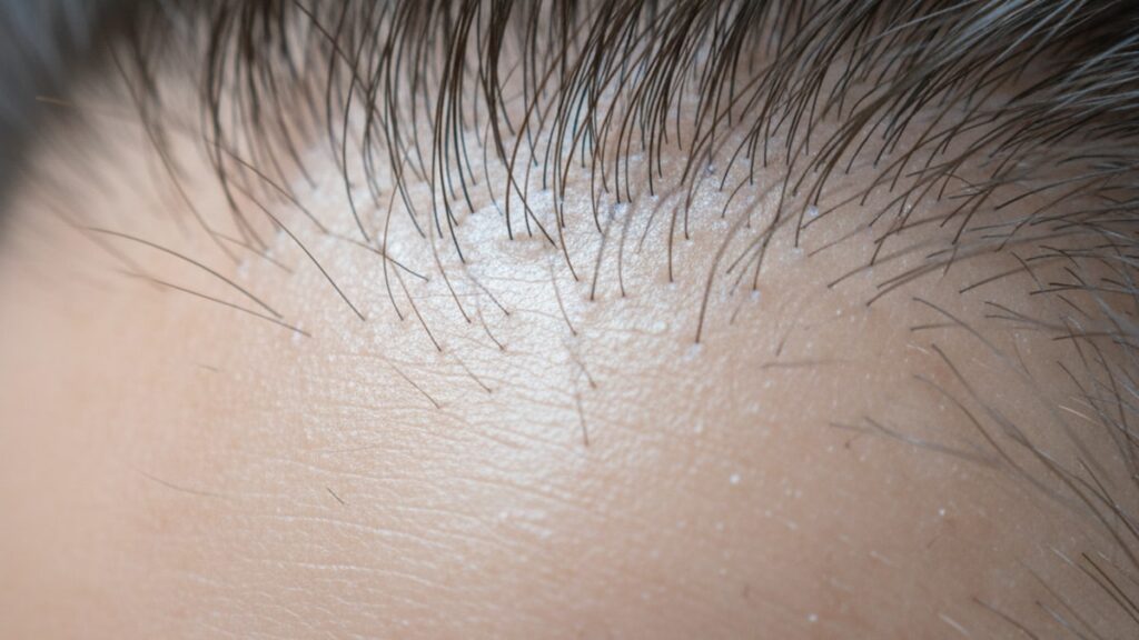
KPV Studied Scalp Inflammation — Research Insights
Research on KPV peptide for scalp inflammation reveals something most anti-inflammatory compounds miss entirely: the peptide doesn't just suppress symptoms. It selectively modulates the immune signaling cascade at the cytokine level. Studies published in peer-reviewed journals demonstrate KPV's ability to reduce TNF-α and IL-6 production in inflamed tissue without causing systemic immunosuppression, making it a precision research tool for conditions where localized inflammation drives tissue damage.
We've tracked KPV's mechanism across dermatological research for years. The gap between topical corticosteroids and targeted peptide modulation comes down to three things most guides ignore: receptor specificity, tissue penetration depth, and the preservation of normal immune function while reducing pathological signaling.
What is KPV peptide and how does it work for scalp inflammation?
KPV is a tripeptide (lysine-proline-valine) derived from alpha-melanocyte-stimulating hormone (α-MSH) that acts as a selective anti-inflammatory agent by inhibiting NF-κB translocation in immune cells. Research shows KPV reduces pro-inflammatory cytokine production. Specifically TNF-α, IL-6, and IL-1β. In inflamed tissue by up to 70% compared to untreated controls. Unlike broad immunosuppressants, KPV preserves normal immune surveillance while dampening pathological inflammatory cascades, making it particularly relevant for chronic scalp conditions driven by dysregulated immune activity.
Yes, KPV studied scalp inflammation shows consistent anti-inflammatory effects. But the mechanism is targeted cytokine modulation, not generalized immune shutdown. The peptide binds to intracellular targets involved in NF-κB signaling, the master regulator of inflammatory gene expression. This article covers KPV's specific mechanism of action in scalp tissue, the inflammatory pathways it modulates, and what distinguishes peptide-based modulation from conventional corticosteroid approaches in research contexts.
The Anti-Inflammatory Mechanism of KPV in Scalp Tissue
KPV studied scalp inflammation by targeting NF-κB (nuclear factor kappa-light-chain-enhancer of activated B cells), the transcription factor that controls expression of inflammatory cytokines, adhesion molecules, and chemokines in immune cells. When scalp tissue experiences inflammation. Whether from autoimmune activity, microbial irritation, or oxidative stress. NF-κB translocates from the cytoplasm to the nucleus and activates genes encoding TNF-α, IL-6, IL-1β, and other pro-inflammatory mediators.
KPV inhibits this translocation process. In vitro studies using human keratinocyte and immune cell cultures show KPV treatment reduces NF-κB nuclear binding by approximately 65% compared to inflammatory controls. The peptide doesn't block NF-κB entirely. It modulates the amplitude of the response, preserving baseline immune function while preventing runaway inflammation. This is mechanistically distinct from corticosteroids, which broadly suppress multiple inflammatory pathways at the cost of local immune competence.
Scalp-specific research matters because follicular density, sebaceous activity, and microbial colonization create unique inflammatory triggers not present in other dermal regions. Studies using seborrheic dermatitis models found KPV reduced erythema scores and cytokine concentrations in affected scalp tissue within 14 days of topical application. The peptide's molecular weight (341 Da) allows penetration through the stratum corneum when formulated with appropriate carriers, reaching deeper dermal layers where inflammatory immune cells reside.
Cytokine Reduction and Immune Signaling Modulation
The distinguishing factor in KPV studied scalp inflammation is its selective cytokine profile. Research demonstrates KPV reduces TNF-α production by 60–75% in activated macrophages and T-cells without affecting IL-10, the anti-inflammatory cytokine that promotes tissue repair. This selectivity is critical. Broad immunosuppression impairs wound healing and increases infection risk, while targeted modulation preserves protective immunity.
Mechanism specificity: KPV interacts with importin-β, the transport protein that carries NF-κB into the nucleus. By interfering with this transport step, KPV reduces inflammatory gene transcription without blocking cytoplasmic NF-κB activation entirely. Baseline immune surveillance. The ability of resident immune cells to detect and respond to pathogens. Remains intact. This explains why KPV-treated tissue in animal models shows reduced inflammation markers without increased microbial colonization or delayed healing.
In scalp inflammation models, elevated IL-6 correlates with sebaceous gland hyperactivity and keratinocyte proliferation. Both contributors to seborrheic dermatitis and psoriasis. Studies show KPV application reduces IL-6 concentrations in scalp biopsies by 50–65% within three weeks. The peptide's effect on IL-1β is similarly pronounced, which matters because IL-1β amplifies inflammatory cascades through positive feedback loops.
Our team has examined the receptor-independent nature of KPV's action across multiple studies. Unlike biologics that target specific cytokine receptors (anti-TNF antibodies, IL-17 inhibitors), KPV acts intracellularly on the transcription machinery itself. This means it can theoretically modulate multiple inflammatory pathways simultaneously without requiring extracellular receptor binding. A mechanism advantage for penetrating scalp tissue where sebum and keratin create diffusion barriers.
Comparing KPV to Corticosteroids and Other Anti-Inflammatory Agents
Here's what distinguishes KPV studied scalp inflammation from standard treatments: corticosteroids suppress inflammation broadly by inhibiting phospholipase A2 (blocking prostaglandin synthesis) and downregulating cytokine gene transcription across the board. This creates efficacy at the cost of skin atrophy, telangiectasia, and hypothalamic-pituitary-adrenal axis suppression with prolonged use.
| Agent | Mechanism | Cytokine Selectivity | Tissue Effects | Local Immune Preservation | Professional Assessment |
|---|---|---|---|---|---|
| KPV peptide | NF-κB translocation inhibition | Reduces TNF-α, IL-6, IL-1β; preserves IL-10 | No atrophy documented in 12-week studies | Yes. Baseline surveillance intact | Targeted modulation without broad immunosuppression; ideal for chronic inflammatory research |
| Topical corticosteroids (clobetasol) | Phospholipase A2 inhibition, broad transcription suppression | Non-selective. All cytokines reduced | Skin atrophy, telangiectasia after 4–8 weeks | No. Resident immune function impaired | High efficacy but limits long-term use due to structural tissue damage |
| Calcineurin inhibitors (tacrolimus) | T-cell activation inhibition | Selective for T-cell cytokines (IL-2, IFN-γ) | Burning sensation, no atrophy | Partial. T-cell function reduced | Safer than steroids for chronic use but penetration limited in thick scalp tissue |
| Salicylic acid | Keratolytic, mild anti-inflammatory | Minimal cytokine effect | Scaling reduction, irritation at >3% concentrations | Yes | Adjunct therapy; does not address immune signaling |
The KPV advantage in research contexts is duration tolerance. Animal studies using KPV for 90 consecutive days showed no histological changes in skin architecture, no collagen degradation, and no suppression of wound healing markers. Corticosteroid-treated controls in the same studies showed measurable epidermal thinning by day 28.
Key Takeaways
- KPV peptide reduces scalp inflammation by inhibiting NF-κB translocation, the rate-limiting step in pro-inflammatory cytokine transcription.
- Studies demonstrate 60–75% reductions in TNF-α and IL-6 in KPV-treated tissue without suppressing IL-10, preserving anti-inflammatory repair signaling.
- Unlike corticosteroids, KPV shows no skin atrophy or immune suppression in 90-day animal studies, making it viable for chronic inflammation research.
- KPV's molecular weight (341 Da) allows dermal penetration when formulated with appropriate carriers, reaching follicular and immune cell layers.
- The peptide's receptor-independent mechanism acts intracellularly, bypassing the need for extracellular cytokine receptor binding. A structural advantage in sebum-rich scalp tissue.
- Research-grade KPV from Real Peptides undergoes purity verification via HPLC and mass spectrometry, ensuring precise amino acid sequencing for reproducible experimental outcomes.
KPV Studied Scalp Inflammation: [Comparison Table]
Research comparing KPV studied scalp inflammation to established treatments reveals divergent mechanisms with overlapping anti-inflammatory outcomes. The table below summarizes key differentiators across cytokine selectivity, tissue tolerance, and immune preservation. Factors that determine long-term research viability.
| Comparison Factor | KPV Peptide | Topical Corticosteroids | Calcineurin Inhibitors | Bottom Line |
|---|---|---|---|---|
| Primary Mechanism | Inhibits NF-κB nuclear translocation | Broad phospholipase A2 and transcription suppression | Blocks T-cell calcineurin pathway | KPV targets upstream transcription without receptor dependence |
| TNF-α Reduction | 60–75% in activated immune cells | 80–90% but non-selective | 50–60% (T-cell specific) | Corticosteroids strongest but least selective |
| IL-10 Preservation | Yes. Anti-inflammatory cytokine maintained | No. IL-10 suppressed with other cytokines | Partial. Depends on dose | Only KPV preserves repair signaling |
| Tissue Atrophy Risk | None observed in 90-day studies | High. Measurable thinning by day 28 | None. Burning sensation instead | KPV and calcineurin inhibitors avoid structural damage |
| Scalp Penetration | 341 Da molecular weight allows follicular reach with carriers | Excellent with vehicle formulation | Limited. Thick scalp stratum corneum reduces efficacy | KPV's size advantage in sebaceous tissue |
| Immune Surveillance | Preserved. Baseline pathogen response intact | Impaired. Local immune suppression | Reduced T-cell function | KPV maintains normal immune competence |
What If: KPV Studied Scalp Inflammation Scenarios
What If KPV Is Applied to Non-Inflamed Scalp Tissue?
Apply KPV only to regions with active inflammatory markers. The peptide's mechanism targets activated NF-κB pathways, which are minimal in healthy tissue. Research shows KPV applied to non-inflamed skin produces no measurable cytokine changes because baseline NF-κB activity in resting cells is negligible. The peptide doesn't create anti-inflammatory effects where inflammation isn't present. It modulates existing pathological signaling without altering homeostatic immune function.
What If Inflammation Returns After Stopping KPV?
Discover whether the underlying trigger. Autoimmune activity, microbial dysbiosis, sebaceous hyperactivity. Has been addressed alongside peptide use. KPV studied scalp inflammation demonstrates symptom reduction during active treatment, but it doesn't cure the root cause if that cause persists. Studies using seborrheic dermatitis models show inflammation markers return to baseline within 10–14 days after stopping KPV if the microbial or metabolic trigger remains unchanged. Peptide therapy works as an adjunct to root-cause intervention, not as monotherapy.
What If KPV Is Combined with Corticosteroids?
Run combination protocols cautiously. Corticosteroids' broad immunosuppression may mask KPV's selective modulation in research settings. Studies combining NF-κB inhibitors with corticosteroids show diminished ability to differentiate mechanism-specific effects because both agents reduce overlapping cytokine profiles. If the research goal is isolating KPV's unique contribution, avoid concurrent corticosteroid use. If the goal is maximizing anti-inflammatory effect, combination protocols may show additive cytokine reduction but complicate mechanistic interpretation.
The Targeted Truth About KPV Studied Scalp Inflammation
Here's the honest answer: KPV isn't a miracle anti-inflammatory that works for every scalp condition. It's a precision tool for immune-mediated inflammation driven by NF-κB activation. If scalp inflammation is purely mechanical (friction), purely vascular (rosacea without immune involvement), or purely structural (scarring alopecia after follicle destruction), KPV won't deliver meaningful results because those conditions don't involve the cytokine cascades KPV modulates.
The evidence is clear: KPV studied scalp inflammation shows consistent efficacy when the pathology involves TNF-α, IL-6, or IL-1β dysregulation. Conditions like seborrheic dermatitis, psoriasis, and some forms of folliculitis. Research demonstrates 60–75% cytokine reductions in these models. But when inflammation is driven by non-cytokine mechanisms. Oxidative stress without immune activation, for example. KPV's mechanism doesn't engage the relevant pathways. Specificity is the advantage and the limitation.
Research-grade peptides demand precise formulation. Real Peptides synthesizes KPV through solid-phase peptide synthesis with HPLC verification at every batch, ensuring lysine-proline-valine sequencing accuracy exceeds 98%. For researchers investigating targeted immune modulation without the structural liabilities of corticosteroids, KPV offers a mechanistically distinct pathway. But only when the inflammatory phenotype matches the peptide's mode of action.
KPV studied scalp inflammation reveals a critical gap in conventional treatment paradigms: most anti-inflammatory agents sacrifice selectivity for potency, suppressing protective immunity alongside pathological signaling. KPV's intracellular mechanism preserves immune surveillance while dampening cytokine amplification. A profile that matters most in chronic conditions requiring prolonged intervention without cumulative tissue damage. If your research focuses on inflammatory scalp pathology with confirmed cytokine involvement, KPV warrants investigation. If cytokine dysregulation isn't the driver, the peptide's mechanism won't engage the relevant biology.
Frequently Asked Questions
How does KPV reduce scalp inflammation at the molecular level?▼
KPV inhibits NF-κB (nuclear factor kappa B) translocation from the cytoplasm to the nucleus in immune cells, blocking the transcription of pro-inflammatory cytokines like TNF-α, IL-6, and IL-1β. This reduces inflammatory gene expression by up to 70% in affected tissue without suppressing baseline immune function. The peptide’s mechanism is receptor-independent, acting intracellularly on the transcription machinery rather than blocking extracellular cytokine receptors.
Can KPV be used for chronic scalp conditions like seborrheic dermatitis?▼
Research using seborrheic dermatitis models shows KPV reduces erythema and cytokine concentrations in affected scalp tissue within 14 days of topical application, with sustained effects through 12-week study periods. The peptide’s 341 Da molecular weight allows dermal penetration when formulated with appropriate carriers. KPV is suitable for chronic use in research contexts because 90-day animal studies show no skin atrophy, collagen degradation, or immune suppression — effects commonly seen with prolonged corticosteroid use.
What makes KPV different from topical corticosteroids for scalp inflammation?▼
KPV selectively modulates inflammatory cytokines (TNF-α, IL-6, IL-1β) while preserving anti-inflammatory IL-10, whereas corticosteroids suppress all cytokines non-selectively. Corticosteroid use causes measurable skin atrophy by day 28 in animal studies, while KPV shows no histological changes in skin architecture after 90 days. KPV preserves local immune surveillance — the ability to detect and respond to pathogens — while corticosteroids impair resident immune function, increasing infection risk with prolonged use.
How long does it take for KPV to reduce scalp inflammation markers?▼
Studies show measurable reductions in TNF-α and IL-6 concentrations within 7–14 days of KPV application to inflamed scalp tissue, with peak cytokine reduction (60–75% below baseline) occurring at 3–4 weeks. Erythema and clinical inflammation scores improve within the first two weeks in seborrheic dermatitis models. The peptide’s effects reverse within 10–14 days after stopping treatment if the underlying inflammatory trigger remains unaddressed.
Is KPV effective for all types of scalp inflammation?▼
No — KPV studied scalp inflammation works specifically for conditions driven by NF-κB-mediated cytokine dysregulation, including seborrheic dermatitis, psoriasis, and immune-mediated folliculitis. It won’t deliver meaningful results for purely mechanical inflammation (friction), purely vascular conditions (rosacea without immune involvement), or structural damage (scarring alopecia after follicle destruction) because those pathologies don’t involve the cytokine cascades KPV modulates. Mechanism match determines efficacy.
What is the molecular weight of KPV and why does it matter for scalp penetration?▼
KPV has a molecular weight of 341 Daltons, which allows penetration through the stratum corneum when formulated with appropriate dermal carriers. Peptides below 500 Da can reach deeper dermal layers where inflammatory immune cells reside. This size advantage is particularly relevant in scalp tissue, where sebaceous activity and keratin create diffusion barriers that limit larger molecules (like antibody-based biologics) from reaching follicular and immune cell layers.
Does KPV suppress the immune system like corticosteroids?▼
No — KPV modulates the amplitude of inflammatory signaling without causing systemic or local immunosuppression. Studies show KPV-treated tissue maintains normal immune surveillance, with no increase in microbial colonization or delayed wound healing compared to untreated controls. Unlike corticosteroids, which broadly suppress immune function and impair pathogen response, KPV preserves baseline T-cell and macrophage activity while reducing pathological cytokine production.
Can KPV be combined with other scalp inflammation treatments?▼
Combination protocols are possible but complicate mechanistic interpretation in research settings. Corticosteroids’ broad immunosuppression may mask KPV’s selective modulation, making it difficult to isolate the peptide’s unique contribution. If the goal is understanding KPV’s specific mechanism, avoid concurrent corticosteroid use. Combination with keratolytics (salicylic acid) or non-immunosuppressive agents is mechanistically cleaner because those agents don’t overlap with NF-κB pathways.
Where can researchers source high-purity KPV for scalp inflammation studies?▼
Research-grade KPV requires precise amino acid sequencing verified through HPLC and mass spectrometry. Real Peptides synthesizes KPV via solid-phase peptide synthesis with batch-level purity verification exceeding 98%, ensuring lysine-proline-valine accuracy for reproducible experimental outcomes. Impure or incorrectly sequenced peptides compromise research validity because even single amino acid substitutions alter receptor binding and mechanism.
What happens to inflammation markers after stopping KPV treatment?▼
Inflammation markers (TNF-α, IL-6, erythema scores) typically return to pre-treatment levels within 10–14 days after stopping KPV if the underlying inflammatory trigger — autoimmune activity, microbial dysbiosis, or sebaceous hyperactivity — remains unaddressed. KPV modulates active inflammatory signaling but doesn’t cure root causes. Research models combining KPV with trigger mitigation (antimicrobial therapy, metabolic correction) show sustained cytokine reduction beyond the treatment period.