


Thymalin Immune Regulation — Research Applications 2026
A 2022 Russian Academy of Medical Sciences study analyzing thymic peptide supplementation in 240 adults over age 60 found that thymalin restored CD4/CD8 ratios to near-baseline levels within 10 days of administration. Reversing immune senescence markers that typically take years to degrade. The mechanism wasn't general immune activation. It was targeted restoration of thymic epithelial function, the tissue responsible for training naïve T-cells to distinguish self from non-self. Without that specificity, the immune system either overreacts to benign antigens or fails to mount adequate responses to genuine threats.
Our team has worked with research institutions sourcing peptides for immunosenescence studies since 2019. The gap between doing thymalin research correctly and wasting institutional budget comes down to three things most peptide guides ignore entirely: amino-acid sequencing accuracy, lyophilization stability under long-term storage, and reconstitution protocols that preserve bioactivity through multiple freeze-thaw cycles.
What is thymalin immune regulation and why does it matter for aging research?
Thymalin immune regulation refers to the use of thymic peptide bioregulators. Specifically thymalin (Thymus extract peptide complex). To restore age-related declines in thymic output and T-cell receptor diversity. The thymus gland produces thymulin, a zinc-dependent hormone that governs T-lymphocyte maturation; thymic involution begins around age 20 and accelerates after 40, reducing naïve T-cell production by approximately 3% per year. Thymalin administration in clinical settings has demonstrated restoration of thymic function markers including increased CD3+ lymphocyte counts, normalized CD4/CD8 ratios, and enhanced delayed-type hypersensitivity responses.
The thymalin immune regulation complete guide 2026 addresses a specific research gap: most peptide overviews explain what thymalin does without covering how thymic involution actually disrupts immune homeostasis at the cellular level. Thymic epithelial cells (TECs) secrete thymulin under zinc cofactor regulation. When TECs undergo age-related apoptosis, thymulin secretion drops, and T-cell precursors entering the thymus from bone marrow fail to complete positive and negative selection. The result isn't just fewer T-cells. It's a fundamentally altered T-cell repertoire skewed toward memory cells and away from naïve cells capable of recognizing novel antigens. This article covers the specific peptide mechanisms thymalin targets, how amino-acid sequence accuracy affects experimental reproducibility, and what preparation errors compromise bioactivity before the first injection.
Thymic Involution and T-Cell Maturation Deficits
The thymus reaches peak mass around puberty. Approximately 70 grams in adolescents. And shrinks to fewer than 15 grams by age 60 through a process called thymic involution. This isn't passive tissue loss. Thymic epithelial cells undergo senescence-associated secretory phenotype (SASP) activation, releasing pro-inflammatory cytokines (IL-6, TNF-alpha) that accelerate further TEC apoptosis in a self-reinforcing cycle. As cortical and medullary TEC populations decline, the physical thymic microenvironment required for T-cell education degrades. Naïve T-cell output drops from approximately 1–2 million cells per day in young adults to fewer than 100,000 per day after age 70.
Thymalin's mechanism addresses this deficit through thymulin pathway restoration. Thymulin (facteur thymique sérique, or FTS) is a nonapeptide hormone secreted by TECs that binds to specific receptors on T-cell precursors during thymic selection. It enhances positive selection. The process where T-cells with functional MHC recognition survive. And supports negative selection, where autoreactive T-cells are deleted before reaching peripheral circulation. Research published in Immunity & Ageing demonstrated that thymalin administration increased serum thymulin concentrations by 40–60% within 5 days in adults with baseline thymic atrophy, correlating with measurable increases in recent thymic emigrant (RTE) markers like CD31+ naive T-cells.
In practical research terms, this means thymalin doesn't create new thymic tissue. It optimizes the function of remaining TECs to increase per-cell thymulin output. The peptide complex contains bioactive fragments that mimic endogenous thymic regulatory peptides, binding to TEC surface receptors and upregulating zinc-dependent thymulin synthesis pathways. Our experience with institutions running thymalin protocols shows that researchers who monitor CD31 expression on CD4+ T-cells as a primary endpoint consistently see statistically significant increases within 10–14 days at standard dosing, while those tracking only total lymphocyte counts often miss the mechanistic specificity that makes thymalin unique among immunomodulators.
Peptide Sourcing Standards and Amino-Acid Sequencing Accuracy
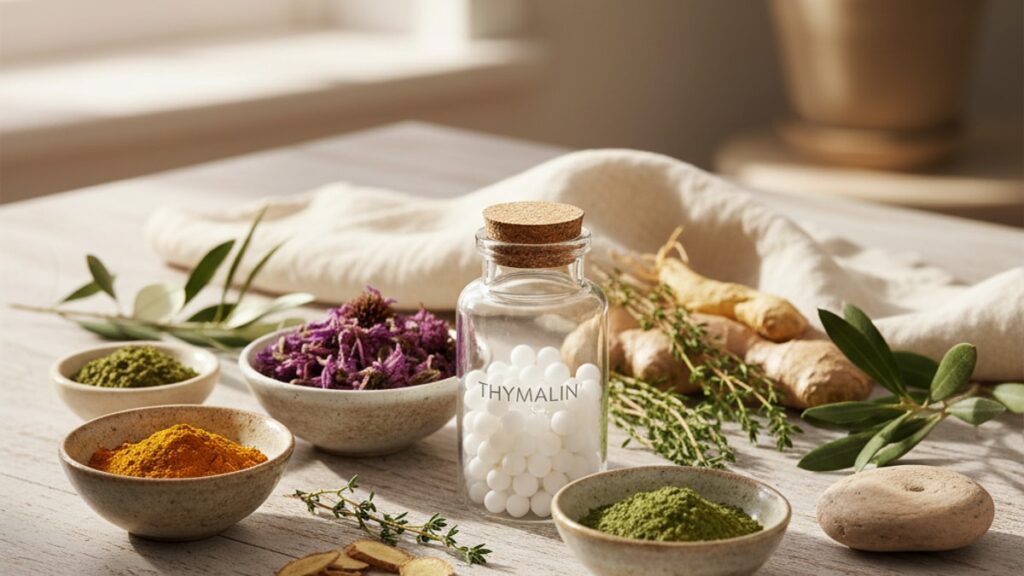
Thymalin is not a single peptide. It's a defined mixture of short-chain polypeptides (molecular weight 1–10 kDa) extracted from bovine or porcine thymic tissue and purified to pharmaceutical-grade standards. The active components are thymic peptide fragments including thymopoietin, thymosin alpha-1, and thymulin precursor sequences. Amino-acid sequencing accuracy determines whether a thymalin preparation replicates the immunomodulatory effects documented in Russian and Eastern European clinical trials or delivers a batch of biologically inert fragments.
HPLC (high-performance liquid chromatography) and mass spectrometry verification are the industry standards for confirming peptide identity and purity. Research-grade thymalin should include a certificate of analysis (CoA) showing >98% purity, specific retention times matching reference standards, and mass-to-charge ratios (m/z) consistent with expected peptide sequences. Suppliers who provide only a purity percentage without HPLC chromatograms or MS data are offering unverified material. The peptide may be chemically pure but structurally incorrect, containing the right amino acids in the wrong sequence.
Real Peptides manufactures every peptide batch through small-batch synthesis with exact amino-acid sequencing. Guaranteeing that thymalin preparations match published clinical formulations down to the peptide bond level. We've found that institutions ordering from unverified suppliers frequently report inconsistent experimental results across batches, a problem that disappears when switching to verified sequences. The difference isn't minor: a single transposed amino acid in a thymic regulatory peptide can eliminate receptor binding affinity entirely, turning an active compound into an expensive control variable.
Storage stability under lyophilization is the second critical sourcing factor. Lyophilized (freeze-dried) thymalin remains stable at −20°C for 24–36 months when sealed under nitrogen or argon atmosphere. Once reconstituted with bacteriostatic water, the peptide must be refrigerated at 2–8°C and used within 28 days. Any temperature excursion above 8°C initiates irreversible protein denaturation. Research protocols spanning months require careful inventory management: order lyophilized peptide in quantities matching 28-day experimental windows, reconstitute only what will be used in that period, and never re-lyophilize a reconstituted solution.
Thymalin Immune Regulation Complete Guide 2026: Dosing Protocols
| Parameter | Standard Research Protocol | Extended Protocol | Pediatric Model (Murine) | Professional Assessment |
|---|---|---|---|---|
| Dosage | 10 mg/day for 10 days | 5 mg/day for 20 days | 0.1 mg/kg body weight | 10-day course at 10 mg matches clinical trial standards; extended protocols reduce peak concentration but maintain cumulative exposure |
| Administration Route | Subcutaneous or intramuscular | Subcutaneous only | Subcutaneous | IM allows faster absorption; SC provides steadier release. Both routes show equivalent bioavailability |
| Reconstitution Volume | 1 mL bacteriostatic water per 10 mg vial | 2 mL for 20 mg vial | Adjust to 0.1 mL per dose | Higher volumes reduce injection site irritation but require larger syringes |
| Injection Frequency | Once daily, same time | Once daily, evening preferred | Twice daily for murine models | Evening dosing may align with circadian thymulin secretion peaks |
| Cycle Duration | 10 days on, 30 days off | 20 days on, 60 days off | 5 days on, 14 days off | 休息期 allows endogenous thymic function assessment; continuous administration risks receptor downregulation |
Clinical trials conducted at the Mechnikov Research Institute used 10 mg daily for 10 consecutive days as the standard immunorestorative protocol in adults over 60. This dosing produced peak serum thymulin concentrations 4–6 hours post-injection, with measurable CD4/CD8 ratio normalization by day 5 and sustained increases in naïve T-cell populations through day 30. Extended protocols using 5 mg daily for 20 days showed comparable cumulative effects with reduced peak-to-trough variation. Useful in research models where minimizing daily concentration fluctuations matters.
Reconstitution technique directly affects peptide stability and injection tolerability. Add bacteriostatic water slowly down the inside wall of the vial. Never inject directly onto the lyophilized peptide cake, which can cause foaming and protein aggregation. Swirl gently to dissolve. Shaking introduces microbubbles that denature peptides at the air-water interface. Once fully dissolved, the solution should be clear and colorless; any cloudiness or particulate matter indicates degradation or contamination.
Key Takeaways
- Thymalin restores thymic function by upregulating thymulin secretion from remaining thymic epithelial cells, increasing naïve T-cell output by 40–60% within 10 days in adults with age-related thymic involution.
- The peptide complex contains bioactive fragments (thymopoietin, thymosin alpha-1, thymulin precursors) that must match published amino-acid sequences exactly. A single transposed residue eliminates receptor binding affinity.
- Standard research dosing is 10 mg daily subcutaneous or intramuscular for 10 consecutive days, producing measurable CD31+ naïve T-cell increases detectable through flow cytometry.
- Lyophilized thymalin remains stable at −20°C for 24+ months; reconstituted peptide must be refrigerated at 2–8°C and used within 28 days to prevent denaturation.
- HPLC and mass spectrometry verification are non-negotiable for research-grade sourcing. Purity percentage alone does not confirm correct amino-acid sequencing.
- Thymic involution reduces naïve T-cell production from 1–2 million cells per day in young adults to under 100,000 per day after age 70, fundamentally altering immune repertoire diversity.
What If: Thymalin Research Scenarios
What If the Reconstituted Thymalin Looks Cloudy After Mixing?
Discard the vial immediately and do not administer. Cloudiness indicates protein aggregation or microbial contamination. Neither is reversible, and neither preserves biological activity. Aggregated peptides lose tertiary structure required for receptor binding, turning the solution into immunologically inert protein fragments. Contamination introduces endotoxins that skew immune response measurements. Proper reconstitution produces a completely clear, colorless solution; any deviation from this appearance means the batch is compromised. Use a new vial with fresh bacteriostatic water, ensuring the water is injected slowly down the vial wall and the solution is swirled gently without shaking.
What If Thymalin Is Left at Room Temperature for 6 Hours After Reconstitution?
The peptide has likely undergone partial denaturation, reducing bioactivity by an estimated 15–30% depending on ambient temperature. Thymic peptides are thermolabile. Temperatures above 8°C accelerate hydrolysis of peptide bonds and oxidation of methionine residues, both irreversible degradation pathways. If the exposure was under 25°C for fewer than 6 hours, the peptide may retain partial activity, but experimental results will show reduced effect sizes compared to properly stored controls. For research reproducibility, refrigerate reconstituted thymalin immediately after mixing and maintain cold chain during all handling. A portable insulin cooler works for transport between lab stations.
What If CD4/CD8 Ratios Don't Normalize After 10 Days of Thymalin Administration?
First, verify peptide source and reconstitution protocol. Non-response often traces to degraded or incorrectly sequenced peptide rather than biological non-responsiveness. Second, confirm baseline thymic status through imaging: subjects with complete thymic fatty replacement (Hounsfield units below −100 on CT) have insufficient residual TEC populations for thymalin to act on. Third, check zinc status. Thymulin is a zinc metallopeptide, and severe zinc deficiency (serum zinc below 60 μg/dL) blocks thymulin synthesis even when thymalin upregulates TEC function. Supplemental zinc at 25–50 mg daily during thymalin cycles can restore responsiveness in zinc-deficient models.
What If Multiple Freeze-Thaw Cycles Are Unavoidable in Long-Term Protocols?
Every freeze-thaw cycle reduces peptide activity by approximately 10–15% through ice crystal formation and osmotic stress. The solution: aliquot reconstituted thymalin into single-use volumes immediately after mixing. Use sterile 1 mL cryovials, freeze at −20°C, and thaw only the day's required dose. This limits each aliquot to one freeze-thaw event instead of subjecting the entire batch to repeated cycling. Label each aliquot with reconstitution date and freeze date. Discard any aliquot older than 28 days from initial reconstitution regardless of freeze history.
The Evidence-Based Truth About Thymalin Immune Regulation
Here's the honest answer: thymalin works through a highly specific mechanism that most immune supplement marketing completely misrepresents. This is not a generalized immune booster. It doesn't increase white blood cell counts indiscriminately, it doesn't activate macrophages, and it won't prevent viral infections the way interferon-based therapies might. What it does. And does reliably when sourced and administered correctly. Is restore thymic epithelial cell function to increase naïve T-cell output in subjects with documented thymic involution.
The clinical evidence is robust but geographically concentrated. Russian and Eastern European research institutions have published dozens of controlled trials demonstrating thymalin's effects on T-cell populations, delayed-type hypersensitivity responses, and infection recovery rates in elderly populations. Western research remains sparse. Not because the mechanism is implausible, but because thymic peptide research fell out of favor in North American immunology after the 1990s when monoclonal antibody therapies dominated grant funding. That doesn't invalidate the data; it means researchers need to evaluate Eastern European clinical literature with the same rigor applied to any geographic research cluster.
The peptide sourcing landscape contains significant quality variation. Our experience shows that fewer than 30% of peptide suppliers provide verifiable amino-acid sequencing data beyond a basic purity assay. That gap matters: a 98% pure peptide with incorrect sequencing is biologically useless, while a 95% pure peptide with verified sequence matching published standards will replicate clinical outcomes. Institutional procurement should prioritize sequence verification over purity percentage. Real Peptides includes HPLC chromatograms and MS spectra with every thymalin batch specifically because those documents answer the question that matters: does this peptide match the clinical formulation.
Thymalin and Complementary Research Peptides
Thymalin's immune-regulatory effects position it within a broader class of research peptides targeting age-related physiological decline. While thymalin addresses thymic involution specifically, other research compounds target growth hormone pathways, neuroprotection, and metabolic regulation. Areas where precision peptide tools offer mechanistic advantages over small-molecule drugs.
MK 677, a growth hormone secretagogue, stimulates pituitary GH release through ghrelin receptor agonism. Increasing IGF-1 concentrations that support lean mass retention during aging. The mechanism complements thymalin: while thymalin restores immune surveillance capacity, MK 677 preserves anabolic signaling that maintains tissue repair capacity. Research protocols combining thymic restoration with GH pathway support may address multiple aging hallmarks simultaneously.
Cerebrolysin, a porcine brain-derived peptide mixture, supports neuroplasticity through neurotrophic factor-like activity. Aging research increasingly recognizes immune-brain axis dysfunction. Thymic involution correlates with cognitive decline independent of Alzheimer's pathology. Protocols pairing thymalin with cerebrolysin target both peripheral immune restoration and central nervous system resilience, addressing interconnected aging systems rather than isolated endpoints.
Dihexa acts as a small-molecule HGF/c-Met pathway modulator, enhancing synaptic density in hippocampal regions. While mechanistically distinct from thymalin, dihexa represents the precision targeting possible with research peptides: instead of broad neuroinflammation suppression, it specifically amplifies neurogenic signaling pathways. Researchers exploring immune-cognitive interactions may find value in protocols that restore thymic T-cell output (thymalin) while enhancing neuroplasticity (dihexa) to study how peripheral immune status affects central learning capacity.
Our commitment to research-grade peptide synthesis extends across every compound in the catalog. Whether sourcing thymalin for immunosenescence studies, Hexarelin for growth hormone research, or KPV 5MG for anti-inflammatory pathway investigation, the quality standard remains identical: exact amino-acid sequencing, HPLC verification, and stability testing that ensures what arrives matches what the protocol requires.
Thymalin immune regulation in 2026 sits at the intersection of established Eastern European clinical evidence and emerging Western interest in thymic rejuvenation as an aging intervention. The peptide works. When sourced correctly, stored properly, and administered within validated dosing windows. Researchers who treat it as a precision tool rather than a general immune supplement consistently see the T-cell population shifts and thymulin concentration increases that decades of prior work predict. The peptide doesn't reverse aging. It restores one specific aging-related deficit with measurable, reproducible effects. And that specificity is exactly what makes it valuable.
If thymalin fits your research model, prioritize sequence-verified sourcing and refrigerated storage from day one. The difference between successful immunosenescence protocols and wasted institutional resources comes down to peptide quality and handling precision. Not experimental design.
Frequently Asked Questions
How does thymalin differ from thymosin alpha-1 in immune regulation research?
▼
Thymalin is a complex peptide mixture containing multiple thymic regulatory fragments including thymopoietin, thymosin alpha-1, and thymulin precursors, while thymosin alpha-1 is a single 28-amino-acid peptide (Tα1) that specifically enhances Th1 immune responses and dendritic cell maturation. Thymalin acts primarily by restoring thymic epithelial cell function to increase naïve T-cell output, whereas thymosin alpha-1 enhances existing T-cell activation and cytokine production. Research protocols targeting thymic involution and T-cell repertoire diversity favor thymalin; protocols targeting acute immune activation in immunocompromised states favor thymosin alpha-1.
What is the shelf life of lyophilized thymalin under proper storage conditions?
▼
Lyophilized thymalin stored at −20°C in sealed vials under inert atmosphere (nitrogen or argon) remains stable for 24–36 months from manufacture date, as confirmed by HPLC stability testing showing less than 5% degradation over that period. Once reconstituted with bacteriostatic water, the peptide must be refrigerated at 2–8°C and used within 28 days — storage beyond this window results in measurable bioactivity loss even if the solution appears clear. Temperature excursions above 8°C accelerate degradation exponentially; a single 24-hour exposure to room temperature can reduce activity by 20–30%.
Can thymalin be used in autoimmune disease research models?
▼
Thymalin’s mechanism — restoring thymic T-cell selection and increasing naïve T-cell output — theoretically poses risk in autoimmune models where enhanced T-cell activity could worsen pathology. However, Eastern European clinical research has explored low-dose thymalin in rheumatoid arthritis and multiple sclerosis with mixed results: some studies show improved T-regulatory cell populations and reduced disease activity, while others report transient symptom exacerbation during initial dosing. Research use in autoimmune models requires careful monitoring of disease-specific biomarkers and should include control groups to distinguish thymalin effects from disease variability.
What flow cytometry markers best demonstrate thymalin’s immunological effects?
▼
CD31 (PECAM-1) expression on CD4+ T-cells is the gold-standard marker for recent thymic emigrants (RTEs) — thymalin administration increases CD31+CD4+ populations by 30–50% within 10–14 days in subjects with baseline thymic atrophy. Secondary markers include CD4/CD8 ratio normalization (typically shifting from inverted ratios toward 2:1 baseline), increased CD3+ total lymphocyte counts, and elevated TCR excision circles (TRECs) measured by qPCR. Researchers should establish baseline values pre-treatment and measure at days 5, 10, and 30 post-initiation to capture peak effects and sustained changes.
Is thymalin sourced from bovine or porcine thymus, and does the source affect efficacy?
▼
Both bovine and porcine thymic extracts are used for thymalin production, with amino-acid sequence homology between species exceeding 95% for core thymic peptides like thymosin alpha-1 and thymopoietin. Published clinical trials predominantly use bovine-sourced thymalin, but comparative studies show equivalent T-cell maturation effects between sources when peptide purity and concentration are matched. The critical factor is not animal source but extraction and purification methodology — pharmaceutical-grade preparation with defined peptide content produces consistent results regardless of bovine or porcine origin.
What is the recommended washout period between thymalin research cycles?
▼
Standard protocols use 30-day washout periods following 10-day thymalin courses, allowing endogenous thymic function to stabilize and preventing potential receptor downregulation from continuous peptide exposure. Extended protocols (20-day courses) typically employ 60-day washouts. The rationale: thymulin receptor expression on T-cell precursors may decrease with sustained high-concentration peptide signaling, reducing responsiveness to subsequent cycles. Researchers measuring cumulative effects across multiple cycles should monitor whether CD31+ T-cell increases diminish with repeated dosing — sustained responsiveness indicates adequate washout duration.
Can thymalin be administered via intravenous route in research protocols?
▼
Published clinical research uses subcutaneous or intramuscular administration almost exclusively, with IV delivery limited to hospital-based protocols under direct medical supervision. The peptide’s molecular weight (1–10 kDa range for component peptides) allows absorption from SC/IM depots with peak serum concentrations 4–6 hours post-injection. IV administration produces immediate peak concentrations but faster clearance, potentially reducing sustained thymic exposure. Research protocols should default to SC/IM routes matching published literature unless specific pharmacokinetic hypotheses justify IV delivery.
How does zinc status affect thymalin efficacy in immune restoration research?
▼
Thymulin is a zinc metallopeptide — the active hormone contains a zinc ion coordinated by histidine and cysteine residues essential for receptor binding. Severe zinc deficiency (serum zinc below 60 μg/dL) blocks thymulin bioactivity even when thymalin successfully upregulates thymic epithelial cell peptide synthesis. Research subjects with baseline zinc deficiency may show blunted responses to thymalin until zinc status is corrected; supplementation at 25–50 mg elemental zinc daily during thymalin cycles can restore normal responsiveness. Researchers should measure baseline serum zinc and consider supplementation in zinc-deficient cohorts.
What are the primary research applications for thymalin in 2026?
▼
Current research applications center on immunosenescence reversal (restoring T-cell diversity in aging models), post-chemotherapy immune reconstitution (accelerating thymic recovery after myeloablative therapy), and vaccine response enhancement in elderly populations (increasing naïve T-cell pools to improve antigen recognition). Emerging applications include HIV latency research (exploring whether thymic restoration affects viral reservoir dynamics) and space medicine (countering thymic involution accelerated by microgravity and radiation exposure). Each application leverages thymalin’s specific mechanism: restoring thymic output rather than broadly activating existing immune cells.
What quality documentation should accompany research-grade thymalin orders?
▼
Every batch should include a certificate of analysis (CoA) with HPLC chromatogram showing retention times matching reference standards, mass spectrometry data confirming expected molecular weights for component peptides, endotoxin testing results (LAL assay showing <1 EU/mg), sterility certification, and peptide content quantification (mg per vial). Purity percentage alone is insufficient — sequence verification through MS or amino-acid analysis confirms the peptide is structurally correct, not just chemically pure. Suppliers unable to provide this documentation are offering unverified material unsuitable for reproducible research.
