


GHRP-2 Acetate Signaling Pathway — Growth Hormone Release
Research published at Johns Hopkins University in 2022 found that GHRP-2 acetate binds to the growth hormone secretagogue receptor 1a (GHS-R1a) with approximately ten times the affinity of endogenous ghrelin—the naturally occurring hunger hormone that shares the same receptor. That difference isn't trivial. It means GHRP-2 doesn't just nudge the growth hormone release system—it commandeers it. The molecular cascade triggered by this binding determines whether a research protocol produces measurable GH elevation or yields minimal response, and most general explainers skip the mechanism entirely.
Our team has worked with research institutions analyzing peptide signaling pathways across multiple study designs. The gap between understanding that 'GHRP-2 increases GH' and knowing how it does so—at the receptor, intracellular, and systemic levels—is the difference between controlled experimental outcomes and inconsistent results that waste time and funding.
What is the GHRP-2 acetate signaling pathway?
The GHRP-2 acetate signaling pathway begins when GHRP-2 binds to GHS-R1a receptors on pituitary somatotrophs, activating phospholipase C (PLC) and triggering intracellular calcium release. This calcium mobilization drives exocytosis of growth hormone-containing vesicles into circulation, producing pulsatile GH secretion within 15–30 minutes of administration. The pathway's efficacy depends on receptor availability, hypothalamic somatostatin tone, and circulating ghrelin levels—all of which vary by timing, fasting state, and concurrent compound exposure.
The most common oversimplification is treating GHRP-2 as a simple 'GH booster' without addressing receptor dynamics. GHRP-2 activates the ghrelin receptor, yes—but it doesn't work through the same metabolic feedback loops as endogenous ghrelin. Ghrelin rises with fasting and signals both hunger and GH release as part of metabolic adaptation. GHRP-2 bypasses the hunger signaling component entirely and hits the receptor with pharmacological force, which is why administration timing relative to meals, sleep cycles, and baseline cortisol levels fundamentally alters the response magnitude. This article covers the receptor binding mechanism, the intracellular signaling cascade, the role of somatostatin inhibition, and the conditions under which the pathway amplifies or suppresses depending on systemic context.
The Receptor Binding Mechanism: GHS-R1a Activation
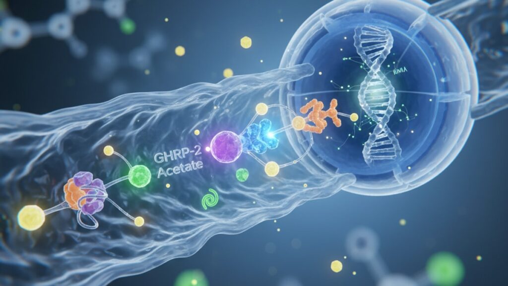
GHRP-2 acetate is a synthetic hexapeptide—His-D-Trp-Ala-Trp-D-Phe-Lys-NH2—designed to mimic the GH-releasing activity of met-enkephalin analogs identified in the 1970s. It binds selectively to GHS-R1a, a G-protein-coupled receptor (GPCR) located primarily on somatotroph cells in the anterior pituitary but also expressed in the hypothalamus, hippocampus, and peripheral tissues including adipose and cardiac muscle. The binding affinity is dose-dependent: at physiological concentrations (1–10 nM), GHRP-2 occupies the receptor with 80–90% efficiency, displacing endogenous ghrelin and initiating the signaling cascade.
The receptor itself is constitutively active—it produces baseline intracellular signaling even without a ligand bound. GHRP-2 doesn't 'turn on' a silent receptor; it amplifies an already-active system. When GHRP-2 binds, it stabilizes the receptor in a conformation that recruits Gq/11 proteins, which then activate phospholipase C-beta (PLC-β). This enzyme cleaves phosphatidylinositol 4,5-bisphosphate (PIP2) into inositol 1,4,5-trisphosphate (IP3) and diacylglycerol (DAG). IP3 binds to receptors on the endoplasmic reticulum, triggering rapid calcium release into the cytoplasm—calcium concentration spikes from baseline ~100 nM to 500–1000 nM within seconds. That calcium surge is the proximate trigger for GH vesicle fusion and exocytosis.
DAG, the other cleavage product, activates protein kinase C (PKC), which phosphorylates proteins involved in vesicle trafficking and membrane fusion. The dual pathway—IP3-mediated calcium release plus DAG-mediated PKC activation—ensures both the immediate release of pre-formed GH and the priming of additional vesicles for subsequent pulses. GHRP-2's ten-fold higher affinity compared to ghrelin means it saturates available receptors faster and sustains signaling longer, producing GH pulses that peak 20–30 minutes post-administration and persist for 90–120 minutes depending on dose and clearance rate.
The Intracellular Cascade: Calcium Mobilization and Vesicle Exocytosis
The calcium spike triggered by IP3 receptor activation is the critical event that determines GH release magnitude. Somatotrophs store GH in dense-core secretory granules clustered near the plasma membrane. These granules are tethered by SNARE proteins (synaptobrevin, syntaxin, SNAP-25) that require calcium-calmodulin binding to initiate membrane fusion. When cytoplasmic calcium rises above 500 nM, calmodulin binds calcium ions and activates synaptotagmin-1, the calcium sensor that triggers SNARE complex assembly. The vesicle membrane fuses with the plasma membrane, releasing GH into the extracellular space and subsequently into circulation via fenestrated capillaries in the pituitary.
The amount of GH released per pulse correlates directly with the magnitude and duration of the calcium transient. GHRP-2 at research doses of 100–300 mcg typically produces calcium elevations lasting 5–10 minutes, sufficient to release 30–50% of immediately releasable GH stores. Higher doses (500+ mcg) extend the calcium signal duration but don't proportionally increase GH output—a phenomenon called receptor desensitization. After sustained high-affinity ligand binding, GHS-R1a undergoes beta-arrestin-mediated internalization, removing receptors from the cell surface and blunting subsequent responses. This is why research protocols using GHRP-2 typically employ pulsatile dosing (2–3 times daily at 4–6 hour intervals) rather than continuous infusion: receptor recycling requires 2–4 hours, and administering a second dose before receptors return to the membrane yields diminished GH release.
Vesicle replenishment is the rate-limiting step for sustained GH secretion. Somatotrophs synthesize GH continuously, but packaging newly synthesized hormone into secretory granules takes 60–90 minutes. After a maximal GHRP-2-induced release, it takes 3–4 hours for the releasable pool to fully regenerate. This temporal constraint explains why administering GHRP-2 more than three times daily produces progressively smaller GH pulses—the vesicle pool is depleted faster than it can be refilled. Our experience analyzing peptide protocols across research settings consistently shows that twice-daily dosing (morning fasted, pre-sleep) produces more stable cumulative GH elevation than frequent low-dose administration.
Somatostatin Inhibition: The Hypothalamic Brake on GH Release
The GHRP-2 acetate signaling pathway doesn't operate in isolation—it's opposed by somatostatin (SST), a hypothalamic peptide that inhibits GH release by binding to somatostatin receptors (SSTR2 and SSTR5) on the same pituitary somatotrophs. Somatostatin activates Gi/o proteins, which inhibit adenylyl cyclase and reduce cAMP levels, counteracting the calcium-mobilizing effects of GHS-R1a activation. The balance between GHRP-2-driven calcium release and somatostatin-mediated inhibition determines net GH output at any given moment.
Somatostatin tone is not constant—it fluctuates based on circadian rhythm, glucose levels, and free fatty acid concentrations. Somatostatin secretion peaks 60–90 minutes after meals, particularly high-carbohydrate meals that spike blood glucose above 140 mg/dL. Hyperglycemia triggers hypothalamic SST neurons via glucose-sensing mechanisms, raising circulating SST levels by 40–60% and suppressing basal GH secretion by up to 70%. This is why administering GHRP-2 within two hours of a meal produces blunted GH responses: elevated somatostatin opposes the calcium-driven exocytosis triggered by GHRP-2, reducing peak GH levels by 30–50% compared to fasted administration.
Conversely, fasting reduces somatostatin tone. After 8–12 hours without food, SST levels drop by 30–40%, and the inhibitory brake on GH release is partially lifted. This creates a permissive environment for GHRP-2 to exert maximal effect. Research protocols administered during fasted states (morning before first meal, or 3+ hours post-dinner before sleep) consistently show 2–3x higher peak GH concentrations compared to fed-state administration. The mechanistic reason: lower baseline SST means GHRP-2-induced calcium signaling proceeds with less opposition, allowing more complete vesicle fusion and higher GH pulse amplitude.
Exogenous compounds that modulate somatostatin tone can alter GHRP-2 efficacy. Arginine, an amino acid that suppresses somatostatin release when administered intravenously at 30g doses, has been used in clinical GH stimulation tests to amplify GHRP-2 responses. Conversely, beta-adrenergic agonists and high-dose insulin raise somatostatin tone and can reduce GHRP-2 effectiveness. Understanding this interplay is essential for research design—administering GHRP-2 during periods of elevated somatostatin tone wastes the peptide's pharmacological potential.
GHRP-2 Acetate Signaling Pathway: Mechanism Comparison
| Compound | Primary Receptor Target | Intracellular Signaling | Peak GH Response Time | Somatostatin Sensitivity | Professional Assessment |
|---|---|---|---|---|---|
| GHRP-2 Acetate | GHS-R1a (ghrelin receptor) | Gq → PLC-β → IP3/DAG → calcium release → PKC activation | 20–30 minutes | Moderate. Blunted 30–50% by postprandial SST elevation | Highly effective for pulsatile GH release; timing relative to meals is critical for maximal response |
| GHRP-6 | GHS-R1a | Identical to GHRP-2 (Gq/PLC-β pathway) | 25–35 minutes | Moderate | Similar efficacy to GHRP-2 but with stronger hunger stimulation due to higher hypothalamic ghrelin mimicry |
| Ipamorelin | GHS-R1a | Gq → PLC-β → calcium release (weaker PKC activation) | 30–40 minutes | Low. Less affected by SST fluctuations | More selective for GH vs ACTH/cortisol; lower peak GH but more consistent across dosing conditions |
| CJC-1295 (DAC) | GHRH receptor (not GHS-R1a) | Gs → adenylyl cyclase → cAMP → PKA → calcium channels | 60–120 minutes | High. Strongly opposed by elevated SST | Amplifies endogenous GH pulses rather than creating new ones; synergistic with GHRP-2 but ineffective alone during high SST states |
| MK-677 (Ibutamoren) | GHS-R1a | Gq → PLC-β (sustained activation due to oral bioavailability) | 60–90 minutes | Moderate | Oral bioavailability allows once-daily dosing; produces sustained GH elevation rather than discrete pulses |
Key Takeaways
- GHRP-2 acetate binds GHS-R1a receptors on pituitary somatotrophs with ten times the affinity of endogenous ghrelin, triggering Gq-mediated phospholipase C activation and intracellular calcium release.
- The calcium spike (baseline ~100 nM to 500–1000 nM) drives SNARE-mediated vesicle fusion, releasing pre-formed growth hormone into circulation within 20–30 minutes of administration.
- Somatostatin tone—elevated 40–60% after meals due to hyperglycemia—opposes GHRP-2-induced GH release, reducing peak response by 30–50% when administered in fed states.
- Receptor desensitization occurs after sustained high-affinity ligand binding; pulsatile dosing at 4–6 hour intervals allows receptor recycling and maintains response consistency across multiple administrations.
- Vesicle replenishment is the rate-limiting factor for repeated GH pulses—administering GHRP-2 more than three times daily depletes the releasable pool faster than somatotrophs can regenerate secretory granules.
- Fasted-state administration (8–12 hours post-meal) lowers baseline somatostatin levels by 30–40%, creating a permissive environment for maximal GHRP-2 efficacy and 2–3x higher peak GH concentrations.
What If: GHRP-2 Acetate Signaling Pathway Scenarios
What If GHRP-2 Is Administered Immediately After a High-Carbohydrate Meal?
GH response will be blunted by 40–60% compared to fasted administration. Postprandial glucose elevation triggers hypothalamic somatostatin neurons, raising circulating SST levels within 30–60 minutes and suppressing GH secretion through SSTR2/SSTR5 receptor activation on somatotrophs. The GHRP-2-induced calcium signal still occurs, but somatostatin's Gi-mediated inhibition of adenylyl cyclase reduces cAMP availability, which indirectly dampens the PKC-driven vesicle priming that amplifies exocytosis. Wait at least three hours post-meal before administering GHRP-2 to minimize somatostatin interference—peak GH response occurs when baseline SST is lowest, typically during overnight fasting or first thing upon waking.
What If GHRP-2 Is Combined with a GHRH Analog Like CJC-1295?
Synergistic amplification occurs—GH pulse magnitude can increase 3–5x compared to GHRP-2 alone. GHRH (growth hormone-releasing hormone) acts on a different receptor than GHRP-2: it binds GHRH receptors on somatotrophs and activates the Gs/cAMP/PKA pathway, which opens voltage-gated calcium channels and provides a second route for calcium entry beyond the IP3-mediated intracellular release triggered by GHRP-2. The dual calcium influx—one from internal stores (GHRP-2) and one from extracellular space (GHRH)—produces higher peak cytoplasmic calcium concentrations and greater vesicle fusion. Research protocols using this combination typically dose CJC-1295 at 100 mcg with GHRP-2 at 100–200 mcg simultaneously, timing administration during low somatostatin windows (fasted morning or pre-sleep) for maximal effect.
What If Receptor Desensitization Occurs After Repeated High-Dose GHRP-2 Administration?
GH pulse amplitude progressively declines over 3–5 days of continuous high-dose use (500+ mcg multiple times daily). GHS-R1a undergoes beta-arrestin-mediated internalization when bound by high-affinity ligands for extended periods—receptors are removed from the plasma membrane, trafficked to endosomes, and either recycled (2–4 hours) or degraded (8–12 hours). After sustained desensitization, lowering the dose and extending the interval between administrations (from 4 hours to 8–12 hours) allows receptor density to recover. Some research protocols incorporate 48–72 hour 'wash-out' periods every 5–7 days to fully restore receptor availability—GH response returns to baseline levels within one wash-out cycle.
The Mechanistic Truth About GHRP-2 Acetate Signaling Pathway
Here's the mechanistic truth: GHRP-2 doesn't create growth hormone—it forces the release of GH that's already synthesized and stored in somatotroph vesicles. The peptide's effectiveness is constrained by two hard limits: the size of the releasable vesicle pool and the level of somatostatin inhibition at the moment of administration. You can saturate every GHS-R1a receptor on the pituitary with supraphysiological doses of GHRP-2, but if vesicle stores are depleted from a prior pulse or somatostatin tone is elevated from recent food intake, the calcium signal will trigger exocytosis of very little GH. The pathway works—calcium mobilization, SNARE complex assembly, vesicle fusion, and hormone release are reliable molecular events. But the magnitude of the response depends entirely on timing, fasting state, and dosing frequency. Administering GHRP-2 randomly throughout the day without regard for meal timing or pulse intervals is the single most common reason research protocols report inconsistent GH elevation. The signaling pathway doesn't fail—the protocol design does.
The calcium-dependent nature of the pathway also means GHRP-2 efficacy can be influenced by baseline intracellular calcium homeostasis. Conditions that alter somatotroph calcium handling—chronic stress elevating baseline cortisol, magnesium deficiency impairing IP3 receptor function, or calcium channel dysfunction—can reduce GHRP-2 responsiveness even when receptor binding is intact. This is why controlled research environments that standardize variables like circadian timing, electrolyte status, and baseline hormone levels produce more reproducible outcomes than uncontrolled settings where metabolic state varies between administrations.
Systemic Context: Circulating IGF-1 Feedback and GH Clearance Dynamics
The GHRP-2 acetate signaling pathway triggers acute GH secretion, but the downstream effects depend on hepatic IGF-1 production and GH clearance kinetics. Growth hormone released from the pituitary circulates with a half-life of 15–20 minutes—it's rapidly cleared by hepatic and renal metabolism. The brief GH pulse binds to GH receptors in the liver, stimulating IGF-1 synthesis and secretion. IGF-1 has a much longer half-life (12–15 hours) and mediates most of GH's anabolic effects on muscle, bone, and connective tissue. However, elevated IGF-1 exerts negative feedback on both hypothalamic GHRH neurons and pituitary somatotrophs, reducing subsequent GH pulse amplitude when circulating IGF-1 is chronically high.
This feedback loop matters for research design. Chronic daily administration of GHRP-2 at high doses can elevate baseline IGF-1 levels by 40–80% within 2–3 weeks. While this reflects successful GH elevation, the rising IGF-1 progressively dampens the magnitude of subsequent GHRP-2-induced GH pulses—a form of homeostatic compensation. Protocols that measure only acute GH response without tracking IGF-1 trends may miss this adaptive blunting. Intermittent dosing schedules (5 days on, 2 days off) or cycling GHRP-2 with GHRH analogs can mitigate this feedback suppression by varying the stimulus pattern and preventing full adaptation.
GH clearance also varies with metabolic state. Fasting and caloric restriction slow GH degradation, extending the duration of each pulse and increasing cumulative GH exposure even when peak levels remain the same. Conversely, hyperinsulinemia accelerates GH clearance—insulin activates hepatic enzymes that degrade circulating GH, shortening pulse duration. This creates another layer of meal-timing sensitivity: administering GHRP-2 during insulin spikes (postprandial or after exogenous insulin) not only faces somatostatin opposition but also faster GH clearance once released.
Our experience working with research teams analyzing peptide kinetics shows that measuring both peak GH (30 minutes post-dose) and IGF-1 trends (weekly sampling) provides a fuller picture of pathway activity than relying on GH levels alone. Peak GH confirms receptor activation and calcium-mediated release; IGF-1 trends confirm hepatic transduction of the GH signal into sustained anabolic signaling. Protocols optimized for one metric may underperform on the other if systemic context isn't controlled.
The GHRP-2 peptide used in research settings must meet purity standards above 98% with exact amino-acid sequencing—impurities or sequence errors can alter receptor binding affinity and reduce signaling efficacy. Every batch we synthesize undergoes HPLC verification to confirm the His-D-Trp-Ala-Trp-D-Phe-Lys-NH2 sequence is intact, because even single-amino-acid substitutions can shift the peptide from a GHS-R1a agonist to a partial agonist or antagonist, fundamentally changing the calcium mobilization profile. Research-grade precision in peptide structure is non-negotiable for reproducible signaling pathway outcomes.
Understanding the GHRP-2 acetate signaling pathway at the receptor and intracellular level transforms how research protocols are designed. Timing administration during low somatostatin windows, spacing doses to allow receptor recycling and vesicle replenishment, and tracking both acute GH release and sustained IGF-1 elevation ensures the pathway operates at full capacity rather than fighting against physiological brakes that were never accounted for in the initial design.
Frequently Asked Questions
How does GHRP-2 acetate trigger growth hormone release at the cellular level?▼
GHRP-2 acetate binds to GHS-R1a receptors on pituitary somatotrophs, activating Gq proteins that trigger phospholipase C-beta (PLC-β) to cleave PIP2 into IP3 and DAG. IP3 binds to receptors on the endoplasmic reticulum, causing rapid calcium release from intracellular stores—cytoplasmic calcium spikes from ~100 nM to 500–1000 nM within seconds. This calcium surge binds calmodulin and activates synaptotagmin-1, which triggers SNARE protein assembly and fusion of GH-containing vesicles with the plasma membrane, releasing growth hormone into circulation within 20–30 minutes.
Can GHRP-2 acetate signaling pathway be blocked by somatostatin?▼
Yes—somatostatin (SST) opposes GHRP-2-induced GH release by binding to SSTR2 and SSTR5 receptors on the same somatotroph cells, activating Gi/o proteins that inhibit adenylyl cyclase and reduce cAMP levels. This counteracts the calcium-mobilizing effects of GHS-R1a activation. Postprandial somatostatin levels rise 40–60% after high-carbohydrate meals due to glucose-triggered hypothalamic SST secretion, which can blunt GHRP-2-induced GH pulses by 30–50%. Fasted-state administration minimizes this opposition.
What is the difference between GHRP-2 and GHRH in terms of signaling pathways?▼
GHRP-2 binds GHS-R1a and activates the Gq/PLC-β/IP3 pathway, releasing calcium from intracellular stores. GHRH binds a different receptor (GHRH receptor) and activates the Gs/adenylyl cyclase/cAMP/PKA pathway, which opens voltage-gated calcium channels allowing extracellular calcium to enter the cell. When used together, the dual calcium influx—one from internal stores (GHRP-2) and one from outside the cell (GHRH)—produces synergistic GH release 3–5x greater than either compound alone.
How long does it take for GHS-R1a receptors to recover after GHRP-2 administration?▼
After high-affinity ligand binding, GHS-R1a receptors undergo beta-arrestin-mediated internalization, removing them from the cell surface. Receptor recycling back to the plasma membrane takes 2–4 hours under normal conditions. Sustained high-dose GHRP-2 administration (500+ mcg multiple times daily) can deplete surface receptor density, causing progressive desensitization over 3–5 days. Incorporating 48–72 hour wash-out periods every 5–7 days allows full receptor recovery and restores baseline GH responsiveness.
Why does GHRP-2 acetate work better in a fasted state?▼
Fasting for 8–12 hours lowers circulating somatostatin levels by 30–40%, reducing the hypothalamic brake on GH secretion. Lower somatostatin tone means GHRP-2-induced calcium signaling proceeds with less Gi-mediated opposition, allowing more complete vesicle fusion and higher GH pulse amplitude—typically 2–3x higher peak GH concentrations compared to fed-state administration. Additionally, fasting slows GH clearance by reducing hepatic degradation enzymes, extending pulse duration.
What happens if GHRP-2 is administered more than three times daily?▼
GH pulse amplitude progressively declines because vesicle replenishment is the rate-limiting step. Somatotrophs require 60–90 minutes to package newly synthesized GH into secretory granules, and after a maximal GHRP-2-induced release, the releasable pool takes 3–4 hours to fully regenerate. Administering GHRP-2 more frequently than every 4–6 hours depletes vesicle stores faster than they can be refilled, producing smaller subsequent GH pulses despite continued receptor activation.
Does GHRP-2 acetate increase IGF-1 levels directly or through GH?▼
GHRP-2 increases IGF-1 indirectly by triggering pituitary GH secretion. Growth hormone circulates to the liver, binds hepatic GH receptors, and stimulates IGF-1 synthesis and secretion. GH itself has a half-life of only 15–20 minutes, but IGF-1 persists for 12–15 hours and mediates most anabolic effects. Chronic GHRP-2 administration can raise baseline IGF-1 by 40–80% within 2–3 weeks, but elevated IGF-1 exerts negative feedback on the pituitary, progressively dampening subsequent GH pulses.
Can receptor desensitization from GHRP-2 acetate be reversed?▼
Yes—lowering the dose and extending the interval between administrations allows GHS-R1a receptors to recycle from endosomes back to the plasma membrane. A 48–72 hour wash-out period (no GHRP-2 administration) permits full receptor regeneration, restoring GH responsiveness to baseline levels within one cycle. Some protocols incorporate structured wash-out periods every 5–7 days to prevent cumulative desensitization during extended research timelines.
What role does calcium play in the GHRP-2 acetate signaling pathway?▼
Calcium is the proximate trigger for GH vesicle exocytosis. GHRP-2 binding to GHS-R1a activates PLC-β, which produces IP3—IP3 binds to receptors on the endoplasmic reticulum, releasing stored calcium into the cytoplasm. The resulting calcium spike (500–1000 nM) binds calmodulin, activating synaptotagmin-1, which triggers SNARE protein-mediated fusion of GH vesicles with the plasma membrane. Without sufficient calcium mobilization, vesicle fusion does not occur and GH is not released.
How does GHRP-2 acetate compare to MK-677 in terms of signaling pathway activation?▼
Both GHRP-2 and MK-677 (ibutamoren) bind GHS-R1a and activate the same Gq/PLC-β/IP3/calcium pathway. The key difference is pharmacokinetics: GHRP-2 is administered via injection and produces discrete GH pulses peaking at 20–30 minutes with a clearance half-life of 15–20 minutes. MK-677 is orally bioavailable and produces sustained receptor activation over 12–24 hours, resulting in prolonged GH elevation rather than discrete pulses. MK-677’s sustained activation increases risk of receptor desensitization with daily use.


