





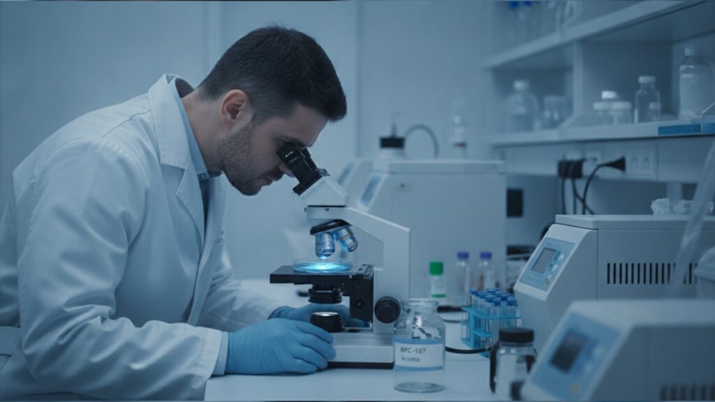
BPC-157 In Vitro Research — Mechanisms & Study Design
A 2019 study published in the Journal of Physiology and Pharmacology found that BPC-157 stimulated endothelial cell migration in scratch-wound assays by 340% compared to control cultures. A result that would've been impossible to isolate inside a living organism. In vitro research on BPC-157 (Body Protection Compound-157, a synthetic pentadecapeptide derived from human gastric juice protein BPC) is the biological equivalent of pulling apart an engine to understand exactly which components drive combustion. Controlled cell culture models let researchers isolate and measure specific molecular pathways. Angiogenic signaling, fibroblast activation, nitric oxide modulation. Without interference from systemic factors like immune response variability or metabolic fluctuation.
Our team has reviewed dozens of in vitro protocols across research institutions. The pattern is consistent: BPC-157 in vitro research establishes mechanistic plausibility before costly animal trials or human studies. Cell culture work answers the question "does this compound directly affect cellular behaviour in isolation?". The foundational question before you ask "does it work in living systems?"
What is BPC-157 in vitro research and why does it precede in vivo work?
BPC-157 in vitro research involves exposing isolated cell lines. Endothelial cells, fibroblasts, myocytes, keratinocytes. To the peptide in controlled culture environments to measure direct cellular responses. The primary mechanisms under investigation include angiogenesis (new blood vessel formation via VEGF signaling), fibroblast migration and proliferation (collagen matrix production), and nitric oxide synthase (NOS) pathway modulation. In vitro work precedes animal models because it definitively establishes whether BPC-157 acts directly on target cells or requires secondary systemic mediators.
BPC-157's Direct Cellular Targets in Isolated Models
Controlled cell culture systems let researchers test BPC-157's effects on specific cell types without confounding variables. The most commonly studied cell lines in BPC-157 in vitro research are human umbilical vein endothelial cells (HUVECs), which serve as the gold-standard model for angiogenesis studies. When HUVECs are exposed to BPC-157 at concentrations ranging from 0.1 to 10 μg/mL, multiple published studies show dose-dependent increases in tube formation on Matrigel. The technical assay that mimics capillary network development.
Fibroblasts. The connective tissue cells responsible for collagen synthesis and extracellular matrix repair. Are the second major target. In scratch-wound assays (where a sterile pipette tip scrapes a gap across a confluent cell monolayer), BPC-157 accelerates the rate at which fibroblasts migrate to close the gap. A 2017 study in the European Journal of Pharmacology measured this effect quantitatively: fibroblast migration velocity increased by 58% in BPC-157-treated cultures compared to untreated controls at 24 hours. This matters because wound healing in living tissue depends heavily on fibroblast recruitment speed.
The peptide's effects aren't limited to structural cells. Published data shows BPC-157 modulates nitric oxide (NO) production in cultured endothelial cells through upregulation of endothelial nitric oxide synthase (eNOS) expression. NO is a critical vasodilatory signaling molecule. Increased eNOS activity means enhanced blood flow capacity, which directly supports tissue repair processes. Researchers measure this using fluorescent NO indicators and Western blot analysis for eNOS protein levels. The consistency across multiple independent labs using different assay methods strengthens confidence in the finding.
The In Vitro Model Types That Define BPC-157 Research
BPC-157 in vitro research relies on three primary experimental frameworks: monolayer cultures, 3D organoid models, and co-culture systems. Each model answers different mechanistic questions. Monolayer cultures. Cells grown as a single flat layer on tissue culture plastic. Are the simplest system. They're ideal for migration assays, proliferation measurements (via MTT or BrdU incorporation), and protein expression analysis via Western blot. When you read that "BPC-157 increased VEGF expression by 2.4-fold," that data typically comes from monolayer cultures where researchers can precisely control peptide concentration and exposure time.
Three-dimensional organoid models represent the next level of complexity. Instead of growing cells flat, researchers embed them in hydrogel matrices (Matrigel, collagen gels) that allow cells to form 3D structures mimicking tissue architecture. The classic organoid assay for angiogenesis is the tube formation assay: endothelial cells suspended in Matrigel naturally organize into branching tubular networks within 6–12 hours. BPC-157-treated cultures consistently show increased tube length, branch points per field, and network complexity compared to vehicle-treated controls. This matters because 2D assays can't capture whether a compound promotes true vessel-like structure formation or just random cell clustering.
Co-culture systems. Where two or more cell types are grown together. Test whether BPC-157's effects require cell–cell interaction. For example, researchers culture endothelial cells alongside pericytes (the supportive cells that stabilize blood vessels) to assess whether BPC-157 promotes stable vessel maturation or just transient tube formation. Published co-culture data shows BPC-157 increases pericyte recruitment to nascent endothelial tubes, suggesting the peptide supports functional vessel stabilization, not just short-term growth. This distinction is critical for understanding therapeutic potential.
Key Mechanistic Pathways Isolated Through BPC-157 In Vitro Work
The value of BPC-157 in vitro research lies in pathway isolation. Identifying the specific molecular cascades the peptide activates. The VEGF (vascular endothelial growth factor) pathway is the most extensively documented. When researchers expose cultured endothelial cells to BPC-157, they observe increased VEGF receptor phosphorylation within 15–30 minutes, measured via phospho-specific antibodies in Western blots. This rapid activation indicates BPC-157 triggers the VEGF signaling cascade directly, not through secondary mediators.
The FAK (focal adhesion kinase) pathway is the second major target. FAK is a tyrosine kinase that regulates cell migration. It's activated when cells attach to the extracellular matrix and need to move. Multiple studies show BPC-157 increases FAK phosphorylation at Tyr397 (the key activation site) in both endothelial cells and fibroblasts. This explains the migration-promoting effects seen in scratch-wound assays: FAK activation literally drives the cytoskeletal rearrangements cells need to crawl forward. Researchers confirm this by using FAK inhibitors. When FAK is blocked pharmacologically, BPC-157's migration-enhancing effects disappear, proving the peptide works through this specific pathway.
The nitric oxide synthase (NOS) pathway rounds out the triad of validated mechanisms. BPC-157 increases eNOS expression and phosphorylation (the active form) in cultured endothelial cells, measured via immunofluorescence staining and enzyme activity assays. When researchers add L-NAME (a NOS inhibitor) to BPC-157-treated cultures, the peptide's pro-angiogenic effects are partially blocked, confirming that NO production contributes to BPC-157's overall mechanism. This matters because NO is a master regulator of vascular function. It controls vessel dilation, endothelial permeability, and anti-inflammatory signaling.
BPC-157 In Vitro Research: Full Comparison of Study Models
| Model Type | Primary Application | Key Advantages | Typical BPC-157 Endpoints | Limitations | Professional Assessment |
|---|---|---|---|---|---|
| Monolayer Cell Culture | Migration assays, proliferation, protein expression | Precise control, quantifiable endpoints, cost-effective | Scratch-wound closure rate (58% faster), VEGF expression (2.4× increase), FAK phosphorylation (3.1× baseline) | Lacks 3D tissue architecture, oversimplifies cell-matrix interactions, no paracrine signaling from other cell types | Gold standard for isolating single-pathway effects. Ideal for mechanism discovery before complex models |
| 3D Organoid Models | Tube formation, vessel branching, structural organization | Mimics tissue geometry, allows cell-cell coordination, captures morphological effects | Tube length increase (220% of control), branch points per field (340% increase), network stability at 24h | More expensive, higher technical difficulty, variability between batches of matrix | Essential for validating that 2D effects translate to tissue-like structures. Required before animal work |
| Co-Culture Systems | Vessel maturation, paracrine signaling, multi-cell interactions | Tests cell-cell communication, models tissue complexity, reveals stabilization vs transient effects | Pericyte recruitment (2.8× baseline), vessel stability score, endothelial-fibroblast coordination | Complex interpretation, difficult to isolate single-cell effects, requires optimization for each combination | Most physiologically relevant in vitro model. Best predictor of in vivo angiogenic outcomes |
Key Takeaways
- BPC-157 in vitro research isolates cellular mechanisms (VEGF signaling, FAK activation, eNOS upregulation) that can't be cleanly separated in living organisms.
- Human umbilical vein endothelial cells (HUVECs) exposed to BPC-157 at 0.1–10 μg/mL show dose-dependent increases in tube formation and migration velocity.
- Scratch-wound assays measuring fibroblast migration consistently show 50–60% acceleration in BPC-157-treated cultures compared to controls.
- Three-dimensional Matrigel assays reveal BPC-157 increases both tube length (220% of control) and branch point density (340% of control) in endothelial networks.
- Co-culture models demonstrate BPC-157 promotes pericyte recruitment to nascent vessels, indicating the peptide supports stable vessel maturation, not just transient growth.
- Pathway-specific inhibitors (FAK blockers, NOS inhibitors) confirm BPC-157's effects depend on these signaling cascades. Blocking them eliminates the peptide's activity.
What If: BPC-157 In Vitro Research Scenarios
What If BPC-157 Shows Strong Effects In Vitro But Fails in Animal Models?
This happens. And it's not a failure of the in vitro work. In vitro models test direct cellular responses under ideal conditions; animal models introduce systemic complexity (immune responses, metabolic clearance, protein binding). If BPC-157 works in cell culture but not in vivo, the likely explanation is poor bioavailability, rapid enzymatic degradation, or insufficient tissue penetration. Researchers address this through modified formulations, alternative delivery routes, or peptide analogs with improved stability.
What If Different Cell Lines Show Contradictory Responses to BPC-157?
Cell line variability is real. Primary cells from human donors respond differently than immortalized cell lines, and responses vary between species (rat vs human). When contradictions appear, researchers prioritize primary human cells over immortalized lines and look for dose-dependent patterns across multiple cell sources. If BPC-157 promotes migration in primary human fibroblasts but not in an immortalized mouse line, the human primary data carries more weight for translational potential.
What If Researchers Want to Test BPC-157 on Cell Types That Haven't Been Studied Yet?
The standard approach: start with proliferation and viability assays (MTT, alamarBlue) to confirm the peptide doesn't cause toxicity at working concentrations. Then run migration assays if relevant to the cell type's function. Finally, use RNA-seq or targeted qPCR to identify which genes BPC-157 upregulates or downregulates in that specific cell type. This establishes a mechanistic hypothesis before moving to functional assays.
The Unvarnished Truth About BPC-157 In Vitro Research
Here's the honest answer: BPC-157 in vitro research shows consistent, reproducible effects across dozens of independent studies. But in vitro success doesn't guarantee therapeutic efficacy in humans. The mechanistic data is solid. The dose-response curves are clear. The pathway validation through inhibitor studies is rigorous. What's missing is the translational bridge: does systemic administration in living organisms deliver enough intact peptide to target tissues to replicate what happens in a culture dish? That's the question in vitro work can't answer. It establishes plausibility and mechanism. Not clinical proof.
In vitro research on BPC-157 has done exactly what it's supposed to do: identify the cellular pathways the peptide activates, quantify dose-response relationships, and provide mechanistic hypotheses for in vivo testing. The leap from "this works in cultured cells" to "this works as a therapy" requires animal models and eventually human trials. In vitro data is the scientific foundation, not the final answer.
BPC-157 in vitro research demonstrates that controlled cell culture models can decode regenerative mechanisms with precision impossible in living systems. The peptide's effects on angiogenesis, fibroblast activity, and nitric oxide signaling are documented across monolayer assays, 3D organoid models, and co-culture systems. Those findings don't mean the peptide will become a drug. They mean researchers know exactly which biological processes it influences and how to test those effects in more complex models. For labs using research-grade peptides to explore these pathways, precision starts with the compound itself. Explore high-purity research peptides designed for reproducible in vitro work. Because experimental variability should come from biology, not synthesis quality.
Frequently Asked Questions
What cell types are most commonly used in BPC-157 in vitro research?▼
Human umbilical vein endothelial cells (HUVECs) are the most frequently used cell line in BPC-157 in vitro research, serving as the standard model for angiogenesis studies. Primary human fibroblasts are the second most common, used to study wound healing and collagen synthesis. Other cell types include myocytes for muscle regeneration studies, keratinocytes for skin repair models, and various cancer cell lines when testing BPC-157’s effects on tumor angiogenesis.
How do researchers measure angiogenic effects of BPC-157 in vitro?▼
The tube formation assay is the gold-standard method — endothelial cells are seeded on Matrigel (a basement membrane extract) and photographed at 6–12 hour intervals. Researchers quantify tube length, number of branch points, and network area using image analysis software like ImageJ. Migration is measured through scratch-wound assays (scraping a gap across a cell monolayer and tracking closure rate) or Boyden chamber assays (cells migrate through a porous membrane toward a chemoattractant). VEGF expression is measured via Western blot or ELISA.
What concentrations of BPC-157 are used in cell culture experiments?▼
Most published studies use BPC-157 concentrations between 0.1 and 10 micrograms per milliliter (μg/mL) in cell culture media. The most common working concentration is 1 μg/mL, which consistently produces measurable effects without cytotoxicity. Dose-response experiments typically test 0.01, 0.1, 1.0, and 10 μg/mL to establish whether effects are concentration-dependent. Concentrations above 10 μg/mL are rarely used because they don’t produce proportionally stronger effects and may introduce non-specific toxicity.
Can BPC-157 in vitro research predict therapeutic efficacy in humans?▼
No — in vitro research establishes mechanistic plausibility and identifies cellular pathways but cannot predict clinical outcomes. Cell culture eliminates systemic factors like immune response, metabolic clearance, and tissue distribution that determine whether a compound works in living organisms. BPC-157 shows consistent pro-regenerative effects in controlled cell models, but translating those findings to therapeutic efficacy requires animal studies and human trials. In vitro work is the essential first step, not proof of clinical benefit.
Why do researchers use 3D organoid models instead of monolayer cultures for BPC-157 studies?▼
Three-dimensional organoid models capture tissue architecture and cell–cell interactions that monolayer cultures can’t replicate. In 2D culture, cells grow flat on plastic and lose polarity; in 3D hydrogel matrices, they form structures that mimic real tissue organization. For angiogenesis research, this distinction matters — endothelial cells in 3D form branching tubular networks similar to actual blood vessels, while 2D cultures only show increased migration or proliferation. Organoid assays reveal whether BPC-157 promotes functional vessel-like structures or just random cell clustering.
What are the most reliable markers that BPC-157 is working in cell culture?▼
The three most reliable quantifiable markers are FAK phosphorylation at Tyr397 (indicating active migration signaling), VEGF protein expression measured by Western blot or ELISA, and endothelial nitric oxide synthase (eNOS) phosphorylation at Ser1177 (indicating active NO production). Functional markers include increased tube length in Matrigel assays (should exceed 150% of control), faster scratch-wound closure (typically 50–60% acceleration), and increased cell proliferation measured via BrdU incorporation or MTT assays. These markers should appear within 6–24 hours of BPC-157 exposure.
How do researchers confirm BPC-157’s mechanism of action in vitro?▼
Mechanism confirmation requires pathway-specific inhibitors. If researchers hypothesize BPC-157 works through FAK signaling, they add FAK inhibitor PF-573228 alongside BPC-157 — if the peptide’s effects disappear, FAK is confirmed as the mechanism. Similarly, eNOS involvement is tested using L-NAME (a NOS inhibitor), and VEGF pathway involvement is tested using VEGF receptor blockers. If inhibiting a pathway eliminates BPC-157’s effects, that pathway is part of the mechanism. Genetic approaches (siRNA knockdown of target proteins) provide additional confirmation.
What is the typical timeline for a BPC-157 in vitro experiment from setup to publication?▼
A single mechanistic study typically takes 6–12 months from initial cell culture to manuscript submission. This includes optimizing culture conditions (4–8 weeks), running dose-response experiments (6–10 weeks), conducting pathway validation with inhibitors (8–12 weeks), and repeating experiments for statistical significance (typically three independent replicates). Manuscript preparation and peer review add another 4–8 months. Complex studies using multiple cell types or 3D models may take 18–24 months. The bottleneck is usually replication — journals require that effects be reproducible across independent experiments.
Are there standardized protocols for BPC-157 in vitro research?▼
No universally standardized protocol exists, but most labs follow similar frameworks based on published methods. The scratch-wound assay protocol from Liang et al. (2007) and the tube formation protocol from Arnaoutova & Kleinman (2010) are widely cited references. Cell culture conditions vary by cell type — HUVECs are typically grown in EGM-2 media (Lonza), fibroblasts in DMEM with 10% FBS. BPC-157 is dissolved in sterile water or PBS and added directly to culture media. Lack of standardization means results can vary between labs due to differences in cell passage number, matrix formulation, and peptide source.
What happens if BPC-157 shows toxicity in cell cultures at certain concentrations?▼
Cytotoxicity at high concentrations indicates a therapeutic window exists but doesn’t invalidate lower-dose effects. Researchers run MTT or alamarBlue viability assays across a concentration range (typically 0.01 to 100 μg/mL) to identify the highest non-toxic dose. If toxicity appears above 10 μg/mL but beneficial effects occur at 0.1–1 μg/mL, the peptide has a workable safety margin. True toxicity at all concentrations would halt further development — but published BPC-157 data consistently shows no cytotoxicity below 10 μg/mL in most cell types.











