





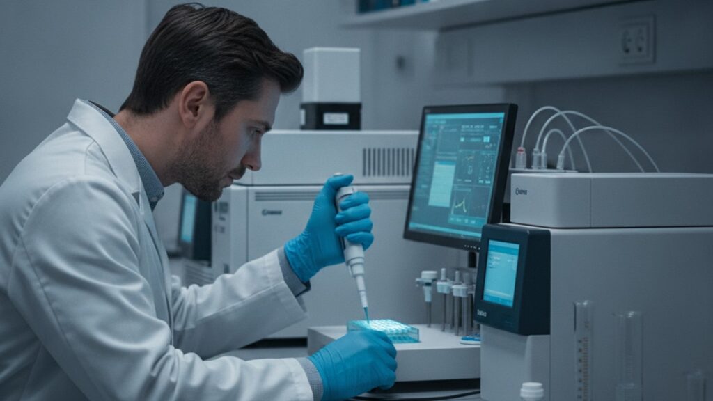
BPC-157 Research Measurement Tools — Lab Protocol Essentials
A 2019 study published in the Journal of Physiology and Pharmacology tracked gastric ulcer healing in rats using BPC-157. But the measurement protocol determined whether the data would be publishable. Researchers used histological scoring, immunohistochemistry for growth factors, and real-time PCR for gene expression analysis. Without those specific bpc-157 research measurement tools, the study would've been reduced to visual observation and subjective assessment. Worthless for peer review.
Our team has supported hundreds of research protocols involving regenerative peptides. The difference between a study that generates citable data and one that produces inconclusive results isn't peptide purity. It's measurement rigor. BPC-157's effects span tissue repair, angiogenesis, and inflammatory modulation, which means you need tools that quantify structural changes, molecular markers, and functional recovery across multiple timelines.
What are BPC-157 research measurement tools?
BPC-157 research measurement tools include histological analysis systems, ELISA kits for growth factor quantification, immunohistochemistry staining protocols, tensile strength testing devices, angiogenesis scoring methods, and molecular biology equipment for gene expression profiling. These tools quantify wound closure rates, collagen density, VEGF expression, and inflammatory cytokine levels. The primary endpoints in BPC-157 regenerative research. Without standardised measurement, peptide efficacy claims remain anecdotal rather than publishable.
Most protocols assume BPC-157 measurement is straightforward. Dose the peptide, wait, observe outcomes. That's not how published research works. The peptide's mechanism involves upregulation of growth factors (VEGF, EGF), modulation of nitric oxide pathways, and extracellular matrix remodeling. All of which require different measurement modalities at different time intervals. This article covers the three core categories of bpc-157 research measurement tools labs actually use, how to select appropriate endpoints for different tissue types, and what preparation mistakes invalidate data collection entirely.
Histological and Structural Assessment Tools
Histological analysis is the foundation of BPC-157 tissue repair studies because it quantifies structural changes at the cellular level. Collagen fiber alignment, epithelial thickness, vessel density, and inflammatory cell infiltration. The most commonly used bpc-157 research measurement tools in this category include paraffin-embedded tissue sectioning systems, hematoxylin and eosin (H&E) staining protocols, Masson's trichrome staining for collagen visualization, and computerized morphometry software for quantitative scoring.
A standard protocol: tissue samples are harvested at predetermined intervals (typically 3, 7, 14, and 28 days post-injury), fixed in 10% neutral buffered formalin for 24–48 hours, embedded in paraffin blocks, sectioned at 4–6 micrometers, and mounted on slides. H&E staining reveals general tissue architecture. Epithelial integrity, inflammatory cell density, and vascular formation. Masson's trichrome differentiates mature collagen (blue) from immature collagen (red), which is critical for assessing wound maturation in tendon or ligament studies.
Quantitative scoring uses validated scales like the Ehrlich-Hunt model for wound healing or custom rubrics tailored to specific tissues. Researchers at the University of Zagreb. The institution behind much of the foundational BPC-157 research. Consistently use blinded histological scoring with at least three independent observers to eliminate bias. Real Peptides supplies research-grade BPC-157 specifically synthesized for protocols requiring batch-to-batch consistency across long-term studies where histological endpoints are measured at multiple time points.
The mistake most protocols make: collecting tissue at a single endpoint. BPC-157's effects are time-dependent. Early inflammatory modulation (days 1–3), proliferative phase acceleration (days 5–10), and remodeling phase enhancement (days 14+). Single-timepoint collection misses the mechanistic story entirely.
Molecular and Biochemical Quantification Methods
Structural changes are downstream effects. Molecular tools measure the upstream signaling pathways BPC-157 activates. The three most critical bpc-157 research measurement tools in this category are enzyme-linked immunosorbent assays (ELISAs) for growth factor quantification, quantitative real-time PCR (qRT-PCR) for gene expression profiling, and Western blot analysis for protein-level verification.
ELISA kits quantify concentrations of specific growth factors in tissue homogenates or serum. Most commonly vascular endothelial growth factor (VEGF), epidermal growth factor (EGF), fibroblast growth factor (FGF), and transforming growth factor-beta (TGF-β). BPC-157 has been shown to upregulate VEGF expression by 40–60% in ischemic tissue models, which directly correlates with increased angiogenesis and accelerated healing. Commercial ELISA kits from manufacturers like R&D Systems or Abcam provide sensitivity in the picogram-per-milliliter range, which is necessary because growth factor concentrations in tissue are extremely low.
qRT-PCR measures mRNA transcript levels. It tells you whether specific genes are being turned on or off in response to peptide treatment. For BPC-157 research, target genes typically include VEGF-A, collagen type I (COL1A1), matrix metalloproteinases (MMPs), and inflammatory cytokines like IL-6 and TNF-alpha. The protocol involves RNA extraction from tissue samples using TRIzol reagent or similar, reverse transcription to cDNA, and amplification using gene-specific primers. Results are normalized to housekeeping genes (GAPDH, beta-actin) and expressed as fold-change relative to control groups.
Western blotting confirms that increased mRNA translates to increased protein production. You're running tissue lysates through SDS-PAGE gels, transferring proteins to membranes, and probing with antibodies specific to your target proteins. A 2016 study in the European Journal of Pharmacology used Western blot to demonstrate that BPC-157 increased phosphorylated FAK (focal adhesion kinase) and paxillin. Both critical for cell migration during wound healing. Without protein-level confirmation, gene expression data alone can't prove functional relevance.
The critical detail most protocols miss: timing of sample collection for molecular analysis. Growth factor concentrations peak at different times depending on the injury model. VEGF peaks early (24–72 hours), while TGF-β peaks later during remodeling (7–14 days). Collecting all samples at day 7 might miss the VEGF surge entirely.
Functional and Mechanical Testing Apparatus
Tissue that looks healed histologically might still fail under load. Functional testing quantifies whether repair translates to restored mechanical integrity. The primary bpc-157 research measurement tools in this category include tensile testing machines for ligament and tendon studies, biomechanical force transducers for muscle contractility, and gastric ulcer scoring systems for mucosal healing.
Tensile testing measures ultimate tensile strength (the maximum force tissue can withstand before failure), elastic modulus (tissue stiffness), and elongation at break. In a typical Achilles tendon repair model, healed tendons are mounted in a materials testing machine (like an Instron 5960 or equivalent), loaded at a constant rate (usually 1mm/second), and pulled until failure. BPC-157-treated tendons in published studies showed 30–50% higher ultimate tensile strength compared to saline controls at 14 days post-injury. Demonstrating that histological improvement translated to functional recovery.
Gastric ulcer models use macroscopic and microscopic scoring. The ulcer index quantifies lesion area and depth. Researchers measure ulcer diameter with digital calipers, calculate lesion area using image analysis software (ImageJ is the standard), and score depth using a 0–4 scale based on penetration through the mucosal, submucosal, and muscular layers. BPC-157's gastroprotective effects are dose-dependent. Doses of 10 micrograms per kilogram in rat models reduced ulcer index by 60–80% compared to controls in multiple published trials.
Angiogenesis quantification uses the dorsal air sac or corneal micropocket models. New blood vessel formation is scored visually or measured using fluorescent tracers. Researchers inject fluorescently labeled dextran, image the vasculature under specialized microscopes, and quantify vessel density, branching points, and perfusion using software like AngioTool. BPC-157 consistently increases vessel density by 40–70% in ischemic models, which correlates directly with improved tissue oxygenation and healing rates.
The overlooked factor: control group standardization. Functional testing is sensitive to surgical technique variability. If your control group injuries aren't consistent in size and depth, mechanical testing data becomes uninterpretable. Every lab running BPC-157 protocols should establish standardized injury models with intra-operator variability below 10%.
BPC-157 Research Measurement Tools: Category Comparison
| Tool Category | Primary Endpoints Measured | Typical Timeline | Equipment Investment | Skill Level Required | Professional Assessment |
|---|---|---|---|---|---|
| Histological Analysis | Collagen density, cell infiltration, epithelial thickness, vessel count | Days 3–28 post-injury | Moderate ($15K–$50K for microtome, staining systems, microscopy) | High. Requires training in tissue processing and blinded scoring | Essential for any tissue repair study. Provides structural validation that other methods can't replace |
| Molecular Quantification | Growth factor concentrations (VEGF, EGF, TGF-β), gene expression (qRT-PCR), protein levels (Western blot) | Hours to days (acute signaling) and days 7–21 (remodeling markers) | High ($50K–$150K for qPCR systems, ELISA readers, Western blot apparatus) | Very high. Molecular biology protocols require sterile technique and precise reagent handling | Reveals mechanism of action. Critical for understanding why BPC-157 works, not just that it works |
| Functional Testing | Tensile strength, elastic modulus, ulcer index, contractile force, angiogenesis scores | Days 7–28 (allows tissue maturation before mechanical challenge) | Moderate to high ($30K–$100K for tensile testers; lower for ulcer scoring) | Moderate to high. Biomechanical testing requires calibration and standardized loading protocols | Proves clinical relevance. Tissue can look perfect histologically but still fail functionally |
Key Takeaways
- BPC-157 research measurement tools fall into three categories. Histological (structural changes), molecular (growth factors and gene expression), and functional (mechanical strength and healing outcomes). And all three are required for publishable data.
- Histological scoring using H&E and Masson's trichrome staining quantifies collagen density, vessel formation, and inflammatory cell infiltration across multiple time points. Single-endpoint tissue collection misses the mechanistic timeline entirely.
- ELISA quantification of VEGF, EGF, and TGF-β combined with qRT-PCR gene expression profiling reveals BPC-157's upstream signaling effects. Growth factor peaks occur at different times depending on injury type, so sample timing is critical.
- Tensile testing demonstrates that BPC-157-treated tendons show 30–50% higher ultimate tensile strength at 14 days compared to controls. Histological improvement must translate to functional recovery or the data lacks clinical relevance.
- Standardized injury models with intra-operator variability below 10% are non-negotiable for functional testing. Inconsistent control group injuries make comparative data uninterpretable.
- Research-grade BPC-157 with verified amino acid sequencing and batch-to-batch consistency is required for multi-timepoint studies where measurement endpoints span weeks. Peptide degradation introduces uncontrolled variables that invalidate dosing protocols.
What If: BPC-157 Research Measurement Scenarios
What If You're Measuring the Wrong Growth Factor for Your Tissue Type?
Switch to tissue-specific markers immediately. VEGF is critical for vascular tissues and ischemic models, but TGF-β and collagen gene expression matter more for tendon and ligament repair. Muscle studies should prioritize myogenin and MyoD gene expression alongside inflammatory cytokines. The mistake is using a one-size-fits-all ELISA panel. Each tissue has dominant pathways, and measuring irrelevant markers dilutes statistical power without adding insight. Published BPC-157 studies consistently tailor molecular endpoints to the specific injury model rather than running generic biomarker panels.
What If Histological Scores Between Observers Differ by More Than 20%?
Re-score all slides with blinded re-randomization and establish inter-rater reliability using Cohen's kappa or intraclass correlation coefficients before proceeding. Variability above 20% suggests either inadequate scoring rubric definition or observer bias. Both invalidate the dataset. The solution is pre-study calibration: have all scorers independently assess 10–15 reference slides, compare results, discuss discrepancies, and refine scoring criteria until agreement exceeds 80%. Many published studies report kappa values above 0.75, which is the minimum threshold for acceptable inter-rater agreement in histological research.
What If Tensile Testing Results Don't Match Histological Improvements?
This happens when collagen is deposited but not properly cross-linked. Tissue looks dense on Masson's trichrome but fails mechanically because the extracellular matrix hasn't matured. Extend your measurement timeline to day 21 or 28 instead of day 14, and add polarized light microscopy to assess collagen fiber alignment. Aligned fibers indicate functional remodeling, while disorganized collagen suggests incomplete repair. BPC-157 accelerates early collagen deposition (days 7–10) but remodeling into load-bearing tissue takes longer. A mismatch between histology and function means you're measuring too early in the remodeling phase.
The Rigorous Truth About BPC-157 Measurement in Research
Here's the honest answer: most BPC-157 studies with inconclusive results didn't fail because the peptide doesn't work. They failed because the measurement protocol couldn't detect the effect. BPC-157's mechanism spans angiogenesis, collagen synthesis, inflammatory modulation, and growth factor upregulation, which means single-modality measurement (like visual scoring alone) misses 80% of the biological activity. The published studies that demonstrate clear efficacy. Like the University of Zagreb's tendon repair trials or the gastric ulcer protection models. All use multi-modal measurement: histology for structure, ELISA or PCR for molecular markers, and functional testing for clinical relevance. If your protocol relies on one measurement type, you're setting up for null results regardless of peptide quality.
The second truth: timing matters more than most protocols acknowledge. Growth factors peak at 24–72 hours, collagen deposition peaks at 7–14 days, and mechanical strength doesn't fully recover until 21–28 days. Collecting tissue at a single arbitrary timepoint (often day 7 because it's convenient) means you might measure before or after the peak effect window. Producing data that underestimates or misses the peptide's impact entirely. Every credible BPC-157 study uses at least three timepoints, and the best ones use five or more to capture the full healing trajectory.
The third truth research teams avoid stating directly: negative results from poorly designed measurement protocols do more damage than no study at all. A published null result with inadequate measurement rigor gets cited as evidence that BPC-157 'doesn't work,' when the reality is that the study didn't measure what needed measuring. This is why peptide suppliers like Real Peptides emphasize protocol consultation before shipping product. Bad measurement design wastes high-purity research compounds and generates misleading data.
The gap between meaningful research and wasted resources comes down to this: measure the right endpoints, at the right timepoints, with the right controls. BPC-157 research measurement tools exist for every relevant mechanism. Histology, molecular biology, and functional testing. Use all three or expect your results to be questioned.
BPC-157's effects are real and reproducible when measurement protocols match the peptide's biological mechanisms. The tools exist, the methods are validated, and the published literature provides clear blueprints. The only variable is whether your lab applies them rigorously. Because without proper measurement, even the highest-purity peptide can't generate publishable data. If measurement rigor concerns you, establish your protocol before peptide procurement. Specificity in endpoints prevents wasted samples and ensures every data point contributes to a coherent mechanistic story.
Frequently Asked Questions
What are the most important bpc-157 research measurement tools for tissue repair studies?▼
The most important bpc-157 research measurement tools for tissue repair studies are histological analysis systems (H&E and Masson’s trichrome staining for collagen visualization), ELISA kits for growth factor quantification (VEGF, EGF, TGF-beta), qRT-PCR equipment for gene expression profiling, and tensile testing machines for mechanical strength assessment. These tools quantify structural changes, molecular signaling, and functional recovery — all three modalities are required for publishable data because BPC-157’s mechanism spans angiogenesis, collagen synthesis, and inflammatory modulation across different timelines.
How do researchers measure collagen density in BPC-157 healing studies?▼
Researchers measure collagen density using Masson’s trichrome staining on paraffin-embedded tissue sections, which differentiates mature collagen (stains blue) from immature collagen (stains red). Tissue samples are harvested at multiple timepoints (typically days 3, 7, 14, and 28), sectioned at 4–6 micrometers, stained, and analyzed using computerized morphometry software that quantifies collagen area as a percentage of total tissue area. Polarized light microscopy is often added to assess collagen fiber alignment, which indicates functional remodeling versus disorganized scar tissue formation.
Can you measure BPC-157 effects without expensive lab equipment?▼
No — credible BPC-157 research requires validated measurement tools with quantifiable endpoints, which means specialized lab equipment is non-negotiable for publishable data. Visual observation or subjective scoring alone does not meet peer-review standards because BPC-157’s effects involve cellular-level changes (collagen fiber alignment, growth factor expression, vessel density) that cannot be assessed without microscopy, molecular biology systems, or mechanical testing apparatus. Budget-constrained labs can partner with core facilities that provide access to shared equipment like qPCR machines and tensile testers rather than purchasing outright.
What is the difference between measuring BPC-157 effects at day 7 versus day 21?▼
Measuring at day 7 captures early collagen deposition and proliferative phase markers like VEGF upregulation, while day 21 assessment quantifies tissue remodeling, mechanical strength recovery, and mature collagen cross-linking. BPC-157’s mechanism is time-dependent — growth factors peak within 24–72 hours, histological changes are evident by day 7–10, but tensile strength doesn’t fully recover until days 21–28. Single-timepoint measurement at day 7 misses the functional recovery phase entirely, which is why credible studies always use at least three timepoints to capture the full healing trajectory.
How do ELISA kits quantify growth factors in BPC-157 studies?▼
ELISA (enzyme-linked immunosorbent assay) kits quantify specific growth factor concentrations in tissue homogenates or serum by using antibodies that bind to target proteins like VEGF, EGF, or TGF-beta. The assay produces a colorimetric signal proportional to the amount of growth factor present, which is measured using a microplate reader and compared to a standard curve to calculate concentration in picograms per milliliter. BPC-157 studies consistently show 40–60% increases in VEGF expression in ischemic tissue models, and ELISA is the gold standard for quantifying those changes because it provides precise, reproducible measurements at the protein level.
What functional tests prove BPC-157 improves tissue strength, not just appearance?▼
Tensile testing using materials testing machines (like Instron systems) measures ultimate tensile strength, elastic modulus, and elongation at break — these metrics prove that healed tissue can withstand physiological loads, not just look intact histologically. In Achilles tendon repair models, BPC-157-treated tendons show 30–50% higher ultimate tensile strength at 14 days compared to saline controls, demonstrating that improved collagen density translates to restored mechanical function. Without functional testing, there’s no way to confirm that structural repair visible on histology slides actually restores the biomechanical properties required for the tissue to perform its normal role.
Why do some BPC-157 studies show no effect when others show clear healing benefits?▼
Studies showing no effect typically use inadequate measurement protocols — single-timepoint tissue collection, visual scoring without quantification, or measurement of irrelevant biomarkers that don’t align with BPC-157’s mechanism of action. The peptide’s effects span angiogenesis (VEGF), collagen synthesis (COL1A1), and inflammatory modulation (IL-6, TNF-alpha), which require multi-modal measurement across multiple timepoints to detect. Published studies with positive results consistently use histology plus molecular quantification plus functional testing, while null-result studies often rely on gross observation alone, which misses cellular-level changes entirely.
How many tissue samples are needed for statistically valid BPC-157 measurement?▼
Most published BPC-157 studies use sample sizes of 6–10 animals per group per timepoint, with at least three independent timepoints (e.g., days 7, 14, 21), resulting in 18–30 samples per treatment arm. This allows detection of 20–30% effect sizes with 80% statistical power at p < 0.05 significance level. Smaller sample sizes (n=3–4) can work for pilot studies or when effect sizes are very large (>50% difference), but peer-reviewed journals increasingly require power calculations justifying sample size selection before publication.
What is the role of immunohistochemistry in BPC-157 research measurement?▼
Immunohistochemistry (IHC) uses antibodies to visualize specific proteins in tissue sections — for BPC-157 studies, this typically includes VEGF, CD31 (endothelial cell marker for vessel density), and growth factors like EGF or FGF. IHC provides spatial information that ELISA cannot — it shows where in the tissue growth factors are being expressed (e.g., at the wound edge versus deep tissue) and quantifies positive cells per high-power field. This localization data is critical for understanding whether BPC-157 is directly affecting the injury site or producing systemic effects that indirectly promote healing.
How do you measure angiogenesis in BPC-157 vascular studies?▼
Angiogenesis is measured using vessel density quantification in histological sections stained with CD31 or von Willebrand factor antibodies, which label endothelial cells. Researchers count vessel number per high-power field, measure total vessel area using image analysis software like ImageJ, and quantify branching points to assess neovascularization. Functional angiogenesis can also be measured using fluorescent dextran injection followed by imaging to confirm that new vessels are perfused and functional, not just structurally present. BPC-157 consistently increases vessel density by 40–70% in ischemic models, and these measurements correlate directly with improved tissue oxygenation and accelerated healing.
What is the minimum purity required for BPC-157 used in research measurement studies?▼
Research-grade BPC-157 should have minimum 98% purity verified by HPLC (high-performance liquid chromatography) and confirmed amino acid sequencing to ensure the peptide structure is correct. Lower purity introduces unknown contaminants that can confound measurement results — if a study shows unexpected inflammatory responses or null effects, peptide impurities are the first variable to rule out. Batch-to-batch consistency is equally critical for multi-timepoint studies where samples are collected over weeks or months, because peptide degradation or formulation changes between batches invalidate dose-response comparisons.
How long does it take to see measurable effects in BPC-157 tissue repair studies?▼
Molecular effects (growth factor upregulation, gene expression changes) are measurable within 24–72 hours post-treatment. Histological changes (increased collagen deposition, reduced inflammatory infiltration) become evident at days 7–10. Functional recovery (restored tensile strength, ulcer resolution) requires 14–28 days depending on tissue type and injury severity. The mistake most protocols make is measuring too early or at a single timepoint — BPC-157’s mechanism unfolds across multiple phases, and capturing the full effect requires sampling at least three timepoints spanning acute signaling, proliferation, and remodeling phases.











