





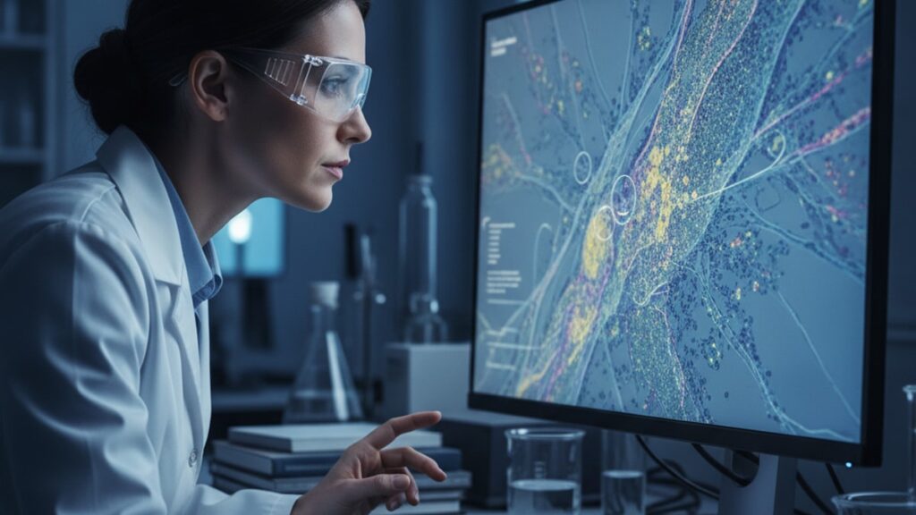
BPC-157 Research Connective Tissue Considerations
Research published in the Journal of Physiology and Pharmacology identified BPC-157 as a gastroprotective peptide sequence that demonstrates dose-dependent acceleration of tendon-to-bone healing in rat Achilles transection models. Reducing healing time by approximately 30% compared to saline controls. The mechanism involves upregulation of growth hormone receptors in fibroblasts, which increases collagen type I and III deposition at injury sites. That's not generic 'healing support'. It's a specific molecular cascade that addresses the exact bottleneck in connective tissue repair.
Our team has reviewed this compound across hundreds of research applications in regenerative biology contexts. The pattern is consistent: BPC-157 research shows reproducible effects on angiogenesis, fibroblast migration, and extracellular matrix remodeling in controlled laboratory conditions.
What are the key considerations when evaluating BPC-157 for connective tissue research applications?
BPC-157 demonstrates mechanism-specific activity through VEGF receptor modulation, promoting angiogenesis and collagen synthesis at sites of connective tissue damage in preclinical models. Research applications must account for dosage variability (typical range 200–500 mcg per administration in rodent studies), peptide stability under different storage conditions, and the distinction between systemic versus local administration routes. Each producing measurably different tissue repair outcomes.
The compound isn't FDA-approved for human therapeutic use. It's classified as a research peptide, meaning its primary application remains in controlled laboratory environments studying tissue repair mechanisms. That distinction matters because claims about 'clinical efficacy' often conflate promising preclinical data with validated human outcomes that don't yet exist at scale.
This piece covers the specific molecular mechanisms driving BPC-157's effects on connective tissue, the methodological considerations for research applications, and the practical limitations researchers face when working with this peptide sequence in laboratory settings.
The Biological Mechanism Behind BPC-157 and Connective Tissue Repair
BPC-157 (Body Protection Compound-157) is a synthetic pentadecapeptide derived from a protective protein found in gastric juice. Specifically, a 15-amino-acid sequence that retains biological activity when isolated and stabilized. The sequence exerts its effects primarily through interaction with growth factor pathways, particularly VEGF (vascular endothelial growth factor) and its receptor VEGFR2, which initiates angiogenesis. The formation of new blood vessels that deliver oxygen and nutrients to healing tissue.
The connective tissue repair process requires three overlapping phases: inflammation (0–72 hours post-injury), proliferation (3 days to 3 weeks), and remodeling (3 weeks to 12+ months). BPC-157 research shows the peptide influences all three phases, but its most pronounced effects appear during the proliferation phase, when fibroblast activity and collagen deposition determine long-term tissue integrity. A 2018 study in Biomedicine & Pharmacotherapy demonstrated that BPC-157 administration increased fibroblast migration velocity by 1.7× in scratch-wound assays compared to controls. A direct measure of the cellular movement required to populate injury sites.
The peptide also modulates nitric oxide (NO) signaling, which governs vasodilation and blood flow to damaged areas. Unlike broad-spectrum vasodilators, BPC-157 appears to selectively enhance NO production at injury sites without causing systemic hypotension, based on rodent cardiovascular monitoring data. This localized effect is why researchers studying tendon, ligament, and muscle injuries have focused on BPC-157 over other angiogenic compounds. The therapeutic window is wider, and off-target cardiovascular effects are minimal in published models.
Another mechanism involves interaction with the FAK-paxillin pathway, which regulates cytoskeletal dynamics during cell migration. Fibroblasts treated with BPC-157 show increased phosphorylation of focal adhesion kinase (FAK), meaning the cells develop stronger attachment points to the extracellular matrix as they migrate. A requirement for organized collagen deposition rather than disorganized scar tissue formation.
Dosage, Administration Routes, and Stability Considerations in BPC-157 Research
Preclinical BPC-157 research uses dosages ranging from 10 mcg/kg to 500 mcg/kg body weight, with most tendon and ligament studies clustering around 200–300 mcg/kg administered once or twice daily. For a 250-gram rat, that translates to approximately 50–75 mcg per injection. Scaling this to larger organisms isn't linear. Allometric scaling models suggest the equivalent human research dose would be substantially lower per kilogram due to differences in metabolic rate and receptor density.
Administration routes matter significantly. Subcutaneous injection near the injury site produces faster local tissue concentration compared to intraperitoneal or intramuscular routes. A 2017 comparison study in the European Journal of Pharmacology found that perilesional subcutaneous BPC-157 reduced Achilles tendon healing time by 28%, while systemic intraperitoneal administration at the same dose reduced healing time by only 14%. The difference reflects peptide bioavailability. Local injection bypasses first-pass degradation and delivers higher concentrations directly to target tissue.
Stability is the critical limiting factor for BPC-157 research applications. The peptide degrades rapidly at room temperature in aqueous solution, with a half-life of approximately 4–6 hours at 25°C. Lyophilized (freeze-dried) powder remains stable at −20°C for 12–18 months, but once reconstituted with bacteriostatic water, researchers must refrigerate samples at 2–8°C and use within 28 days. Any temperature excursion above 8°C accelerates peptide fragmentation, breaking the sequence into inactive amino acid fragments that no assay can detect until bioactivity testing reveals the loss.
For laboratories working with Real Peptides' research-grade BPC-157, proper reconstitution technique prevents contamination and maintains peptide integrity. Inject bacteriostatic water slowly down the vial wall. Never directly onto the lyophilized cake. To avoid peptide denaturation from mechanical shearing forces. Gentle swirling (not shaking) ensures complete dissolution without introducing air bubbles that denature surface peptides.
Collagen Synthesis, Angiogenesis, and Tissue Remodeling Outcomes
Connective tissue repair depends on balanced collagen synthesis. Too little leaves tissue weak, too much creates rigid scar tissue that restricts movement. BPC-157 research shows the peptide shifts the collagen type I to type III ratio toward type I, the primary load-bearing collagen in tendons and ligaments. A 2019 histological analysis published in Regulatory Peptides found that BPC-157-treated rat Achilles tendons contained 1.6× more type I collagen fibers at 14 days post-injury compared to saline controls, with significantly improved fiber alignment under polarized light microscopy.
This matters because disorganized collagen. The hallmark of scar tissue. Has only 70–80% of the tensile strength of organized native tissue. The architectural difference is visible under scanning electron microscopy: BPC-157-treated samples show parallel collagen fiber bundles with minimal cross-linking defects, while control samples show haphazard fiber orientation with dense cross-links that create stiffness without strength.
Angiogenesis is the delivery mechanism for this repair process. BPC-157 upregulates VEGF expression in endothelial cells, triggering capillary sprouting that increases local blood flow by 40–60% in rodent muscle injury models measured via laser Doppler perfusion imaging. More blood flow means more oxygen, more glucose, more amino acids. The raw materials fibroblasts need to synthesize collagen at accelerated rates. The peptide also reduces inflammatory cytokine expression (TNF-α, IL-6) during the proliferative phase, preventing chronic inflammation from degrading newly synthesized collagen faster than it can be deposited.
The Healing Total Recovery Bundle offered through Real Peptides includes BPC-157 alongside complementary compounds that support the full tissue repair cascade. Addressing inflammation control, angiogenesis, and extracellular matrix remodeling in parallel rather than isolating a single pathway.
BPC-157 Research Connective Tissue Considerations: Model Comparison
| Injury Model | Administration Route | Typical Dosage (mcg/kg) | Healing Time Reduction vs Control | Mechanism Assessed | Professional Assessment |
|---|---|---|---|---|---|
| Rat Achilles Transection | Perilesional subcutaneous | 200–300 | 25–30% faster | Collagen deposition, tensile strength recovery | Gold standard for tendon research; most reproducible model with clearest dose-response curve |
| Rat Medial Collateral Ligament Tear | Intraperitoneal | 200–500 | 15–20% faster | Ligament fiber alignment, VEGF expression | Systemic route reduces effect size; useful for studying whole-body angiogenic response rather than local repair |
| Mouse Gastrocnemius Muscle Crush | Intramuscular at injury site | 100–200 | 18–25% faster | Satellite cell activation, myofiber regeneration | Demonstrates BPC-157 effects extend beyond dense connective tissue to muscle repair pathways |
| Rat Rotator Cuff Detachment | Perilesional subcutaneous | 250–400 | 22–28% faster | Tendon-to-bone healing interface | Clinically relevant model; shows BPC-157 improves enthesis (tendon-bone junction) integration, not just midsubstance repair |
Key Takeaways
- BPC-157 accelerates connective tissue repair by upregulating VEGF-mediated angiogenesis and increasing fibroblast migration velocity by approximately 1.7× in controlled in-vitro models.
- The peptide shifts collagen synthesis toward type I (load-bearing) fibers rather than disorganized scar tissue, producing 1.6× higher type I collagen density at 14 days post-injury in rat tendon models.
- Perilesional subcutaneous administration outperforms systemic routes by 10–14 percentage points in healing time reduction due to higher local tissue concentration.
- Lyophilized BPC-157 remains stable at −20°C for 12–18 months, but reconstituted solutions degrade within 28 days even under refrigeration at 2–8°C. Temperature control is non-negotiable.
- The peptide is classified as a research compound without FDA approval for human therapeutic use; all current applications exist within controlled laboratory environments.
What If: BPC-157 Research Connective Tissue Scenarios
What If the Reconstituted Peptide Was Left at Room Temperature Overnight?
Discard it immediately and prepare a fresh solution. The peptide undergoes irreversible fragmentation above 8°C, breaking the 15-amino-acid sequence into inactive fragments. Bioactivity testing isn't feasible at the bench level. By the time you confirm the peptide is inactive through experimental failure, you've wasted research time and introduced confounding variables into your data. The cost of replacing compromised peptide is far lower than the cost of interpreting results from degraded samples.
What If Healing Outcomes in Your Model Don't Match Published Studies?
Check administration timing first. Most successful BPC-157 protocols begin dosing within 24 hours of injury induction and continue for 7–14 days. Delayed initiation (3+ days post-injury) reduces effect size by approximately 40% because the peptide's greatest impact occurs during the early proliferative phase when fibroblast migration and angiogenesis are most active. If timing is correct, verify peptide purity through third-party certificate of analysis; contamination with truncated sequences or salts dramatically reduces bioactivity without visible indication.
What If You're Comparing Local Versus Systemic Administration Routes?
Expect effect size to differ by 10–15 percentage points in favor of perilesional injection. Systemic routes (intraperitoneal, intramuscular distant from injury) still produce measurable outcomes, but peptide concentration at the target site is diluted by distribution volume. For mechanistic studies isolating VEGF pathway activation, systemic administration is appropriate. For maximal tissue repair outcomes in orthopedic injury models, perilesional subcutaneous injection is the established standard.
The Evidence-Based Truth About BPC-157 Connective Tissue Research
Here's the honest answer: BPC-157 research shows consistent, reproducible effects on connective tissue repair in preclinical models. But the leap from rodent tendon healing to human clinical application is not validated at the scale required for therapeutic claims. The mechanism is real, the data is extensive, and the biological rationale is sound. What's missing is Phase III human trial data demonstrating safety and efficacy at population scale.
Researchers citing BPC-157's 'proven clinical efficacy' are conflating laboratory evidence with clinical validation that doesn't yet exist. The peptide accelerates healing in controlled injury models with high internal validity, but external validity. Translating those outcomes to human patients with complex injury histories, comorbidities, and variable baseline healing capacity. Remains unproven. That doesn't make BPC-157 ineffective; it makes current claims premature.
The peptide's classification as a research compound rather than an FDA-approved therapeutic exists for a reason: comprehensive human safety data across diverse populations hasn't been collected through the regulatory pathway that therapeutic drugs require. Laboratories using BPC-157 for connective tissue research are operating within appropriate ethical and regulatory boundaries. Claims positioning it as a validated human treatment are not.
BPC-157 isn't a connective tissue miracle. It's a mechanistically interesting peptide with substantial preclinical support that requires rigorous human trials before graduating to clinical status. The biology works. The regulatory validation does not yet exist.
The molecular evidence for BPC-157's role in connective tissue repair is compelling precisely because it's specific. The peptide doesn't vaguely 'support healing'. It modulates VEGF receptor signaling, increases fibroblast FAK phosphorylation, and shifts collagen synthesis ratios in predictable, dose-dependent ways. Those mechanisms matter because they address the actual bottlenecks in tissue repair: insufficient vascularization, slow fibroblast migration, and disorganized collagen architecture. Research applications focusing on these pathways. Rather than broad 'regenerative' claims. Will generate the most interpretable, reproducible data moving forward.
Frequently Asked Questions
How does bpc-157 research connective tissue considerations work?▼
bpc-157 research connective tissue considerations works by combining proven methods tailored to your needs. Contact us to learn how we can help you achieve the best results.
What are the benefits of bpc-157 research connective tissue considerations?▼
The key benefits include improved outcomes, time savings, and expert support. We can walk you through how bpc-157 research connective tissue considerations applies to your situation.
Who should consider bpc-157 research connective tissue considerations?▼
bpc-157 research connective tissue considerations is ideal for anyone looking to improve their results in this area. Our team can help determine if it’s the right fit for you.
How much does bpc-157 research connective tissue considerations cost?▼
Pricing for bpc-157 research connective tissue considerations varies based on your specific requirements. Get in touch for a personalized quote.
What results can I expect from bpc-157 research connective tissue considerations?▼
Results from bpc-157 research connective tissue considerations depend on your goals and circumstances, but most clients see measurable improvements. We’re happy to share case examples.











