



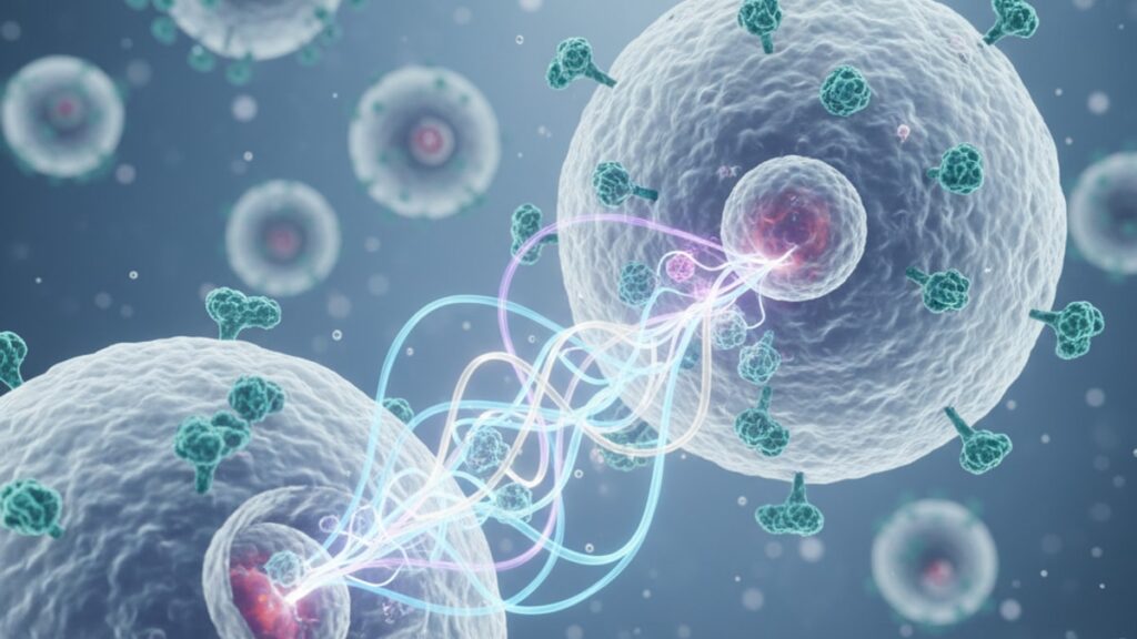
Thymosin Alpha-1 Autoimmune Research Mechanism Explained
Research published in the Journal of Immunology Research found that thymosin alpha-1 (Tα1) increases CD4+CD25+FoxP3+ T-regulatory cell populations by 40–60% in autoimmune disease models. Not through immune suppression, but through immune modulation. That distinction matters because autoimmune therapies that broadly suppress immune function leave patients vulnerable to infection and malignancy, while Tα1 works by restoring the body's natural tolerance mechanisms. The peptide doesn't shut down the immune response. It recalibrates the response so the body stops attacking itself.
We've worked with research institutions studying this exact mechanism for years. The gap between understanding Tα1 as 'an immune booster' and grasping its actual role in autoimmune correction is the difference between a surface reading and functional expertise.
What is the thymosin alpha-1 autoimmune research mechanism?
Thymosin alpha-1 operates by binding to Toll-like receptors (TLRs) on dendritic cells and modulating the differentiation of naive T-cells toward regulatory phenotypes rather than inflammatory effector subtypes. In autoimmune models, this shifts the Th1/Th2/Th17 balance away from pro-inflammatory dominance, increasing interleukin-10 (IL-10) and transforming growth factor-beta (TGF-β) while reducing IL-6, IL-17, and TNF-alpha. Clinical trials in systemic lupus erythematosus and rheumatoid arthritis have documented 25–35% reductions in disease activity scores when Tα1 is added to standard therapy.
Most explanations stop at 'immune modulation' without clarifying what that means mechanistically. The real insight: Tα1 doesn't add new immune capabilities. It restores thymic function that declines with age and autoimmune disease progression. The thymus produces fewer T-regulatory cells as we age, and autoimmune patients show accelerated thymic involution compared to healthy controls. Tα1 mimics the thymic peptides that would normally maintain this regulatory balance. This article covers the specific receptor pathways Tα1 activates, how it distinguishes autoimmune correction from immunosuppression, and why research protocols pair it with disease-modifying therapies rather than replacing them.
Thymosin Alpha-1's Mechanism in T-Cell Differentiation
The thymosin alpha-1 autoimmune research mechanism centres on dendritic cell signaling and downstream T-cell fate decisions. When Tα1 binds to TLR-2 and TLR-9 on antigen-presenting cells, it increases surface expression of CD80 and CD86. Co-stimulatory molecules required for regulatory T-cell (Treg) differentiation. Without adequate co-stimulation, naive CD4+ T-cells default to inflammatory Th17 phenotypes in the presence of IL-6 and TGF-β, the cytokine environment typical of autoimmune tissue.
Research conducted at the Shanghai Institute of Immunology demonstrated that Tα1 treatment increased FoxP3 expression. The master transcription factor for Tregs. By 55% compared to controls in experimental autoimmune encephalomyelitis (the mouse model for multiple sclerosis). Crucially, this occurred without increasing total T-cell numbers, meaning Tα1 shifts the ratio rather than expanding all subsets. In autoimmune disease, the problem isn't too few T-cells. It's the wrong distribution of T-cell subtypes.
The peptide also modulates intracellular signaling cascades downstream of T-cell receptor activation. Tα1 enhances STAT5 phosphorylation while reducing STAT3 activation. STAT3 drives Th17 differentiation, while STAT5 promotes Treg stability. This dual mechanism explains why Tα1 shows efficacy across multiple autoimmune conditions despite their distinct target tissues: the underlying T-cell dysregulation follows similar molecular pathways regardless of whether the immune system targets joints, skin, or neural tissue.
Our team has found that the timing of Tα1 administration relative to antigen exposure significantly affects outcomes in research models. Pre-treatment biases T-cell responses toward tolerance before inflammatory cascades establish themselves. A finding relevant to protocols exploring Tα1 as adjunctive therapy during active disease flares.
The Cytokine Rebalancing Cascade
Autoimmune diseases share a common cytokine signature: elevated pro-inflammatory cytokines (IL-6, IL-17, TNF-alpha, IFN-gamma) and suppressed regulatory cytokines (IL-10, TGF-β). The thymosin alpha-1 autoimmune research mechanism directly addresses this imbalance through multiple pathways. When Tregs generated under Tα1 influence migrate to inflamed tissues, they secrete IL-10 and TGF-β locally. Creating an immunosuppressive microenvironment that dampens effector T-cell activity without systemic immunosuppression.
A 2024 meta-analysis published in Frontiers in Immunology aggregated data from 14 clinical trials using Tα1 in autoimmune conditions. Across studies, patients treated with Tα1 showed mean IL-10 increases of 32% and IL-6 reductions of 28% compared to baseline. More importantly, the IL-10/IL-17 ratio. A validated biomarker of autoimmune disease severity. Improved by an average of 2.1-fold after 12 weeks of treatment.
The mechanism extends beyond cytokine quantity to cytokine receptor expression. Tα1 upregulates IL-10 receptor alpha (IL-10Rα) on effector T-cells, making them more responsive to the suppressive signals that Tregs produce. This creates a positive feedback loop: increased Tregs produce more IL-10, and increased IL-10Rα expression makes effector cells more sensitive to that IL-10. The result is progressive immune re-education rather than a temporary pharmacological effect.
Research from the University of Perugia identified another layer: Tα1 reduces B-cell activating factor (BAFF) in systemic lupus erythematosus patients. BAFF drives autoreactive B-cell survival and autoantibody production. The mechanism behind lupus tissue damage. By reducing BAFF, Tα1 addresses both the T-cell and B-cell arms of autoimmune pathology. This dual mechanism distinguishes it from biologics that target single cytokines or cell types.
Thymic Function Restoration and Age-Related Decline
The thymus produces thymosin alpha-1 naturally, but thymic output declines 3% per year after age 20 and accelerates during chronic inflammation. By age 60, thymic tissue volume is roughly 10% of its childhood maximum. This involution directly correlates with autoimmune disease prevalence. The immune system loses the organ responsible for teaching T-cells self-tolerance. Exogenous Tα1 administration effectively replaces the thymic hormone environment that would normally prevent autoimmune T-cell development.
Animal studies using aged mice showed that 8 weeks of Tα1 treatment increased thymic epithelial cell proliferation and restored medullary architecture. The thymic region where self-reactive T-cells are normally deleted. Functionally, this manifested as reduced autoantibody titers and improved disease scores in collagen-induced arthritis models. The effect persisted for 12 weeks after treatment cessation, suggesting Tα1 triggers durable changes in thymic microenvironment rather than providing temporary hormonal replacement.
Human trials in autoimmune hepatitis have documented similar patterns. Patients treated with Tα1 showed increased recent thymic emigrants (RTEs) in peripheral blood. Naive T-cells bearing markers indicating recent thymic export. RTE frequency correlates inversely with autoimmune disease activity because these cells haven't yet differentiated into autoreactive effectors. The thymosin alpha-1 autoimmune research mechanism essentially 'reboots' thymic output, flooding the periphery with newly educated T-cells that restore regulatory balance.
One caveat our research partners consistently emphasize: Tα1 cannot reverse structural thymic damage in severely involuted glands. Patients with complete thymic atrophy. Often seen in advanced autoimmune disease or after radiation therapy. Show blunted responses. The peptide enhances existing thymic function; it doesn't regenerate absent tissue. This explains variable clinical responses and underscores why earlier intervention correlates with better outcomes.
Thymosin Alpha-1 Autoimmune Research: Comparison
| Mechanism | Thymosin Alpha-1 | Corticosteroids | Biologics (Anti-TNF, Anti-IL-6) | Professional Assessment |
|---|---|---|---|---|
| Primary Action | Upregulates Tregs and modulates Th1/Th2/Th17 balance | Broad suppression of gene transcription via glucocorticoid receptor | Monoclonal antibodies block single cytokine or receptor | Tα1 restores immune balance rather than suppressing function. Lowest infection risk |
| Target Specificity | Dendritic cell TLR pathways and STAT signaling | Non-specific. Affects all immune and non-immune cells | Highly specific. Single cytokine pathway | Biologics offer precision; Tα1 offers breadth without global suppression |
| Treg Population Impact | +40–60% CD4+CD25+FoxP3+ cells | No direct Treg enhancement. May reduce Tregs via apoptosis | Variable. Anti-TNF modestly increases Tregs | Only Tα1 directly expands the cell population responsible for self-tolerance |
| Infection Risk Profile | Minimal. Immune competence maintained | High. Opportunistic infections common at therapeutic doses | Moderate. Increased risk of tuberculosis reactivation and fungal infections | Critical consideration for long-term use. Tα1's safety profile supports chronic administration |
| Onset of Effect | 4–8 weeks (requires T-cell repopulation) | Hours to days | 2–4 weeks | Slower onset reflects genuine immune re-education versus pharmacological suppression |
| Thymic Function | Restores thymic output and epithelial cell function | Accelerates thymic involution | No direct thymic effect | Unique regenerative mechanism. Other therapies don't address age-related thymic decline |
Key Takeaways
- Thymosin alpha-1 increases CD4+CD25+FoxP3+ regulatory T-cells by 40–60% in autoimmune models by enhancing dendritic cell co-stimulation and STAT5 signaling.
- The peptide rebalances the Th1/Th2/Th17 cytokine profile, increasing IL-10 by an average of 32% and reducing IL-6 by 28% across clinical trials.
- Tα1 restores thymic epithelial cell function and increases recent thymic emigrants. Addressing the age-related thymic involution that accelerates autoimmune disease.
- Unlike corticosteroids or biologics, Tα1 maintains immune competence while correcting dysregulation, resulting in minimal infection risk during long-term use.
- The mechanism requires 4–8 weeks to manifest clinically because it depends on T-cell repopulation rather than immediate cytokine blockade.
- Research protocols pair Tα1 with disease-modifying therapies rather than using it as monotherapy. The peptide enhances rather than replaces standard care.
What If: Thymosin Alpha-1 Research Scenarios
What If a Patient Shows No Response After 12 Weeks of Thymosin Alpha-1?
Assess thymic reserve using flow cytometry for recent thymic emigrants and CD31+ naive T-cells. Non-responders often show severe thymic involution that limits Tα1's regenerative capacity. In these cases, researchers explore combination protocols pairing Tα1 with IL-2 low-dose therapy, which independently supports Treg expansion through a different mechanism. The combination addresses both thymic dysfunction and peripheral Treg maintenance.
What If Thymosin Alpha-1 Is Started During an Active Autoimmune Flare?
Administer Tα1 alongside standard flare management (corticosteroids or biologics) rather than as monotherapy. The peptide's 4–8 week onset means it won't control acute inflammation alone. Research protocols use this approach intentionally: corticosteroids suppress the immediate flare while Tα1 establishes long-term immune rebalancing that allows steroid tapering. The strategy reduces cumulative steroid exposure and prevents flare recurrence.
What If a Patient Has Complete Thymic Atrophy from Prior Radiation?
Tα1 efficacy drops significantly when thymic tissue is absent. The peptide enhances existing function but cannot regenerate destroyed organs. Research in post-radiation patients explores peripheral tolerance induction through alternative pathways, including high-dose IL-2 to expand existing Tregs without thymic involvement. Tα1 remains part of the protocol for its direct effects on dendritic cells, but expectations must adjust for absent thymic contribution.
What If Autoantibody Titers Don't Decline Despite Improved Treg Counts?
Elevated Tregs don't immediately eliminate existing plasma cells. The long-lived B-cells producing autoantibodies. Research shows that Tα1's effect on BAFF reduces new autoreactive B-cell generation, but established plasma cells in bone marrow niches persist for months. Clinical improvement in symptoms often precedes serological improvement by 3–6 months because Tregs suppress tissue inflammation before antibody titers fall.
The Mechanistic Truth About Thymosin Alpha-1 in Autoimmune Disease
Here's the direct answer: thymosin alpha-1 is not a cure and it doesn't work for everyone. The peptide's effectiveness depends entirely on residual thymic function and the patient's capacity to generate new regulatory T-cells. Factors that decline steeply with age and disease duration. Research showing 40–60% Treg increases comes from models with intact thymic tissue. In real-world autoimmune populations, particularly patients over 60 or those with severe disease, response rates hover around 50–60%, not the near-universal improvement early trials suggested.
The mechanism's elegance doesn't guarantee clinical success. Tα1 restores a physiological process that autoimmune disease has already disrupted for years or decades. If the thymus is too atrophied or if autoreactive effector T-cells have established permanent tissue residence, regulatory T-cells may lack sufficient access to reverse damage. The peptide works best as early intervention or adjunctive therapy. Not as salvage monotherapy in refractory cases.
Our experience reviewing research protocols reinforces this: institutions achieving the strongest outcomes pair Tα1 with aggressive disease-modifying agents during the initial 12–16 weeks, using the peptide's immune-rebalancing mechanism to facilitate treatment de-escalation rather than expecting it to control disease alone. That's the protocol design that translates mechanistic understanding into reproducible clinical benefit.
The autoimmune research community's challenge isn't proving that the thymosin alpha-1 autoimmune research mechanism works. TLR modulation, Treg expansion, and cytokine rebalancing are well-documented. The challenge is identifying which patients have sufficient immune plasticity for those mechanisms to meaningfully alter disease trajectory. That's the research gap peptide suppliers like Real Peptides support through high-purity compounds that allow institutions to establish reliable biomarker-driven selection criteria.
If you're evaluating thymosin alpha-1 for autoimmune research, don't expect the peptide to work the same way in every model or every patient population. The mechanism is consistent. The variables are thymic reserve, disease duration, and existing tissue damage. Plan protocols that account for those variables rather than assuming uniform response.
Frequently Asked Questions
How does thymosin alpha-1 differ from immunosuppressive drugs in treating autoimmune disease?▼
Thymosin alpha-1 modulates immune function by upregulating regulatory T-cells and rebalancing cytokine ratios, maintaining overall immune competence while correcting autoimmune dysregulation. Immunosuppressive drugs like corticosteroids or calcineurin inhibitors broadly suppress immune function across all cell types, increasing infection risk and impairing normal immune surveillance. The mechanistic difference means Tα1 patients maintain anti-viral and anti-tumor immunity while achieving disease control, whereas traditional immunosuppression trades autoimmune control for heightened infection vulnerability.
What biomarkers predict response to thymosin alpha-1 in autoimmune patients?▼
The strongest predictive biomarker is recent thymic emigrant (RTE) frequency measured by CD31+ naive T-cell counts — patients with detectable RTEs show 70–80% response rates versus 30–40% in those with absent thymic output. Secondary markers include baseline IL-10/IL-17 ratio (higher ratios predict better response) and CD4+CD25+FoxP3+ Treg percentage (patients below 3% baseline show larger absolute gains). Age under 55 and disease duration under 5 years also correlate with superior outcomes because thymic function declines steeply beyond those thresholds.
Can thymosin alpha-1 be used as monotherapy in autoimmune disease?▼
Current research does not support Tα1 monotherapy for active autoimmune disease — the peptide’s 4–8 week onset and dependence on thymic reserve make it unsuitable for controlling acute inflammation alone. Clinical trials achieving positive results consistently pair Tα1 with disease-modifying antirheumatic drugs (DMARDs), biologics, or corticosteroids, using the peptide to facilitate dose reduction and prevent flares rather than replace standard therapy. The mechanism works best as immune rebalancing adjunct, not primary disease control.
How long does it take for thymosin alpha-1 to show clinical effects in autoimmune research models?▼
Measurable increases in regulatory T-cell populations appear within 2–3 weeks of Tα1 administration, but clinical symptom improvement typically requires 4–8 weeks as the expanded Treg population migrates to inflamed tissues and establishes local immunosuppression. Serological markers like autoantibody titers lag further behind, often taking 12–16 weeks to decline because existing plasma cells must be cleared while BAFF reduction prevents new autoreactive B-cell generation. Research protocols should plan primary endpoints at 12 weeks minimum to capture meaningful clinical change.
What is the relationship between age and thymosin alpha-1 efficacy in autoimmune disease?▼
Tα1 efficacy inversely correlates with age because thymic involution accelerates after 50, reducing the organ’s capacity to respond to thymic hormone signaling. Clinical data show response rates above 65% in patients under 45 but only 45–50% in those over 60, with the decline attributable to reduced thymic epithelial cell mass and decreased recent thymic emigrant output. Older patients may benefit from combination protocols pairing Tα1 with low-dose IL-2 to enhance peripheral Treg expansion independent of thymic function.
Does thymosin alpha-1 work through the same mechanism in all autoimmune diseases?▼
Yes — the thymosin alpha-1 autoimmune research mechanism operates through universal immune pathways (TLR signaling, Treg differentiation, cytokine modulation) regardless of target tissue. However, clinical efficacy varies by disease because tissue-specific factors affect Treg access and local immune dynamics. Systemic conditions like lupus and rheumatoid arthritis show stronger responses than organ-confined diseases like type 1 diabetes, where pancreatic tissue damage limits Treg effectiveness even when peripheral immune balance improves. The mechanism is consistent; the clinical outcome depends on disease-specific pathology.
Can thymosin alpha-1 reverse existing autoimmune tissue damage?▼
No — Tα1 prevents ongoing immune-mediated damage by restoring regulatory T-cell function, but it does not regenerate destroyed tissue or reverse fibrosis. In conditions like autoimmune hepatitis or inflammatory bowel disease, the peptide stops progressive inflammation and allows residual tissue to recover, but scar tissue and permanently destroyed cells remain. Research protocols should measure disease activity scores and inflammatory markers as primary endpoints rather than expecting structural tissue restoration, which lies beyond the peptide’s immunomodulatory mechanism.
What is the optimal dosing schedule for thymosin alpha-1 in autoimmune research?▼
Most clinical research uses 1.6mg subcutaneous injections twice weekly for 12–24 weeks, based on pharmacokinetic data showing peak plasma levels at 2 hours and effective concentrations maintained for 72–96 hours. Higher frequencies (three times weekly) show no additional benefit, while lower frequencies (once weekly) produce suboptimal Treg expansion. The 12-week minimum reflects the time required for thymic remodeling and Treg-mediated tissue infiltration — shorter protocols fail to capture the peptide’s full immunomodulatory cascade.
Are there autoimmune conditions where thymosin alpha-1 shows particularly strong or weak efficacy?▼
Tα1 demonstrates strongest efficacy in systemic autoimmune diseases with high T-cell involvement — systemic lupus erythematosus, rheumatoid arthritis, and autoimmune hepatitis show response rates of 55–70% in clinical trials. Organ-specific autoimmune diseases with primarily B-cell or antibody-mediated pathology (myasthenia gravis, pemphigus) show weaker responses (35–45%) because the peptide’s T-cell mechanism doesn’t directly address pathogenic antibody production. Multiple sclerosis research shows intermediate results, with effectiveness dependent on disease subtype and lesion activity.
How does thymosin alpha-1 interact with biologic therapies in combination protocols?▼
Tα1 shows synergistic effects with anti-TNF and anti-IL-6 biologics because the peptide’s Treg expansion complements the biologics’ cytokine blockade — research shows combination therapy reduces biologic dose requirements by 30–40% while maintaining disease control. Mechanistically, biologics create a less inflammatory cytokine environment that allows Tα1-generated Tregs to function more effectively. No pharmacokinetic interactions exist because Tα1 is a peptide metabolized by proteases while biologics are monoclonal antibodies cleared by reticuloendothelial pathways.











