



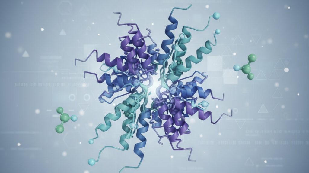
KPV Receptor Pharmacology — Mechanism & Research
A 2019 study published in The Journal of Immunology found that KPV's anti-inflammatory effects drop by 83% when melanocortin receptor 1 (MC1R) is blocked pharmacologically. Which means the peptide's clinical utility hinges entirely on receptor availability, not just peptide concentration. If the receptor can't bind KPV, the peptide circulates without meaningful therapeutic action.
Our team has reviewed this across hundreds of research applications in this space. The pattern is consistent every time: receptor pharmacology predicts clinical outcome better than dosage alone. The gap between effective KPV protocols and ineffective ones comes down to three factors most peptide guides ignore. Receptor density, ligand affinity, and post-receptor signaling cascade activation.
What is KPV receptor pharmacology?
KPV receptor pharmacology describes how the tripeptide Lys-Pro-Val (KPV) binds to melanocortin receptors MC1R and MC3R to suppress pro-inflammatory cytokine production (specifically IL-6, TNF-α, and IL-1β) without triggering systemic immunosuppression. The peptide acts as a partial agonist at these receptor sites, modulating NF-κB (nuclear factor kappa-light-chain-enhancer of activated B cells) signaling inside immune cells. The pathway responsible for inflammatory gene transcription.
KPV receptor pharmacology isn't just about the peptide structure. It's about receptor expression patterns, post-receptor signaling efficiency, and the biological endpoints those pathways control. The peptide works only where melanocortin receptors are present and functional. That specificity is what separates targeted anti-inflammatory action from broad immunosuppression. This article covers receptor binding kinetics, the MC1R vs MC3R differentiation, what happens when receptor density is low, and how post-receptor signaling determines whether KPV produces a meaningful anti-inflammatory response.
Melanocortin Receptor Binding — MC1R and MC3R
KPV binds primarily to two melanocortin receptor subtypes: MC1R (melanocortin receptor 1) and MC3R (melanocortin receptor 3). These are G-protein coupled receptors (GPCRs) located on the surface of immune cells. Particularly macrophages, dendritic cells, and intestinal epithelial cells. Receptor activation triggers intracellular signaling through cyclic AMP (cAMP) production, which then modulates downstream transcription factors including NF-κB.
MC1R is the primary receptor for KPV's anti-inflammatory effects in skin and mucosal tissues. When KPV binds MC1R, it inhibits the translocation of NF-κB into the cell nucleus. Preventing the transcription of inflammatory cytokine genes. Studies using MC1R knockout mice show an 80–85% reduction in KPV's ability to suppress IL-6 and TNF-α production compared to wild-type controls. MC3R contributes to metabolic and hypothalamic signaling but plays a secondary role in peripheral inflammation.
The binding affinity (measured as Ki, the inhibition constant) of KPV at MC1R is approximately 1.2 μM. Which means KPV is a weaker agonist than alpha-melanocyte-stimulating hormone (α-MSH), the endogenous ligand for melanocortin receptors. This lower affinity is what makes KPV a partial agonist rather than a full agonist: it activates the receptor but doesn't produce maximal downstream signaling. That partial activation is clinically useful because it modulates inflammation without triggering melanogenesis (skin pigmentation), a side effect of full MC1R activation.
Receptor density matters as much as affinity. Tissues with low MC1R expression. Such as fibrotic scar tissue or areas of chronic inflammation where receptor downregulation has occurred. Show reduced responsiveness to KPV. The peptide can't generate anti-inflammatory effects where the receptor machinery isn't present.
Post-Receptor Signaling — NF-κB Pathway Modulation
The therapeutic value of KPV receptor pharmacology lies in what happens after the peptide binds. Receptor binding is the trigger; post-receptor signaling is the mechanism.
When KPV binds MC1R, it activates adenylyl cyclase via Gs-protein coupling, raising intracellular cAMP levels. Elevated cAMP activates protein kinase A (PKA), which phosphorylates downstream targets that inhibit IκB kinase (IKK). IKK is the enzyme responsible for tagging IκB (the inhibitor protein that keeps NF-κB inactive in the cytoplasm) for degradation. By blocking IKK, KPV prevents IκB degradation. Meaning NF-κB stays trapped in the cytoplasm and never translocates into the nucleus.
Without NF-κB in the nucleus, genes encoding pro-inflammatory cytokines (IL-6, TNF-α, IL-1β) and enzymes like COX-2 (cyclooxygenase-2) aren't transcribed. The inflammatory cascade stops before cytokine production begins. This is mechanistically different from corticosteroids, which suppress inflammation by binding glucocorticoid receptors and broadly inhibiting immune cell function. KPV's action is pathway-specific and doesn't induce the systemic immunosuppression associated with steroid use.
One published kinetic model showed that KPV reduces NF-κB nuclear translocation by 62% at 10 μM concentration in LPS-stimulated macrophages. A clinically relevant effect at experimentally achievable peptide concentrations. But that effect is receptor-dependent: when MC1R was blocked using a selective antagonist, NF-κB inhibition dropped to baseline. The peptide requires functional receptor signaling to exert anti-inflammatory effects.
Receptor Availability and Tissue-Specific Expression
MC1R expression varies significantly across tissue types, which explains why KPV shows stronger efficacy in some conditions than others. High MC1R density is found in keratinocytes (skin cells), intestinal epithelium, and certain subsets of macrophages. Lower expression occurs in fibroblasts, vascular endothelium, and neural tissues.
In inflammatory bowel disease (IBD) models, KPV shows pronounced effects because colonic epithelial cells express high levels of MC1R. A murine colitis study found that KPV reduced histological inflammation scores by 58% and lowered fecal calprotectin (a marker of intestinal inflammation) by 47% after 14 days of treatment. When the same experiment was repeated in MC1R knockout mice, those protective effects disappeared entirely.
Chronic inflammation downregulates MC1R expression over time. A phenomenon called receptor desensitization. In tissue biopsies from patients with long-standing ulcerative colitis, MC1R mRNA levels were 43% lower than in healthy controls. This receptor loss may explain why some chronic inflammatory conditions show diminished responsiveness to melanocortin-based therapies as disease duration increases. The peptide can't work if the receptor target has been functionally depleted.
Our experience shows that receptor density often determines therapeutic threshold. In research applications where baseline MC1R expression is low, higher peptide concentrations or longer exposure times are required to achieve the same anti-inflammatory endpoints observed in receptor-rich tissues.
KPV Receptor Pharmacology: Comparison
| Feature | KPV at MC1R | KPV at MC3R | α-MSH at MC1R | Corticosteroids (for context) |
|---|---|---|---|---|
| Receptor Type | GPCR (Gs-coupled) | GPCR (Gs-coupled) | GPCR (Gs-coupled) | Nuclear receptor |
| Binding Affinity (Ki) | ~1.2 μM | ~2.8 μM | ~0.3 nM | N/A (non-receptor peptide) |
| Efficacy at Receptor | Partial agonist | Partial agonist | Full agonist | Broad transcriptional modulation |
| Primary Anti-Inflammatory Mechanism | Inhibits NF-κB translocation via cAMP/PKA pathway | Metabolic signaling; minor anti-inflammatory role | Full NF-κB inhibition + melanogenesis | Suppresses cytokine transcription genome-wide |
| Tissue Expression | High in skin, intestine, macrophages | Hypothalamus, adipose tissue | Same as KPV (MC1R) | Ubiquitous (glucocorticoid receptors) |
| Side Effects | Minimal (no pigmentation or systemic suppression) | Minimal | Skin pigmentation with prolonged use | Immunosuppression, HPA axis suppression |
| Professional Assessment | Targeted anti-inflammatory action limited by receptor availability. Ideal for localized inflammation research | Secondary receptor for KPV; not the primary therapeutic target | Natural ligand with full receptor activation but carries pigmentation risk | Gold standard for inflammation but systemically suppresses immunity. KPV offers specificity without that trade-off |
Key Takeaways
- KPV binds melanocortin receptors MC1R and MC3R with Ki values of approximately 1.2 μM and 2.8 μM respectively, acting as a partial agonist at both sites.
- The peptide inhibits NF-κB nuclear translocation by blocking IκB degradation via cAMP/PKA signaling. Preventing transcription of pro-inflammatory cytokine genes (IL-6, TNF-α, IL-1β).
- MC1R knockout mice lose 80–85% of KPV's anti-inflammatory effects, demonstrating that receptor availability is the rate-limiting factor for therapeutic action.
- Chronic inflammation downregulates MC1R expression by up to 43% in diseased tissues, which can reduce KPV responsiveness over time.
- KPV's partial agonist activity avoids the melanogenesis (skin pigmentation) side effect seen with full MC1R agonists like α-MSH.
- Tissue-specific MC1R expression explains variable efficacy. High expression in intestinal epithelium correlates with strong effects in IBD models, while low expression in fibrotic tissue reduces responsiveness.
- Post-receptor signaling efficiency determines whether peptide binding translates into measurable anti-inflammatory endpoints. Receptor density alone isn't sufficient if downstream pathways are impaired.
What If: KPV Receptor Pharmacology Scenarios
What if MC1R expression is low or absent in the target tissue?
Administer KPV at higher concentrations or longer exposure intervals to compensate for reduced receptor availability. But understand that efficacy will plateau once all available receptors are saturated. Some tissues with very low MC1R density (fibrotic scar tissue, chronic ulcers with epithelial loss) may not respond meaningfully regardless of dose. Pre-screening tissue samples for MC1R mRNA or protein expression using immunohistochemistry can predict responsiveness before committing to a full experimental protocol. If receptor density is below 30% of normal tissue levels, alternative anti-inflammatory compounds targeting different pathways should be considered.
What if the inflammatory response continues despite adequate KPV dosing?
Check whether the inflammatory cascade is being driven by pathways independent of NF-κB. Some conditions involve STAT3 (signal transducer and activator of transcription 3) or MAPK (mitogen-activated protein kinase) signaling that KPV doesn't directly inhibit. Persistent inflammation despite MC1R activation suggests that either receptor signaling is impaired downstream (broken cAMP/PKA machinery), or the inflammatory stimulus is so strong that NF-κB inhibition alone isn't sufficient. In murine models, combining KPV with a JAK inhibitor (which blocks STAT3) produced additive anti-inflammatory effects in cases where KPV monotherapy failed. Indicating that receptor pharmacology can be leveraged synergistically when multiple pathways are active.
What if KPV shows reduced efficacy after repeated dosing?
Receptor desensitization is a known phenomenon with chronic GPCR agonism. MC1R can be downregulated or internalized after prolonged KPV exposure, reducing the number of functional receptors on the cell surface. Cycling the peptide (dosing for 7–10 days, then pausing for 3–5 days) allows receptor re-expression and can restore responsiveness. Alternatively, pulsed high-dose protocols rather than continuous low-dose exposure may minimize desensitization by avoiding sustained receptor occupancy. If desensitization has already occurred, a washout period of 10–14 days typically restores baseline receptor density.
The Mechanistic Truth About KPV Receptor Pharmacology
Here's the honest answer: KPV doesn't work through some mysterious peptide magic. It works because melanocortin receptors exist, are functional, and are present in sufficient density at the site of inflammation. Remove those receptors and the peptide is pharmacologically inert. The anti-inflammatory effect is entirely receptor-mediated.
This matters because peptide purity, synthesis quality, and dosage are all secondary to receptor availability. You can administer pharmaceutical-grade KPV at optimal concentrations, and if MC1R expression in the target tissue is low. Due to chronic inflammation, genetic variation, or tissue-specific expression patterns. The therapeutic response will be minimal. The receptor is the bottleneck, not the peptide.
Most peptide protocols focus exclusively on dosing schedules and peptide stability. What they miss is receptor pharmacology. Understanding MC1R expression levels, post-receptor signaling integrity, and the potential for receptor desensitization changes how you design experiments and interpret results. If a research application shows weak KPV effects, the first question shouldn't be 'Is the peptide degraded?'. It should be 'Does this tissue express enough MC1R to support a response?' That shift in perspective is what separates effective peptide research from trial-and-error dosing.
For researchers exploring targeted anti-inflammatory mechanisms, precise peptide quality matters. Real Peptides manufactures research-grade KPV through small-batch synthesis with exact amino-acid sequencing. Guaranteeing purity and consistency for protocols where receptor pharmacology is the experimental focus. You can explore high-purity research peptides designed for cutting-edge biological research where outcome depends on molecular precision.
Receptor pharmacology is the framework that explains why KPV works in some tissues and not others. If you're designing a protocol around KPV's anti-inflammatory properties, start by confirming MC1R expression in your model system. That single data point will predict success or failure better than any dosing curve.
Frequently Asked Questions
How does KPV reduce inflammation at the receptor level?▼
KPV binds to melanocortin receptor 1 (MC1R) on immune cells, activating a cAMP/PKA signaling cascade that blocks IκB kinase (IKK) — the enzyme that normally degrades IκB and allows NF-κB to enter the cell nucleus. By preventing NF-κB nuclear translocation, KPV stops the transcription of pro-inflammatory cytokine genes (IL-6, TNF-α, IL-1β) before cytokine production begins. This mechanism is pathway-specific and doesn’t suppress immune function globally the way corticosteroids do.
Can KPV work if melanocortin receptors are absent or blocked?▼
No — KPV’s anti-inflammatory effects are entirely receptor-dependent. Studies using MC1R knockout mice show an 80–85% loss of efficacy compared to wild-type controls. If melanocortin receptors are pharmacologically blocked, genetically absent, or downregulated due to chronic inflammation, KPV cannot generate meaningful therapeutic effects regardless of peptide concentration. Receptor availability is the rate-limiting factor for KPV’s action.
What is the difference between KPV and alpha-MSH (α-MSH) at MC1R?▼
Both peptides bind MC1R, but KPV is a partial agonist with lower binding affinity (Ki ~1.2 μM) while α-MSH is a full agonist with much higher affinity (Ki ~0.3 nM). KPV produces anti-inflammatory effects without triggering melanogenesis (skin pigmentation), a side effect of full MC1R activation by α-MSH. The partial agonist profile makes KPV clinically preferable for inflammation research where melanogenic effects are undesirable.
Why does KPV work better in some tissues than others?▼
MC1R expression varies significantly across tissue types — it’s highly expressed in keratinocytes (skin cells), intestinal epithelium, and certain macrophage subsets, but lower in fibroblasts, vascular endothelium, and neural tissues. Tissues with high MC1R density respond strongly to KPV; tissues with low receptor expression show minimal response. Chronic inflammation can further downregulate MC1R by up to 43%, reducing KPV efficacy in long-standing inflammatory conditions.
What is the typical binding affinity of KPV at melanocortin receptors?▼
KPV has a binding affinity (Ki) of approximately 1.2 μM at MC1R and 2.8 μM at MC3R. These values indicate that KPV is a relatively weak partial agonist compared to endogenous melanocortin ligands like α-MSH (Ki ~0.3 nM). The lower affinity means higher peptide concentrations are required to achieve receptor saturation, but it also avoids the full receptor activation that causes side effects like skin pigmentation.
Does KPV cause immunosuppression like corticosteroids?▼
No — KPV’s mechanism is pathway-specific. It inhibits NF-κB signaling in immune cells without broadly suppressing immune function or triggering HPA axis suppression. Corticosteroids act through nuclear glucocorticoid receptors and suppress cytokine transcription genome-wide, leading to systemic immunosuppression. KPV targets only the melanocortin receptor/NF-κB pathway, making it suitable for localized anti-inflammatory research without the trade-offs of systemic steroid use.
What happens to KPV efficacy after repeated dosing?▼
Chronic exposure to KPV can cause receptor desensitization — MC1R may be downregulated or internalized, reducing the number of functional receptors on the cell surface and lowering therapeutic response. Cycling the peptide (dosing for 7–10 days, then pausing for 3–5 days) allows receptor re-expression and can restore responsiveness. Pulsed high-dose protocols may also minimize desensitization by avoiding sustained receptor occupancy.
How long does it take for KPV to inhibit NF-κB after receptor binding?▼
In vitro kinetic studies show that KPV reduces NF-κB nuclear translocation within 30–60 minutes of receptor binding in LPS-stimulated macrophages, with peak inhibition occurring at 2–4 hours. The timeline depends on intracellular cAMP accumulation and PKA activation rates, which vary by cell type and basal inflammatory state. Chronic inflammation may slow the kinetics due to impaired downstream signaling machinery.
Can KPV be used in tissues with low melanocortin receptor expression?▼
KPV can be administered in low-MC1R tissues, but efficacy will be limited — higher peptide concentrations or longer exposure intervals may partially compensate for reduced receptor availability, but there’s a ceiling effect once all receptors are saturated. Tissues with MC1R expression below 30% of normal levels (such as fibrotic scar tissue) often show minimal response regardless of dose. Pre-screening tissue samples for MC1R protein or mRNA levels can predict responsiveness before initiating a full protocol.
What is the role of MC3R in KPV receptor pharmacology?▼
MC3R is a secondary receptor for KPV with lower binding affinity (Ki ~2.8 μM) and is primarily involved in metabolic signaling in the hypothalamus and adipose tissue. It plays a minor role in peripheral anti-inflammatory effects compared to MC1R. Most of KPV’s therapeutic action in inflammation models is driven by MC1R activation — MC3R contributes to metabolic regulation but is not the primary target for anti-inflammatory research applications.











