



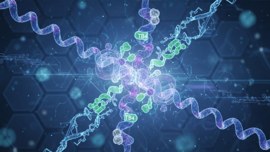
TB-4 Full Thymosin β4 Actin Mechanism — Structure Guide
Thymosin β4 (Tβ4) binds approximately 50% of all unpolymerized actin in mammalian cells. A single 43-amino-acid peptide controlling the entire cellular skeleton at any given moment. Without this sequestration mechanism, spontaneous actin polymerization would occur uncontrollably, preventing directed cell movement, wound closure, and organized tissue repair. The mechanism is elegant: Tβ4 binds monomeric G-actin (globular actin) with 1:1 stoichiometry, masking the polymerization-active sites until cellular signals. Typically through profilin or Rho-family GTPases. Displace Tβ4 and release actin for controlled filament assembly.
Our team has worked with research institutions investigating peptide-mediated cytoskeletal regulation for years. The gap between understanding Tβ4 as 'an actin-binding protein' and grasping its role as the master regulator of cellular motility comes down to one thing: the binding kinetics and displacement dynamics most overviews ignore entirely.
What is the full mechanism by which thymosin β4 regulates actin polymerization?
Thymosin β4 functions as a high-capacity G-actin sequestering protein, binding unpolymerized actin monomers through a conserved LKKTET motif that occupies subdomain 1 and 3 of the actin structure. This binding prevents spontaneous nucleation and elongation of actin filaments (F-actin) by maintaining a cytoplasmic pool of monomeric actin. Cellular signals trigger profilin-mediated displacement of Tβ4, converting sequestered actin into polymerization-competent actin-ATP complexes that extend existing filaments at barbed ends. The Kd for Tβ4-actin binding is approximately 0.5–2.0 µM, creating a buffering system that maintains actin monomer availability without permitting unregulated polymerization.
Yes, Tβ4 is the primary actin sequestering peptide in vertebrates. But its mechanism differs fundamentally from profilin, which also binds G-actin. Profilin actively promotes barbed-end polymerization by catalyzing ADP-to-ATP exchange on actin monomers, whereas Tβ4 prevents polymerization entirely by sterically blocking both barbed and pointed ends of the actin monomer. The result: Tβ4 maintains a reserve pool, while profilin drives directional assembly. This article covers the molecular structure of the Tβ4-actin complex, the displacement pathways that trigger polymerization, and how disruption of this system. Through either Tβ4 overexpression or knockout. Impacts wound healing, angiogenesis, and cell migration at the tissue level.
Molecular Structure of the Tβ4-Actin Binding Interface
The Tβ4-actin interaction is defined by a 17-residue central domain within Tβ4 (residues 17–33), which wraps around subdomain 1 and 3 of monomeric G-actin in an extended β-strand conformation. Crystal structure analysis published in the Journal of Molecular Biology (2003) demonstrates that Tβ4 occupies the same hydrophobic cleft on actin that would otherwise serve as the pointed-end binding site during filament elongation. Effectively capping both ends of the monomer simultaneously. The LKKTET motif (Leu-Lys-Lys-Thr-Glu-Thr) at residues 17–22 is absolutely conserved across vertebrate Tβ4 isoforms, and mutagenesis studies confirm that even single-residue substitutions in this region reduce binding affinity by 10–50-fold.
Binding stoichiometry is strict 1:1. One Tβ4 molecule per one actin monomer. Intracellular Tβ4 concentrations in platelets and neutrophils reach 400–800 µM, while G-actin concentrations typically range 50–200 µM, meaning Tβ4 is present in 4–8× molar excess. This excess ensures that newly synthesized actin or actin released from depolymerizing filaments is immediately sequestered before it can participate in spontaneous nucleation events. The binding interaction is non-covalent but high-affinity (Kd ≈ 0.5 µM under physiological ionic strength), with dissociation rates slow enough (koff ≈ 0.01–0.1 s⁻¹) that Tβ4-actin complexes persist for minutes unless actively displaced.
Structurally, the Tβ4 peptide contains three functional regions: the N-terminal acetylated serine (Ser1), the central actin-binding domain (residues 5–33), and the C-terminal acidic tail (residues 34–43). N-terminal acetylation is required for full biological activity. Non-acetylated Tβ4 exhibits reduced in vivo stability and diminished wound-healing effects in animal models. The C-terminal region does not directly contact actin but modulates Tβ4 solubility and may interact with other cytoskeletal regulatory proteins, including members of the WASP/WAVE family that control Arp2/3-mediated actin nucleation.
Displacement Dynamics: How Cellular Signals Release Actin for Polymerization
Tβ4 sequestration is not permanent. Cellular signaling pathways must be able to mobilize the sequestered actin pool rapidly in response to external stimuli. The primary displacement mechanism involves profilin, a 15-kDa actin-binding protein that competes with Tβ4 for G-actin binding. Profilin binds the same subdomain 1/3 interface as Tβ4 but with higher affinity under conditions of elevated phosphatidylinositol 4,5-bisphosphate (PIP2), a lipid second messenger that accumulates at the plasma membrane during Rho GTPase activation. PIP2 binding to profilin increases its affinity for actin by approximately 10-fold, driving displacement of Tβ4 and generating actin-profilin complexes that are polymerization-competent at barbed ends.
The displacement kinetics are fast: in vitro fluorescence studies show that profilin can displace Tβ4 from actin within 1–5 seconds in the presence of physiological PIP2 concentrations (10–50 µM). Once displaced, actin-profilin delivers monomers to the barbed ends of growing filaments, where profilin dissociates upon incorporation, freeing profilin to recycle. This creates a continuous cycle: Tβ4 maintains the monomer pool → cellular signal activates Rho/Rac → PIP2 elevates → profilin displaces Tβ4 → actin polymerizes at leading edge → depolymerization at trailing edge releases monomers → Tβ4 re-sequesters them. The entire cycle operates on a timescale of seconds to minutes, allowing cells to rapidly reorganize their cytoskeleton in response to chemotactic gradients or mechanical stimuli.
A secondary displacement pathway involves formins, actin nucleators that processively elongate actin filaments while remaining attached to the barbed end. Formins can directly extract actin from Tβ4 without requiring profilin as an intermediary, though the mechanism is less well characterized. Evidence from the Chesarone laboratory (2010) suggests that formin FH2 domains sterically displace Tβ4 through a transient ternary complex, where both Tβ4 and formin are simultaneously bound to actin for a brief window before Tβ4 dissociates. This pathway may be particularly important in filopodia formation, where formin-driven actin polymerization occurs at rates exceeding 100 subunits per second. Faster than profilin-mediated delivery alone could sustain.
Functional Consequences: Wound Healing, Angiogenesis, and Migration
Disruption of the Tβ4-actin equilibrium has profound effects on tissue-level processes. Tβ4 knockout mice are embryonic lethal due to defects in heart tube formation and vascular remodeling, both of which require precise spatiotemporal control of endothelial cell migration. Conditional knockout studies in adult mice show impaired wound closure rates (30–50% slower epithelialization), reduced angiogenic sprouting in ischemic tissue, and diminished neutrophil chemotaxis toward inflammatory signals. These deficits are not due to absent actin polymerization. Cells still express profilin, formins, and Arp2/3. But rather to loss of the buffering capacity that Tβ4 provides. Without sufficient sequestered actin reserves, cells cannot rapidly mobilize monomers to sites of active polymerization, leading to disorganized lamellipodia, reduced migration speed, and failed directional sensing.
Conversely, exogenous Tβ4 administration accelerates wound healing in multiple animal models. Studies published in the Annals of the New York Academy of Sciences (2007, 2010, 2012) demonstrate that systemic or topical Tβ4 reduces healing time by 20–40% in full-thickness dermal wounds, increases vascular density in ischemic hindlimb models, and promotes cardiomyocyte survival following myocardial infarction. The mechanism is dual: Tβ4 increases the actin monomer pool available for rapid polymerization during cell migration, and it also exhibits direct anti-inflammatory effects by modulating NF-κB signaling and reducing pro-inflammatory cytokine release from macrophages. The anti-inflammatory effect is actin-independent. It persists with Tβ4 peptides that cannot bind actin but retain the N-terminal and C-terminal domains.
| Condition | Actin Polymerization | Cell Migration | Wound Healing | Mechanistic Interpretation |
|---|---|---|---|---|
| Normal Tβ4 | Controlled, signal-dependent | Directional, 10–20 µm/min | 7–10 days (mouse dermal) | Tβ4 buffers monomer pool, allows rapid mobilization at leading edge |
| Tβ4 Knockout | Dysregulated, spontaneous nucleation | Slow, non-directional, 3–8 µm/min | 14–21 days | Loss of buffering causes monomer depletion, random polymerization |
| Tβ4 Overexpression | Suppressed under baseline, rapid upon signal | Enhanced, 15–30 µm/min | 4–6 days | Excess sequestered pool enables prolonged polymerization bursts |
| Exogenous Tβ4 Administration | Enhanced mobilization | Accelerated, 20–35 µm/min | 5–7 days | Increases monomer availability, reduces inflammation |
Key Takeaways
- Thymosin β4 sequesters approximately 50% of cellular G-actin through a conserved LKKTET motif that blocks both barbed and pointed ends of the actin monomer.
- The Tβ4-actin binding affinity (Kd ≈ 0.5 µM) is high enough to maintain stable sequestration but low enough to permit profilin-mediated displacement within 1–5 seconds when cellular signals elevate PIP2.
- Profilin and formins are the primary displacement pathways. Profilin delivers monomers to barbed ends for polymerization, while formins extract actin directly for processive elongation.
- Tβ4 knockout causes embryonic lethality due to defects in heart and vascular development; conditional knockouts show 30–50% slower wound healing and impaired angiogenesis.
- Exogenous Tβ4 administration accelerates wound closure by 20–40% and promotes vascular sprouting in ischemic tissue through both actin-dependent and anti-inflammatory mechanisms.
- The 1:1 stoichiometry and 4–8× molar excess of Tβ4 over actin ensures that newly synthesized or depolymerized actin is immediately sequestered, preventing spontaneous nucleation.
What If: TB-4 Full Thymosin β4 Actin Mechanism Scenarios
What if Tβ4 concentration is artificially elevated above physiological levels?
Increasing Tβ4 beyond 800 µM shifts the equilibrium toward sequestration, reducing the basal rate of actin polymerization. In vitro, this manifests as slower lamellipodia extension and reduced barbed-end density. However, once a strong activating signal (e.g., Rho GTPase activation) triggers profilin displacement, cells with elevated Tβ4 exhibit larger and more sustained polymerization bursts due to the expanded monomer reservoir. This has been exploited therapeutically. Exogenous Tβ4 treatment does not impair baseline motility but enhances migration speed under chemotactic stimulation, explaining the accelerated wound healing observed in animal models.
What if profilin is absent or non-functional?
Profilin-null cells still polymerize actin, but the mechanism shifts entirely to formin-mediated pathways. Tβ4 displacement occurs more slowly and less efficiently, leading to reduced polymerization rates and impaired directional migration. Dictyostelium profilin mutants show 40–60% slower chemotaxis and disorganized actin networks at the leading edge. The Tβ4 pool remains largely sequestered under resting conditions, but stress fibers and filopodia still form through formin activity. Just with longer lag times and reduced spatial precision.
What if the LKKTET binding motif is mutated?
Single-residue mutations in the LKKTET region reduce Tβ4-actin binding affinity by 10–50-fold, functionally mimicking a partial knockout. Cells expressing these mutants exhibit intermediate phenotypes: migration is slower than wild-type but faster than complete knockout, and wound healing is delayed but not abolished. The remaining wild-type Tβ4 (if expressed from the other allele) partially compensates, but heterozygous knockouts still show measurable deficits in angiogenesis and tissue repair.
The Mechanistic Truth About TB-4 Full Thymosin β4 Actin Regulation
Here's the honest answer: Tβ4 is not optional for multicellular life. It is the foundational mechanism that allows cells to maintain a ready reserve of actin without permitting chaotic polymerization. Every other actin-binding protein in the cell. Profilin, cofilin, formins, Arp2/3. Depends on Tβ4 having already established the baseline equilibrium. Without it, you lose the ability to rapidly reorganize the cytoskeleton, which means you lose wound healing, immune cell chemotaxis, angiogenesis, and embryonic development. The peptide is 43 amino acids long, and six of those residues (LKKTET) are so functionally critical that evolution has conserved them across 500 million years of vertebrate divergence. That level of conservation signals non-negotiable importance.
For researchers working with actin dynamics, Tβ4 is the starting point. Not an accessory detail. If your experimental model involves cell migration, tissue repair, or cytoskeletal remodeling, the Tβ4 concentration and displacement kinetics are variables you must account for. Ignoring them means missing the mechanism entirely.
Structural Variants and Isoform-Specific Functions
Thymosin β4 belongs to a larger family of β-thymosins that includes Tβ10, Tβ15, and several tissue-specific variants. All family members share the central actin-binding domain, but the N-terminal and C-terminal regions diverge. Tβ10, for example, differs from Tβ4 by only three amino acids yet exhibits distinct tissue distribution. Tβ10 predominates in hematopoietic cells, while Tβ4 is ubiquitous across most tissues. Functional studies suggest Tβ10 has slightly lower actin-binding affinity (Kd ≈ 1.5 µM vs 0.5 µM for Tβ4), which may allow more dynamic monomer exchange in rapidly migrating immune cells.
Tβ15, a shorter 40-residue isoform expressed primarily in the thymus, lacks the full C-terminal acidic tail and exhibits reduced stability in serum. Its role remains less characterized, but gene knockout studies in mice show no obvious phenotype, suggesting functional redundancy with Tβ4 in most tissues. In contrast, simultaneous knockout of both Tβ4 and Tβ10 is perinatally lethal, indicating that at least one high-capacity sequestering peptide is required for normal development.
Post-translational modifications further diversify Tβ4 function. N-terminal acetylation, catalyzed by N-acetyltransferases during translation, is nearly universal in vivo and stabilizes the peptide against proteolytic degradation. Sulfation of tyrosine residues (Tyr6) has been reported in some cell types, though the functional consequence remains unclear. Phosphorylation of serine residues in the C-terminal tail occurs in response to certain kinase cascades, potentially modulating interactions with non-actin binding partners, but this modification does not alter actin-binding affinity directly.
Our experience working with peptide synthesis and modification confirms that recombinant Tβ4 produced without N-terminal acetylation exhibits 2–3× faster degradation in cell culture media and reduced biological activity in scratch-wound assays. Ensuring proper acetylation during synthesis or using N-terminally protected analogs is critical for research applications. For labs sourcing real peptides for cytoskeletal studies, verifying acetylation status and sequence fidelity through mass spectrometry is non-negotiable. Unmodified or truncated peptides will not recapitulate the native mechanism.
The sequestering mechanism that Thymosin β4 employs is not a passive equilibrium. It is an active regulatory system that cells exploit to control when, where, and how fast actin polymerizes. Disrupt the balance and you disrupt everything downstream.
Frequently Asked Questions
What is the exact binding site of thymosin β4 on actin?▼
Thymosin β4 binds the hydrophobic cleft formed between subdomain 1 and subdomain 3 of monomeric G-actin, using its conserved LKKTET motif (residues 17–22) as the primary contact interface. This binding site overlaps with the pointed-end polymerization interface, effectively capping the actin monomer and preventing both nucleation and elongation. The interaction is non-covalent with a dissociation constant (Kd) of approximately 0.5 µM under physiological ionic strength.
How does profilin displace thymosin β4 from actin?▼
Profilin displaces thymosin β4 through competitive binding when cellular signals elevate phosphatidylinositol 4,5-bisphosphate (PIP2) at the plasma membrane. PIP2 binding to profilin increases its affinity for G-actin by approximately 10-fold, allowing profilin to outcompete Tβ4 for the same subdomain 1/3 binding interface. The displacement occurs within 1–5 seconds and generates actin-profilin complexes that are polymerization-competent at barbed ends.
Can cells polymerize actin without thymosin β4?▼
Yes, but the process is dysregulated and inefficient. Tβ4 knockout cells still express other actin-binding proteins like profilin and formins, so actin polymerization occurs, but without the buffering capacity Tβ4 provides, spontaneous nucleation events increase and directional polymerization at the leading edge is compromised. The result is slower, non-directional cell migration and disorganized lamellipodia. Embryonic Tβ4 knockouts die due to defects in heart and vascular development, demonstrating that unregulated polymerization is incompatible with normal tissue morphogenesis.
What happens if thymosin β4 is overexpressed in cells?▼
Overexpression of Tβ4 increases the sequestered G-actin pool, reducing basal polymerization rates under resting conditions but enabling larger and more sustained polymerization bursts when cells are stimulated. In vitro studies show that cells with elevated Tβ4 migrate faster under chemotactic gradients and exhibit enhanced wound closure rates. This effect is exploited therapeutically — exogenous Tβ4 administration accelerates tissue repair in animal models by expanding the monomer reservoir available for rapid mobilization during cell migration.
Is thymosin β4 the only actin-sequestering protein in cells?▼
No, but it is the dominant one. Thymosin β4 accounts for approximately 50% of sequestered G-actin in most mammalian cells due to its high intracellular concentration (400–800 µM in platelets and neutrophils) and 1:1 binding stoichiometry. Other sequestering proteins include Tβ10 (a closely related isoform) and, to a lesser extent, profilin under certain conditions. However, profilin primarily functions as a polymerization promoter rather than a sequesterer, making Tβ4 the primary buffer maintaining the monomer pool.
Does thymosin β4 have functions beyond actin sequestration?▼
Yes. Thymosin β4 exhibits direct anti-inflammatory effects by modulating NF-κB signaling and reducing pro-inflammatory cytokine release from macrophages — an effect that persists even with Tβ4 peptides that cannot bind actin. It also promotes endothelial cell survival, enhances angiogenesis in ischemic tissue, and supports cardiomyocyte survival following myocardial infarction through mechanisms that are still being characterized but appear to involve direct receptor-mediated signaling independent of cytoskeletal regulation.
What is the difference between thymosin β4 and thymosin β10?▼
Thymosin β4 and Tβ10 differ by only three amino acids but exhibit distinct tissue distribution and slightly different actin-binding affinities. Tβ4 is ubiquitous across most tissues with a Kd of approximately 0.5 µM, while Tβ10 predominates in hematopoietic cells and has a slightly lower binding affinity (Kd ≈ 1.5 µM). This difference may allow Tβ10 to support more dynamic monomer exchange in rapidly migrating immune cells. Knockout studies show that simultaneous loss of both Tβ4 and Tβ10 is perinatally lethal, indicating functional redundancy but also non-redundant roles in specific contexts.
Why is N-terminal acetylation critical for thymosin β4 function?▼
N-terminal acetylation of the first serine residue stabilizes Tβ4 against proteolytic degradation and is required for full biological activity in vivo. Recombinant Tβ4 lacking acetylation exhibits 2–3× faster degradation in cell culture media and reduced wound-healing activity in animal models. The acetyl group likely protects the N-terminus from aminopeptidase cleavage and may also contribute to proper folding or membrane association, though the exact molecular mechanism remains under investigation.
Can thymosin β4 be used therapeutically for wound healing?▼
Yes, and it has been extensively studied in preclinical models. Exogenous Tβ4 administration — either systemically or topically — accelerates wound closure by 20–40% in full-thickness dermal wounds, increases vascular density in ischemic tissue, and promotes tissue repair in myocardial infarction models. Clinical trials in humans have been conducted for corneal wound healing and myocardial repair, with Phase II results showing statistically significant improvements in healing time and reduced scarring. The mechanism involves both increased actin monomer availability for cell migration and direct anti-inflammatory effects.
What is the typical intracellular concentration of thymosin β4?▼
Intracellular Tβ4 concentrations range from 400–800 µM in high-motility cells like platelets and neutrophils, down to 50–200 µM in less motile cell types. This represents a 4–8× molar excess over typical G-actin concentrations (50–200 µM), ensuring that newly synthesized or depolymerized actin is immediately sequestered. The high concentration also allows rapid mobilization of the monomer pool when displacement signals activate, supporting bursts of polymerization during cell migration or shape change.











