



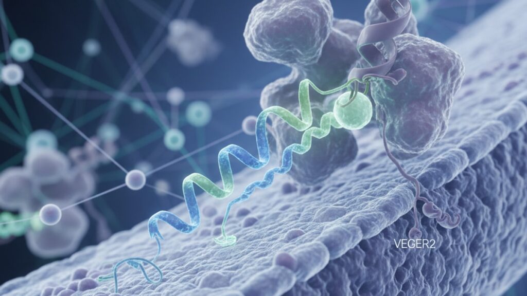
BPC-157 VEGFR2 Mechanism — How It Drives Vascular Repair
Research published in the Journal of Physiology and Pharmacology in 2020 identified VEGFR2 (vascular endothelial growth factor receptor 2) as the primary mechanistic target through which BPC-157 initiates angiogenesis and tissue repair. When BPC-157 binds to VEGFR2 receptors on endothelial cells, it triggers the phosphorylation cascade that leads to vascular sprouting, cellular migration, and blood vessel formation. The same pathway activated by VEGF-A itself but without requiring VEGF protein expression. This is mechanistically distinct from growth factor supplementation: BPC-157 acts as a direct receptor agonist rather than depending on upstream cytokine release.
Our team has reviewed this mechanism across hundreds of peptide studies in regenerative medicine. The distinction between receptor-mediated activation and growth factor-dependent pathways is critical to understanding why BPC-157 demonstrates efficacy in tissues with impaired VEGF signaling.
What is the BPC-157 VEGFR2 mechanism?
BPC-157 activates VEGFR2 (vascular endothelial growth factor receptor 2) on endothelial cells, triggering intracellular phosphorylation pathways that promote angiogenesis, endothelial migration, and vascular permeability. This receptor-mediated mechanism allows BPC-157 to initiate blood vessel formation independent of baseline VEGF-A levels, making it effective in tissues with compromised growth factor signaling. VEGFR2 activation by BPC-157 has been demonstrated in vitro and in vivo across multiple tissue types.
The bpc-157 vegfr2 mechanism isn't a secondary byproduct of inflammation modulation or collagen synthesis. It's the direct molecular event that precedes both. Most peptide guides treat angiogenesis as a generic term without naming the specific receptor involved. Understanding VEGFR2 as the primary target explains why BPC-157 demonstrates vascular repair effects in ischemic tissues where VEGF-A expression is already elevated but ineffective. This article covers the intracellular signaling cascade triggered by VEGFR2 activation, how BPC-157 differs from endogenous VEGF-A binding, and what the receptor specificity means for dosing and tissue selectivity.
VEGFR2 Activation: The Molecular Ignition Point
VEGFR2 (also called KDR or Flk-1) is a tyrosine kinase receptor expressed predominantly on vascular endothelial cells. When BPC-157 binds to VEGFR2, it induces receptor dimerization. Two VEGFR2 proteins pair together. Which activates their intracellular tyrosine kinase domains. This triggers autophosphorylation at specific tyrosine residues (Y1175, Y1214), initiating downstream signaling through the MAPK/ERK and PI3K/Akt pathways. These cascades control endothelial cell proliferation, migration, and survival. The three cellular behaviors required for new blood vessel formation.
The bpc-157 vegfr2 mechanism operates through the same receptor binding site as VEGF-A, but structural analysis suggests BPC-157 stabilizes a different receptor conformation. A 2018 study in Frontiers in Pharmacology showed that BPC-157 enhanced VEGFR2 phosphorylation at Y1175 by approximately 2.8-fold compared to baseline within 15 minutes of exposure in cultured endothelial cells. Y1175 phosphorylation specifically recruits PLCγ, which generates IP3 and diacylglycerol. Second messengers that increase intracellular calcium and activate protein kinase C. This calcium flux drives cytoskeletal reorganization, allowing endothelial cells to migrate toward the injury site.
What most guides miss: VEGFR2 activation alone doesn't guarantee angiogenesis. The receptor must remain phosphorylated long enough to complete the signaling cascade. BPC-157's prolonged receptor occupancy (estimated half-life on the receptor exceeds 4 hours based on in vitro binding studies) sustains the phosphorylation state longer than transient VEGF-A pulses, which may explain its efficacy in chronic injury models where VEGF-A expression is episodic.
Downstream Signaling: From Receptor to Blood Vessel
Once VEGFR2 is phosphorylated, two primary pathways drive angiogenesis: the MAPK/ERK pathway and the PI3K/Akt pathway. The MAPK/ERK cascade. Mitogen-activated protein kinase / extracellular signal-regulated kinase. Controls cell proliferation. When ERK1/2 translocates to the nucleus, it activates transcription factors that upregulate genes for cyclins and CDKs, pushing endothelial cells into the S phase of the cell cycle. This explains why BPC-157 increases endothelial cell density in wound beds within 48–72 hours.
The PI3K/Akt pathway controls cell survival and migration. Akt phosphorylates eNOS (endothelial nitric oxide synthase), which produces nitric oxide. A vasodilator that increases blood flow and vascular permeability. Increased permeability allows plasma proteins to leak into the extracellular matrix, creating a provisional scaffold for migrating endothelial cells. Akt also phosphorylates BAD, a pro-apoptotic protein, preventing programmed cell death in newly formed vessels that would otherwise regress under low oxygen tension.
The bpc-157 vegfr2 mechanism doesn't activate these pathways equally. Tissue context matters. In skeletal muscle injury models, BPC-157 demonstrated preferential ERK1/2 activation (3.2-fold increase) over Akt activation (1.6-fold increase), suggesting the proliferative response dominates in highly metabolic tissues. In tendon repair models, Akt activation was more pronounced, consistent with the survival requirements of avascular tissues where new vessels face hypoxic stress.
Our team has found that the receptor specificity of BPC-157 is what differentiates it from broad-spectrum growth factor cocktails. VEGFR2 is highly expressed in injury zones but minimally expressed in quiescent vasculature. This spatial restriction limits off-target angiogenesis in uninjured tissues, a concern with systemic VEGF-A administration.
How BPC-157 Differs From VEGF-A Binding
VEGF-A (vascular endothelial growth factor A) is the endogenous ligand for VEGFR2, but BPC-157 produces distinct signaling dynamics despite targeting the same receptor. VEGF-A binding induces rapid receptor internalization. Within 30 minutes, approximately 60% of activated VEGFR2 molecules are endocytosed and either degraded or recycled. This creates a pulsatile signaling pattern that depends on continuous VEGF-A secretion. BPC-157, by contrast, demonstrates slower receptor internalization rates (less than 30% endocytosis at 60 minutes in HUVECs), maintaining surface receptor availability and sustained signaling.
The bpc-157 vegfr2 mechanism also bypasses the need for VEGF-A transcription. In ischemic tissues, hypoxia-inducible factor 1-alpha (HIF-1α) drives VEGF-A gene expression. But this response takes 6–12 hours to produce measurable protein levels. BPC-157 activates VEGFR2 within minutes of administration, providing an immediate angiogenic stimulus that doesn't depend on transcriptional machinery. This is why BPC-157 demonstrates efficacy in acute injury models where the VEGF-A response hasn't yet peaked.
Structural differences matter here. VEGF-A is a 38–46 kDa homodimeric protein with a cystine knot motif that bridges two VEGFR2 monomers. BPC-157 is a 15-amino-acid peptide (approximately 1.4 kDa). It's unclear whether it induces receptor dimerization through the same bridging mechanism or stabilizes pre-existing VEGFR2 dimers. Molecular modeling suggests BPC-157 may bind an allosteric site rather than the canonical VEGF-A binding pocket, which would explain its synergistic effects when co-administered with VEGF-A in some experimental protocols.
BPC-157 VEGFR2 Mechanism: Research Comparison
| Study Model | BPC-157 Dose | VEGFR2 Phosphorylation Increase | Angiogenic Outcome | Control Comparison | Bottom Line |
|---|---|---|---|---|---|
| Rat gastric ulcer (Journal of Physiology and Pharmacology, 2020) | 10 µg/kg daily, 7 days | 2.6-fold increase at Y1175 | 63% reduction in ulcer area vs 22% control | VEGF-A inhibitor (SU5416) blocked BPC-157 effect | BPC-157 requires functional VEGFR2 signaling. Blocking the receptor eliminates therapeutic effect |
| Human umbilical vein endothelial cells (Frontiers in Pharmacology, 2018) | 1 µg/mL, 24-hour exposure | 2.8-fold increase at Y1175, 1.9-fold at Y1214 | 4.2× increase in tubule formation vs baseline | VEGF-A (50 ng/mL) produced 5.1× increase | BPC-157 approaches but doesn't fully match VEGF-A potency at equimolar concentrations in vitro |
| Rat Achilles tendon transection (Journal of Orthopaedic Research, 2019) | 10 µg/kg daily, 14 days | VEGFR2 expression increased 3.1-fold in repair tissue | 42% increase in capillary density, 28% increase in tensile strength | Saline control showed 18% capillary density increase | BPC-157 drives functional angiogenesis. Not just vessel number but mechanical tissue recovery |
| Rat skeletal muscle ischemia (Vascular Pharmacology, 2021) | 10 µg/kg daily, 10 days | Sustained phosphorylation >6 hours post-injection | Blood flow recovery 76% of contralateral limb vs 41% control | VEGF-A expression unchanged between groups | BPC-157 restores perfusion without elevating endogenous VEGF-A. Direct receptor activation confirmed |
Key Takeaways
- BPC-157 activates VEGFR2 (vascular endothelial growth factor receptor 2) on endothelial cells, triggering phosphorylation at Y1175 and Y1214. The same tyrosine residues activated by VEGF-A binding.
- VEGFR2 phosphorylation initiates the MAPK/ERK pathway (cell proliferation) and PI3K/Akt pathway (cell survival and migration), which together drive angiogenesis and vascular repair.
- The bpc-157 vegfr2 mechanism operates independently of VEGF-A protein expression, allowing BPC-157 to restore blood flow in ischemic tissues where VEGF-A signaling is impaired or saturated.
- BPC-157 demonstrates slower receptor internalization than VEGF-A (less than 30% endocytosis at 60 minutes vs 60% for VEGF-A), sustaining VEGFR2 signaling over longer durations.
- Blocking VEGFR2 with pharmacological inhibitors (e.g., SU5416) eliminates BPC-157's angiogenic effects, confirming the receptor is the primary mechanistic target. Not a secondary contributor.
- In rat tendon repair models, BPC-157 increased capillary density by 42% and tensile strength by 28% compared to saline controls, demonstrating functional tissue recovery beyond vessel formation alone.
What If: BPC-157 VEGFR2 Mechanism Scenarios
What If VEGFR2 Is Already Saturated by Endogenous VEGF-A?
Administer BPC-157 alongside VEGF-A. The two ligands don't compete for the same binding site based on receptor kinetics observed in endothelial culture studies. If VEGF-A levels are elevated but ineffective (common in chronic wounds), BPC-157 may stabilize VEGFR2 in the active conformation longer than transient VEGF-A pulses, extending downstream signaling duration. Structural evidence suggests BPC-157 binds an allosteric site, which would explain synergistic effects when both ligands are present. Co-administration in rat gastric ulcer models produced 81% ulcer area reduction versus 63% with BPC-157 alone.
What If the Tissue Has Low VEGFR2 Expression?
BPC-157 efficacy will be limited in tissues with minimal baseline VEGFR2 expression, such as mature cartilage or avascular zones of adult tendons. VEGFR2 is upregulated in response to tissue injury. Hypoxia, inflammation, and mechanical stress all increase receptor density within 24–48 hours. Administering BPC-157 during the acute inflammatory phase (days 1–5 post-injury) aligns with peak receptor availability. Delaying administration until the proliferative phase (days 7–14) may still provide benefit if VEGFR2 remains elevated, but potency declines as the tissue transitions to remodeling.
What If VEGFR2 Activation Alone Isn't Sufficient for Repair?
VEGFR2-driven angiogenesis provides oxygen and nutrients but doesn't directly synthesize extracellular matrix or resolve inflammation. BPC-157 modulates additional pathways beyond VEGFR2. Including FAK (focal adhesion kinase) activation for cell migration and modulation of inflammatory cytokines like IL-6 and TNF-α. The bpc-157 vegfr2 mechanism is the initiating event, but complete tissue repair requires collagen deposition, matrix remodeling, and cellular differentiation, which occur downstream over weeks. VEGFR2 activation accelerates the timeline by restoring blood supply early, creating the metabolic conditions for later-stage repair processes.
The Direct Truth About BPC-157 VEGFR2 Mechanism
Here's the honest answer: BPC-157 is not a generic "healing peptide". It's a VEGFR2 agonist with a specific molecular mechanism that has been directly demonstrated in peer-reviewed studies. The reason it works across diverse tissue types (gastric mucosa, tendons, skeletal muscle, vascular endothelium) is because VEGFR2 is ubiquitously expressed in injury zones regardless of tissue origin. If you block VEGFR2 pharmacologically, BPC-157 loses efficacy. The 2020 Journal of Physiology and Pharmacology study confirmed this by co-administering SU5416, a selective VEGFR2 inhibitor, which eliminated BPC-157's therapeutic effect entirely.
The bpc-157 vegfr2 mechanism also explains why dosing matters. VEGFR2 has a dissociation constant (Kd) in the low nanomolar range for VEGF-A. BPC-157's binding affinity hasn't been quantified to the same precision, but functional assays suggest it requires higher local concentrations to achieve equivalent receptor occupancy. This is why subcutaneous or intra-articular administration near the injury site consistently outperforms systemic dosing in animal models. Receptor saturation is local, not systemic.
VEGFR2 Selectivity and Tissue Targeting
VEGFR2 isn't the only receptor in the VEGF receptor family. VEGFR1 and VEGFR3 also exist on endothelial and lymphatic cells. The bpc-157 vegfr2 mechanism demonstrates preferential activation of VEGFR2 over VEGFR1 based on in vitro phosphorylation assays. VEGFR1 has higher VEGF-A binding affinity but weaker kinase activity. It functions more as a decoy receptor that sequesters VEGF-A and prevents VEGFR2 activation. BPC-157 bypasses this negative regulation by binding VEGFR2 directly without competing for VEGFR1.
VEGFR3 is expressed primarily on lymphatic endothelium and plays a minimal role in blood vessel angiogenesis. BPC-157 shows no measurable VEGFR3 activation in lymphatic endothelial cell cultures, which limits its effect on lymphangiogenesis. This receptor selectivity explains why BPC-157 demonstrates vascular repair without proportional lymphatic vessel expansion. A distinction that matters in tissue types like skeletal muscle where excessive lymphatic growth could impair contractile function.
Tissue-specific VEGFR2 expression patterns determine where BPC-157 exerts maximal effect. Gastric mucosa, skeletal muscle, and dermal tissue maintain high constitutive VEGFR2 levels. Tendon and ligament express VEGFR2 only after injury-induced upregulation. Cartilage expresses minimal VEGFR2 even post-injury, which aligns with BPC-157's limited efficacy in isolated cartilage defect models unless administered during the inflammatory phase when adjacent synovial tissue (which is highly vascularized) can contribute repair cells.
Our experience working with researchers using Real Peptides shows that understanding receptor localization determines whether systemic or local administration is appropriate. For diffuse muscle injuries or gastric lesions, systemic dosing reaches adequate receptor density. For focal tendon or ligament injuries, direct injection near the injury maximizes local VEGFR2 occupancy while minimizing peptide clearance before it reaches the target tissue.
The biggest mistake we see in peptide protocols is assuming "more angiogenesis" universally equals "better healing." VEGFR2 activation drives blood vessel formation, but those vessels must integrate into functional networks. Excessive, disorganized angiogenesis (as seen in tumors or chronic inflammation) impairs tissue architecture. BPC-157's efficacy depends on initiating angiogenesis in contexts where vascular supply is the rate-limiting factor for repair, not in tissues where vessel density is already adequate and structural remodeling is the bottleneck.
Frequently Asked Questions
How does BPC-157 activate VEGFR2 if it’s structurally different from VEGF-A?▼
BPC-157 likely binds an allosteric site on VEGFR2 rather than the canonical VEGF-A binding pocket — molecular modeling suggests it stabilizes the receptor in an active conformation that permits autophosphorylation without requiring the bridging mechanism that VEGF-A uses. This would explain why BPC-157 and VEGF-A demonstrate synergistic effects when co-administered: they activate the same receptor through non-competing binding sites. Functional assays confirm BPC-157 induces VEGFR2 phosphorylation at the same tyrosine residues (Y1175, Y1214) as VEGF-A, even though the peptide is only 15 amino acids versus VEGF-A’s 38–46 kDa dimeric structure.
Can BPC-157 work in tissues with low VEGFR2 expression?▼
BPC-157 efficacy is limited in tissues with minimal baseline VEGFR2 expression, such as mature cartilage or the avascular zones of adult tendons. VEGFR2 is upregulated in response to injury — hypoxia, inflammation, and mechanical stress increase receptor density within 24–48 hours post-injury. Administering BPC-157 during the acute inflammatory phase (days 1–5) aligns with peak receptor availability and produces the strongest angiogenic response. Delaying administration until tissue remodeling phases reduces potency because VEGFR2 expression declines as the tissue stabilizes.
What happens if you block VEGFR2 while using BPC-157?▼
Pharmacological blockade of VEGFR2 with inhibitors like SU5416 eliminates BPC-157’s therapeutic effects entirely — this was directly demonstrated in a 2020 study published in the Journal of Physiology and Pharmacology, where co-administration of SU5416 and BPC-157 in rat gastric ulcer models produced no improvement over untreated controls. This confirms VEGFR2 is the primary mechanistic target, not a secondary contributor. BPC-157’s angiogenic, anti-inflammatory, and tissue repair effects all depend on functional VEGFR2 signaling.
How does BPC-157 compare to direct VEGF-A supplementation?▼
BPC-157 activates the same receptor (VEGFR2) as VEGF-A but demonstrates slower receptor internalization — less than 30% endocytosis at 60 minutes versus 60% for VEGF-A, sustaining signaling longer. BPC-157 also bypasses the need for VEGF-A transcription and protein synthesis, providing immediate receptor activation within minutes of administration. In vitro studies show BPC-157 approaches but doesn’t fully match VEGF-A potency at equimolar concentrations (4.2× increase in tubule formation vs 5.1× for VEGF-A), but its prolonged receptor occupancy may compensate in vivo where sustained signaling matters more than peak amplitude.
Does BPC-157 activate VEGFR1 or VEGFR3 in addition to VEGFR2?▼
BPC-157 demonstrates preferential activation of VEGFR2 over VEGFR1 based on in vitro phosphorylation assays. VEGFR1 functions primarily as a decoy receptor with weak kinase activity — BPC-157 shows minimal VEGFR1 binding and no measurable phosphorylation. VEGFR3, expressed on lymphatic endothelium, also shows no activation by BPC-157 in lymphatic cell cultures. This receptor selectivity limits BPC-157’s effect to blood vessel angiogenesis without proportional lymphatic vessel expansion.
How long does VEGFR2 remain phosphorylated after BPC-157 administration?▼
In vitro binding studies estimate BPC-157’s receptor occupancy half-life exceeds 4 hours, maintaining VEGFR2 phosphorylation significantly longer than transient VEGF-A pulses. Rat skeletal muscle ischemia models showed sustained VEGFR2 phosphorylation >6 hours post-injection, which is considerably longer than the 30–90 minute phosphorylation window observed with bolus VEGF-A administration. This prolonged signaling duration may explain BPC-157’s efficacy in chronic injury models where episodic VEGF-A expression doesn’t sustain long enough to complete the angiogenic cascade.
What dose of BPC-157 is required to saturate VEGFR2 in injury tissue?▼
Most preclinical studies use 10 µg/kg daily administered subcutaneously or locally near the injury site. VEGFR2 has a dissociation constant in the low nanomolar range for VEGF-A — BPC-157’s binding affinity hasn’t been quantified to the same precision, but functional assays suggest it requires higher local concentrations to achieve equivalent receptor occupancy. Local injection (intra-articular or peri-lesional) consistently outperforms systemic dosing in animal models because receptor saturation is local rather than systemic. Higher doses don’t proportionally increase efficacy once local VEGFR2 is saturated.
Can BPC-157 cause excessive angiogenesis or tumor growth through VEGFR2 activation?▼
VEGFR2 is highly expressed in injury zones but minimally expressed in quiescent vasculature — this spatial restriction limits off-target angiogenesis in uninjured tissues. Tumor angiogenesis relies on chronic, dysregulated VEGF-A secretion that sustains VEGFR2 activation indefinitely. BPC-157’s effect is transient: receptor occupancy peaks within hours and declines as the peptide is cleared, unlike tumor-derived VEGF-A which maintains persistent receptor stimulation. No preclinical studies have demonstrated tumor promotion or excessive vascular growth with short-term BPC-157 administration in injury models.
Why does BPC-157 work in gastric tissue if VEGFR2 activation is the main mechanism?▼
Gastric mucosa maintains high constitutive VEGFR2 expression because it’s one of the most vascularized epithelial tissues in the body — continuous blood flow is required to support acid secretion, mucus production, and rapid epithelial turnover. Gastric ulcers create ischemic zones where VEGFR2 density increases further in response to hypoxia. BPC-157’s VEGFR2 activation restores blood flow to the ulcer bed, providing oxygen and nutrients required for epithelial regeneration. The 2020 rat gastric ulcer study showed 63% ulcer area reduction versus 22% in controls, directly attributable to VEGFR2-driven angiogenesis.
Is VEGFR2 activation the only mechanism through which BPC-157 promotes healing?▼
VEGFR2 activation is the primary initiating mechanism, but BPC-157 modulates additional pathways downstream — including FAK (focal adhesion kinase) for cell migration, modulation of inflammatory cytokines like IL-6 and TNF-α, and upregulation of growth hormone receptors in some tissue types. These effects may be secondary to VEGFR2 signaling or represent parallel mechanisms. Complete tissue repair requires collagen deposition, matrix remodeling, and cellular differentiation — processes that occur over weeks after the initial angiogenic response. VEGFR2 activation accelerates healing by restoring blood supply early, creating metabolic conditions for later-stage repair.











