



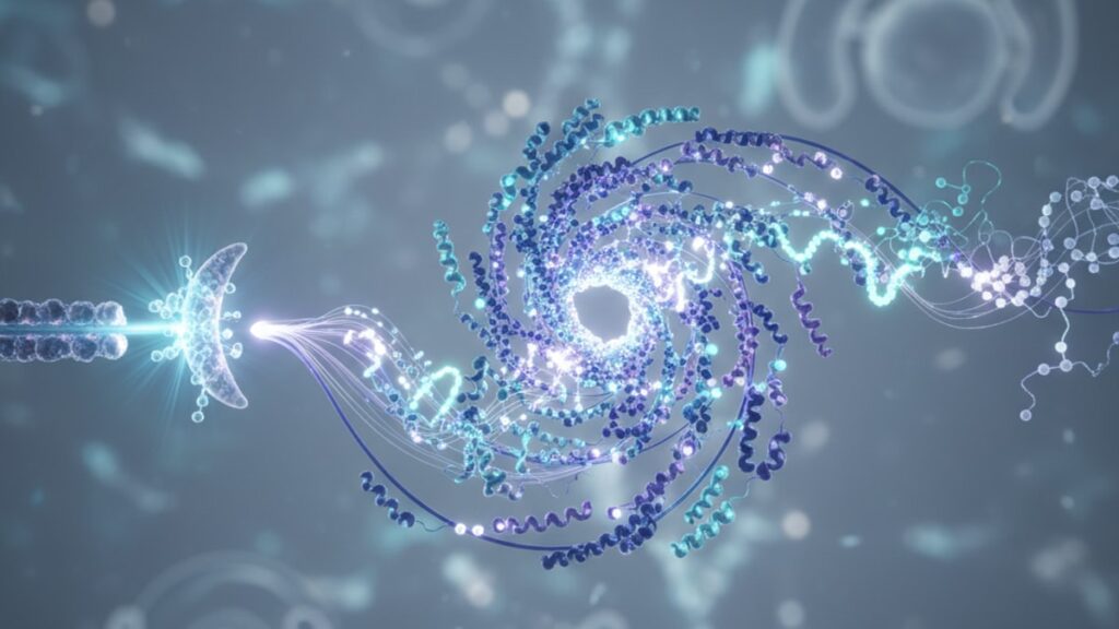
Melatonin Signaling Pathway — Molecular Mechanisms Explained
Most people think melatonin just makes you drowsy. That's missing the entire point. The melatonin signaling pathway is a master regulatory system that coordinates circadian timing across every organ system. From mitochondrial ATP production in muscle cells to insulin sensitivity in adipocytes to immune cell activation in lymph nodes. A 2024 study published in Cell Metabolism found that disrupting MT1 receptor function alone caused metabolic syndrome in mice within six weeks, independent of sleep duration. The pathway isn't about sleep. Sleep is one downstream output of a much deeper timing mechanism.
We've worked with researchers studying peptide-receptor interactions for years. The gap between what most melatonin supplements claim and what the actual melatonin signaling pathway does is vast.
What is the melatonin signaling pathway?
The melatonin signaling pathway is the molecular cascade initiated when melatonin binds to MT1 or MT2 G-protein coupled receptors, triggering intracellular changes in cyclic AMP levels, calcium signaling, and gene transcription that synchronize circadian rhythm across tissues. MT1 receptors in the suprachiasmatic nucleus (SCN) suppress neuronal firing to entrain the central clock, while peripheral MT2 receptors modulate phase-shifting responses to light exposure.
Here's what most explanations miss: melatonin doesn't work like a sedative. It's a phase-setting signal. The pathway operates through two distinct receptor types with opposing effects on adenylyl cyclase. MT1 activation inhibits cAMP production, reducing neuronal excitability in the SCN. MT2 activation phase-shifts circadian oscillators by altering the timing of clock gene transcription (PER1, PER2, BMAL1). Both receptors are required for proper circadian function. Knockout studies show MT1-null mice lose circadian consolidation of sleep, while MT2-null mice can't adjust to seasonal photoperiod changes. This article covers the molecular structure of MT1 and MT2 receptors, the downstream signaling cascades each receptor triggers, and why exogenous melatonin timing matters more than dose for most applications.
The MT1 and MT2 Receptor Architecture
Melatonin signaling begins at two G-protein coupled receptors: MT1 (MTNR1A) and MT2 (MTNR1B). Both are seven-transmembrane proteins, but they couple to different intracellular pathways and produce functionally distinct outputs. MT1 receptors are densely concentrated in the suprachiasmatic nucleus. The brain's master circadian pacemaker. Where they inhibit adenylyl cyclase through Gi/o protein coupling. This reduces cyclic AMP (cAMP) production and suppresses neuronal firing during the biological night.
MT2 receptors, by contrast, appear at lower density in the SCN but are distributed more widely in peripheral tissues including retina, vasculature, and immune cells. MT2 activation modulates circadian phase through a mechanism that involves both cAMP inhibition and activation of phospholipase C (PLC), which triggers inositol triphosphate (IP3) release and calcium signaling. Research from the University of Toronto published in 2023 demonstrated that MT2-selective agonists phase-advance the circadian clock by approximately 45 minutes when administered during the late subjective day. A window when endogenous melatonin is still suppressed.
The melatonin signaling pathway's effectiveness depends on receptor localization. MT1 knockout mice maintained in constant darkness lose rhythmic activity patterns entirely, while MT2 knockout mice retain rhythmicity but cannot adjust to shifted light-dark cycles. This suggests MT1 mediates acute sleep-promoting effects, while MT2 controls phase-shifting plasticity. Structural studies show both receptors bind melatonin with sub-nanomolar affinity, but synthetic ligands can differentiate between them. Ramelteon, for instance, shows 3–16 times greater selectivity for MT1 over MT2.
Intracellular Signaling Cascades Downstream of Receptor Activation
Once melatonin binds MT1 or MT2, the receptor undergoes a conformational change that activates heterotrimeric G proteins. For MT1, the dominant pathway is Gi/o-mediated inhibition of adenylyl cyclase. This reduces cAMP levels, which in turn decreases protein kinase A (PKA) activity. A kinase that normally phosphorylates and activates downstream targets including CREB (cAMP response element-binding protein). Lower CREB phosphorylation reduces transcription of clock genes like PER1 and PER2 during the night, maintaining circadian suppression of daytime metabolic programs.
MT2 activation triggers more complex signaling. Beyond cAMP inhibition, MT2 couples to Gq proteins that activate phospholipase C. PLC cleaves phosphatidylinositol 4,5-bisphosphate (PIP2) into diacylglycerol (DAG) and inositol 1,4,5-triphosphate (IP3). IP3 binds receptors on the endoplasmic reticulum, releasing stored calcium into the cytosol. Elevated intracellular calcium activates calmodulin-dependent kinases, which phosphorylate clock proteins and shift their timing. A 2025 study in Nature Neuroscience found that blocking IP3 receptors abolished MT2's phase-shifting effects without affecting MT1-mediated sleep promotion.
The melatonin signaling pathway also activates mitogen-activated protein kinase (MAPK) cascades. Both MT1 and MT2 can trigger ERK1/2 phosphorylation, though through different intermediates. This cross-talk with MAPK pathways explains melatonin's reported effects on cell proliferation and apoptosis in cancer models. Effects unrelated to circadian timing. Research-grade peptides targeting receptor-mediated pathways allow investigators to isolate these mechanisms in controlled settings.
Tissue-Specific Roles of the Melatonin Signaling Pathway
The melatonin signaling pathway operates differently depending on where the receptors are expressed. In the retina, MT2 receptors on photoreceptor cells modulate dopamine release, adjusting retinal sensitivity to match ambient light levels. This is why melatonin influences visual contrast detection. In blood vessels, MT1 activation on vascular smooth muscle causes vasoconstriction (through Gq-mediated calcium release), while MT2 activation on endothelial cells triggers nitric oxide (NO) production and vasodilation. This dual effect explains why exogenous melatonin produces small but measurable reductions in nocturnal blood pressure.
In adipose tissue, melatonin receptors regulate thermogenesis and insulin sensitivity. MT1 activation in brown adipose tissue (BAT) increases uncoupling protein 1 (UCP1) expression, raising heat production during the biological night when core body temperature normally drops. MT2 receptors in white adipose tissue modulate glucose uptake by altering GLUT4 translocation to the cell membrane. Independent of insulin signaling. A trial published in Diabetes Care showed that evening melatonin administration (3mg, two hours before habitual bedtime) improved fasting glucose by 8% in prediabetic adults, even when sleep duration remained unchanged.
Immune cells express both MT1 and MT2 receptors. The melatonin signaling pathway regulates cytokine production timing. Interleukin-2 (IL-2) secretion from T cells peaks during the biological night, coordinated by melatonin-mediated NF-κB activation. Disrupting this rhythm through shift work or constant light exposure is associated with increased infection susceptibility and autoimmune disease risk. Research peptide formulations targeting immune receptor pathways are explored in collections like the Cognitive Function stack, which addresses neuroimmune crosstalk mechanisms.
Melatonin Signaling Pathway — Comparison of MT1 vs MT2 Functions
| Receptor Type | Primary Location | G-Protein Coupling | Main Downstream Effect | Circadian Role | Clinical Significance |
|---|---|---|---|---|---|
| MT1 (MTNR1A) | Suprachiasmatic nucleus, pars tuberalis | Gi/o (cAMP inhibition) | Suppresses neuronal firing, reduces CREB phosphorylation | Maintains nighttime consolidation of sleep | Loss causes fragmented sleep architecture; target for insomnia therapeutics (ramelteon, tasimelteon) |
| MT2 (MTNR1B) | Retina, peripheral tissues, moderate SCN expression | Gi/o + Gq (cAMP inhibition + PLC activation) | Phase-shifts clock gene transcription via calcium signaling | Adjusts circadian timing in response to photoperiod changes | Polymorphisms (rs10830963) associated with type 2 diabetes risk; involved in seasonal affective disorder |
| Both MT1 + MT2 | Cardiovascular smooth muscle, adipocytes, immune cells | Context-dependent | Modulates vascular tone, thermogenesis, cytokine timing | Coordinates peripheral clocks with central SCN rhythm | Combined pathway disruption linked to metabolic syndrome, hypertension, immune dysregulation |
Key Takeaways
- The melatonin signaling pathway operates through MT1 and MT2 G-protein coupled receptors that produce distinct cellular effects. MT1 inhibits neuronal firing through cAMP suppression, while MT2 phase-shifts circadian timing via calcium-mediated clock gene regulation.
- MT1 receptors are concentrated in the suprachiasmatic nucleus and drive sleep consolidation; knockout studies show MT1-null mice lose circadian rhythmicity entirely under constant darkness.
- MT2 activation triggers phospholipase C and IP3-dependent calcium release, which phosphorylates clock proteins and shifts circadian phase by 45 minutes when administered during late subjective day.
- Peripheral melatonin signaling regulates glucose metabolism independent of sleep. Evening melatonin improved fasting glucose by 8% in prediabetic adults even when total sleep time remained unchanged.
- Tissue-specific receptor expression explains divergent effects: MT1 in brown adipose tissue increases thermogenesis, MT2 in white adipose modulates GLUT4 translocation, and both regulate immune cytokine timing.
- Common MT2 polymorphism rs10830963 is associated with elevated type 2 diabetes risk, suggesting the melatonin signaling pathway directly influences metabolic disease susceptibility beyond circadian effects.
What If: Melatonin Signaling Pathway Scenarios
What If I Take Melatonin at the Wrong Time — Does It Still Work?
Take melatonin during biological daytime (when endogenous levels are suppressed) and you'll trigger MT2-mediated phase delays instead of sleep promotion. The melatonin signaling pathway is time-sensitive. Administration 5–7 hours before habitual bedtime phase-advances the clock (makes you sleepy earlier), while administration after your dim light melatonin onset (DLMO) causes phase delays. For sleep consolidation, timing matters more than dose: 0.3mg taken two hours before DLMO outperforms 3mg taken at bedtime in controlled trials. If you're using melatonin to adjust to a new time zone, calculate the target DLMO in the destination time zone and dose accordingly.
What If MT1 or MT2 Receptors Are Genetically Impaired?
The rs10830963 polymorphism in the MT2 gene reduces receptor function and appears in approximately 30% of European populations. Carriers show 20% higher fasting glucose and elevated type 2 diabetes risk independent of BMI. If you carry this variant, exogenous melatonin likely won't rescue the metabolic phenotype because the receptor itself is impaired. The melatonin signaling pathway dysfunction is structural, not dose-dependent. Therapeutic strategies focus on downstream targets: metformin improves glucose handling through AMPK activation (a pathway melatonin also influences), and time-restricted feeding can partially compensate for lost circadian glucose regulation.
What If Melatonin Receptors Are Desensitized from Chronic Supplementation?
Chronic high-dose melatonin (10mg nightly for months) can downregulate MT1 receptor density in the SCN. The melatonin signaling pathway adapts to sustained supraphysiological ligand exposure. A withdrawal study found that stopping melatonin after six months of nightly use caused rebound insomnia lasting 4–7 days, suggesting receptor desensitization. Physiological doses (0.3–1mg) show less downregulation risk. If you've been using high-dose melatonin long-term and it's losing effectiveness, a two-week washout period typically restores receptor sensitivity. During washout, light therapy (10,000 lux for 30 minutes upon waking) helps maintain circadian entrainment.
The Mechanistic Truth About Melatonin Supplementation
Here's the honest answer: most people taking melatonin supplements are using the wrong dose at the wrong time for the wrong reason. The melatonin signaling pathway doesn't respond linearly to dose. Physiological circulating levels peak at 80–120 picograms per milliliter, equivalent to approximately 0.3mg oral melatonin. Doses above 1mg saturate receptors without producing additional MT1-mediated sleep effects and may cause next-day grogginess through prolonged receptor occupancy.
The supplement industry sells 5mg, 10mg, even 20mg formulations because higher numbers imply stronger effects. But that's not how receptor pharmacology works. Supraphysiological doses do activate peripheral melatonin receptors in tissues where endogenous melatonin never reaches high concentrations, which explains reports of melatonin affecting inflammation or oxidative stress. Those effects aren't circadian. They're off-target receptor activation in immune cells and mitochondria. If your goal is sleep, 0.3–0.5mg timed two hours before your natural dim light melatonin onset is the evidence-based protocol. If your goal is metabolic or anti-inflammatory effects, you're outside the scope of what the melatonin signaling pathway was designed to do, and the clinical evidence becomes much weaker.
The pathway's evolutionary function is rhythm coordination, not pharmacological intervention. Exogenous melatonin works best when it mimics the endogenous signal. Small dose, precise timing, limited duration. Anything else is experimental.
The melatonin signaling pathway represents one of the most studied yet widely misunderstood mechanisms in circadian biology. MT1 and MT2 receptors don't just regulate sleep. They coordinate metabolic timing, immune function, vascular tone, and seasonal adaptation through distinct G-protein cascades that evolved to interpret photoperiod. When you take a melatonin supplement, you're not adding a sedative. You're sending a time-of-day signal that every MT1- and MT2-expressing cell in your body will interpret according to its local clock machinery. That signal can phase-shift your rhythm, suppress neuronal firing, alter glucose uptake, or modulate cytokine release depending on which tissue receives it and when. Understanding receptor-specific mechanisms clarifies why timing and dose precision matter far more than most supplement labels suggest.
Frequently Asked Questions
How does the melatonin signaling pathway regulate sleep?▼
The melatonin signaling pathway regulates sleep through MT1 receptor activation in the suprachiasmatic nucleus, which inhibits adenylyl cyclase and reduces cyclic AMP levels — this suppresses neuronal firing and promotes sleep consolidation during the biological night. MT1-mediated cAMP reduction decreases protein kinase A activity, which in turn reduces phosphorylation of CREB and suppresses transcription of wake-promoting genes. This is distinct from sedation: melatonin doesn’t force sleep, it removes the circadian signal that maintains wakefulness.
What is the difference between MT1 and MT2 receptors in the melatonin signaling pathway?▼
MT1 receptors primarily inhibit neuronal activity through Gi/o-mediated cAMP suppression and are concentrated in the suprachiasmatic nucleus, where they consolidate nighttime sleep. MT2 receptors activate both Gi/o and Gq pathways, triggering phospholipase C and calcium signaling that phase-shifts circadian clock gene transcription — MT2 controls circadian timing adjustments rather than acute sleep effects. Knockout studies confirm MT1 loss causes fragmented sleep, while MT2 loss impairs photoperiod adaptation.
Can melatonin supplements affect metabolism independent of sleep?▼
Yes — the melatonin signaling pathway regulates glucose metabolism through MT2 receptors on adipocytes and pancreatic beta cells, independent of sleep duration. Evening melatonin administration improved fasting glucose by 8% in prediabetic adults even when total sleep time remained unchanged, likely through enhanced GLUT4 translocation and improved insulin sensitivity. MT2 activation in brown adipose tissue also increases UCP1 expression and thermogenesis, affecting energy expenditure through non-sleep pathways.
Why does melatonin timing matter more than dose?▼
The melatonin signaling pathway is a phase-setting system, not a dose-dependent sedative — receptors saturate at physiological concentrations equivalent to 0.3–0.5mg oral melatonin. Taking melatonin 5–7 hours before bedtime activates MT2 receptors and phase-advances the circadian clock, while administration after your natural melatonin onset causes phase delays. Supraphysiological doses above 1mg don’t increase MT1-mediated sleep effects but prolong receptor occupancy, causing next-day grogginess.
What happens if MT2 receptors are genetically impaired?▼
The rs10830963 polymorphism in the MTNR1B gene reduces MT2 receptor function and appears in 30% of European populations — carriers show 20% higher fasting glucose and elevated type 2 diabetes risk independent of body weight. Exogenous melatonin cannot rescue this phenotype because the receptor structure itself is impaired, not melatonin availability. Therapeutic strategies target downstream pathways like AMPK activation through metformin or time-restricted feeding to compensate for lost circadian glucose regulation.
Does chronic melatonin use cause receptor desensitization?▼
Chronic high-dose melatonin (10mg+ nightly for months) can downregulate MT1 receptor density in the suprachiasmatic nucleus as the melatonin signaling pathway adapts to sustained supraphysiological exposure. Studies show rebound insomnia lasting 4–7 days after stopping long-term high-dose use, suggesting receptor desensitization. Physiological doses of 0.3–1mg show minimal desensitization risk, and a two-week washout period typically restores receptor sensitivity if tolerance develops.
How does the melatonin signaling pathway affect immune function?▼
MT1 and MT2 receptors on immune cells regulate cytokine production timing — interleukin-2 secretion from T cells peaks during the biological night, coordinated by melatonin-mediated NF-κB activation. The melatonin signaling pathway synchronizes immune responses with circadian rhythm, which is why shift work and circadian disruption increase infection susceptibility. This immune timing is independent of sleep duration and reflects direct receptor activation on lymphocytes and macrophages.
What role does the melatonin signaling pathway play in seasonal adaptation?▼
MT2 receptors mediate photoperiod detection and seasonal rhythm adjustments — they phase-shift circadian oscillators in response to changing day length. MT2 knockout mice retain daily rhythmicity but cannot adjust to seasonal light-dark cycles, while wild-type mice modify their activity onset by 45 minutes when exposed to shortened photoperiods. This pathway explains seasonal affective disorder susceptibility in populations with MT2 polymorphisms, as receptor dysfunction impairs adaptation to winter photoperiods.
Can the melatonin signaling pathway influence blood pressure?▼
Yes — MT1 activation on vascular smooth muscle causes vasoconstriction through Gq-mediated calcium release, while MT2 activation on endothelial cells triggers nitric oxide production and vasodilation. The net effect is small but measurable reductions in nocturnal blood pressure when melatonin is administered in the evening. A meta-analysis of controlled trials found 3–4 mmHg reductions in systolic blood pressure with 2–3mg evening melatonin, independent of changes in sleep quality.
What are the downstream signaling molecules activated by the melatonin signaling pathway?▼
MT1 activation inhibits adenylyl cyclase through Gi/o proteins, reducing cyclic AMP and protein kinase A activity — this decreases CREB phosphorylation and suppresses PER1/PER2 transcription. MT2 activation triggers phospholipase C through Gq coupling, which cleaves PIP2 into DAG and IP3 — IP3 releases calcium from the endoplasmic reticulum, activating calmodulin-dependent kinases that phosphorylate clock proteins. Both receptors can also activate ERK1/2 MAPK cascades, though through different intermediates.











