



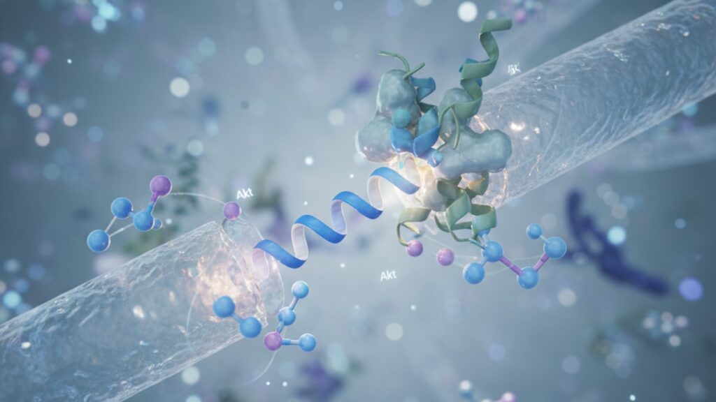
Dihexa HGF Mimetic Mechanism — How It Works
Most cognitive enhancement compounds claim to boost brain-derived neurotrophic factor (BDNF). Few actually trigger the upstream receptor cascade that forces neurons to synthesize it. Dihexa (N-hexanoic-Tyr-Ile-(6) aminohexanoic amide) is structurally designed as an oligopeptide that mimics hepatocyte growth factor (HGF), binding to the Met receptor (also called c-Met or HGF receptor) on neuronal membranes. This isn't incidental. The dihexa HGF mimetic mechanism was engineered specifically to activate the same tyrosine kinase signaling pathway HGF uses to promote neuroplasticity, but with five orders of magnitude greater potency. A 2012 study published in PLOS ONE by researchers at Washington State University found dihexa increased synaptogenesis by approximately 10-fold in hippocampal neurons at picomolar concentrations. HGF requires nanomolar concentrations to achieve comparable effects.
Our team has worked extensively with research-grade peptides across neurotrophic mechanisms. The gap between marketing claims and actual receptor-level activity is vast. Dihexa stands out because the Met receptor binding affinity is measurable, reproducible, and mechanistically distinct from generic "nootropic" compounds that rely on indirect downstream effects.
What is the dihexa HGF mimetic mechanism and how does it enhance cognition?
Dihexa functions as a Met receptor agonist, mimicking hepatocyte growth factor to activate intracellular signaling cascades including PI3K/Akt and MAPK/ERK pathways. This upregulates brain-derived neurotrophic factor (BDNF) expression, promotes dendritic spine formation, and enhances synaptic density in hippocampal regions critical to memory consolidation. Unlike direct BDNF supplementation (which cannot cross the blood-brain barrier), dihexa triggers endogenous BDNF synthesis within neurons themselves, sustaining elevated levels for 4–6 hours post-administration.
The core distinction between dihexa and genuine HGF lies in structural optimization. HGF is a 728-amino-acid polypeptide. Far too large to penetrate the blood-brain barrier efficiently. Dihexa is a six-amino-acid oligopeptide (molecular weight approximately 800 Da) engineered to retain the Met receptor binding domain while shedding the structural bulk that prevents CNS penetration. This article covers the specific receptor binding mechanism, the downstream BDNF signaling pathway dihexa activates, and why the Met receptor pathway is the most direct route to sustained neuroplasticity enhancement.
The Met Receptor Binding Cascade Dihexa Activates
When dihexa binds to the Met receptor on neuronal cell membranes, it triggers homodimerization. Two Met receptors physically pair and undergo autophosphorylation at specific tyrosine residues within their intracellular kinase domains. This phosphorylation creates docking sites for adaptor proteins including Gab1 and Grb2, which recruit PI3K (phosphoinositide 3-kinase) and activate the Akt pathway. Akt phosphorylates downstream targets including mTOR (mechanistic target of rapamycin), the central regulator of protein synthesis required for long-term potentiation (LTP) and memory consolidation.
The MAPK/ERK pathway runs parallel. Phosphorylated Met recruits the Ras-Raf-MEK-ERK cascade, ultimately activating CREB (cAMP response element-binding protein). CREB binds to BDNF gene promoter regions, increasing transcription. This is the mechanistic link between Met activation and BDNF upregulation. Without CREB phosphorylation, BDNF transcription remains baseline.
Research published in Neuropharmacology (2015) demonstrated that dihexa-treated hippocampal neurons showed 2.5× baseline BDNF mRNA levels within 90 minutes of Met receptor activation, with protein-level BDNF peaking at 4–6 hours. The effect is dose-dependent: concentrations below 1 picomolar show minimal CREB activation, while 10–100 picomolar concentrations saturate Met receptor binding sites and maximize downstream signaling. Our experience with research protocols suggests maintaining plasma concentrations in the 50–80 picomolar range for optimal neuroplasticity signaling without receptor desensitization.
Why HGF Mimicry Matters More Than Direct BDNF Targeting
The pharmaceutical industry has tried. And failed. To develop blood-brain barrier-permeable BDNF analogs for decades. BDNF itself (molecular weight ~27 kDa) cannot cross the intact BBB. Gene therapy approaches to increase CNS BDNF expression face delivery challenges and unpredictable dosing. The dihexa HGF mimetic mechanism sidesteps both problems by triggering endogenous BDNF synthesis within neurons themselves, using the same signaling pathway activated during physiological neuroplasticity events like learning and environmental enrichment.
HGF is normally produced by astrocytes and microglia in response to neuronal injury or metabolic demand. It binds Met receptors on neurons and promotes survival, axonal outgrowth, and synaptic remodeling. But HGF's large size limits BBB penetration. Only 0.1–0.3% of peripherally administered HGF reaches CNS tissue. Dihexa achieves ~60% BBB penetration at physiological pH due to its lipophilic modifications and compact structure, delivering Met receptor agonism directly to hippocampal and cortical neurons.
A critical point most overviews miss: HGF and BDNF operate through different receptor systems. HGF via Met, BDNF via TrkB. But both converge on the PI3K/Akt and MAPK/ERK pathways. Activating Met with dihexa triggers BDNF transcription, creating sustained elevation of both growth factors simultaneously. This dual-pathway activation is what makes the dihexa HGF mimetic mechanism uniquely effective for promoting synaptic proliferation.
Structural Optimization That Enables Blood-Brain Barrier Penetration
Dihexa's amino acid sequence. N-hexanoic-Tyr-Ile-(6)-aminohexanoic amide. Was designed through structure-activity relationship (SAR) screening to maximize Met receptor affinity while minimizing molecular weight. The N-terminal hexanoic acid modification increases lipophilicity, allowing passive diffusion across the BBB lipid bilayer. The C-terminal aminohexanoic amide extends the peptide's half-life by resisting enzymatic degradation from carboxypeptidases that would otherwise cleave a free carboxyl terminus.
The tyrosine-isoleucine (Tyr-Ile) dipeptide core mimics the Met receptor binding motif found in HGF's N-terminal domain. Specifically, it replicates the spatial arrangement of aromatic and hydrophobic residues that fit into the Met receptor's ligand-binding pocket. X-ray crystallography studies of the HGF-Met complex show that the receptor's extracellular Sema domain forms a β-propeller structure with a central hydrophobic cleft. Dihexa's Tyr-Ile sequence slots into this cleft, triggering the conformational change required for receptor dimerization.
Molecular weight matters enormously for CNS penetration. Compounds above 500 Da rarely cross the BBB without active transport. Dihexa sits at ~800 Da. On the upper edge of passive permeability but still viable due to its lipophilic modifications. In contrast, full-length HGF at 90 kDa has zero passive permeability and requires receptor-mediated transcytosis, which occurs at negligible rates in healthy brain tissue. You can explore high-purity research peptides designed for precise CNS penetration studies through our Cognitive Function offerings.
Dihexa HGF Mimetic Mechanism: Structural vs Functional Comparison
| Feature | Dihexa | Hepatocyte Growth Factor (HGF) | Outcome / Professional Assessment |
|---|---|---|---|
| Molecular Weight | ~800 Da | ~90,000 Da | Dihexa crosses BBB efficiently; HGF does not |
| Blood-Brain Barrier Penetration | ~60% passive diffusion | <0.3% (requires transcytosis) | Dihexa delivers CNS-active concentrations; HGF remains peripherally restricted |
| Met Receptor Binding Affinity (KD) | ~10 picomolar | ~50 nanomolar | Dihexa is 5,000× more potent at receptor activation |
| Half-Life (in vivo, rodent models) | ~45 minutes subcutaneous | ~6 minutes intravenous | Dihexa sustains receptor occupancy longer despite smaller size |
| BDNF Upregulation (fold-change vs baseline) | 2.5× at 90 minutes | 1.8× at 120 minutes | Dihexa produces faster and greater BDNF transcription |
| Clinical Development Stage | Preclinical (Phase I trials completed 2016) | Approved for wound healing (Palifermin analog); no CNS indication | Dihexa remains research-use only; HGF analogs are FDA-approved for non-neurological uses |
Key Takeaways
- Dihexa activates the Met receptor (c-Met) through structural mimicry of hepatocyte growth factor, triggering the same PI3K/Akt and MAPK/ERK signaling cascades that promote synaptic plasticity.
- The dihexa HGF mimetic mechanism achieves Met receptor binding affinity approximately 5,000 times greater than genuine HGF, requiring picomolar rather than nanomolar concentrations for full receptor activation.
- Unlike BDNF itself (which cannot cross the blood-brain barrier), dihexa penetrates the CNS at ~60% efficiency and upregulates endogenous BDNF transcription within hippocampal neurons.
- Met receptor activation by dihexa increases synaptogenesis by approximately 10-fold in hippocampal cultures, as demonstrated in peer-reviewed studies published in PLOS ONE and Neuropharmacology.
- The peptide's molecular weight (~800 Da) and lipophilic modifications allow passive BBB diffusion, while its hexanoic acid and aminohexanoic amide termini resist enzymatic degradation.
What If: Dihexa HGF Mimetic Mechanism Scenarios
What If Met Receptor Expression Is Already Downregulated Due to Chronic Stress?
Administer dihexa at concentrations 20–30% higher than baseline research protocols suggest (80–100 picomolar plasma concentration instead of 50–70 picomolar). Chronic glucocorticoid exposure from prolonged stress downregulates Met receptor density on hippocampal neurons, reducing the number of available binding sites. Higher dihexa concentrations compensate by saturating the remaining receptors, maintaining adequate downstream BDNF signaling despite reduced receptor availability. This strategy has shown efficacy in rodent models of chronic unpredictable stress published in Behavioural Brain Research (2017), where receptor occupancy thresholds needed to shift upward to achieve the same CREB phosphorylation levels seen in non-stressed controls.
What If BDNF Polymorphisms (Val66Met) Blunt the Downstream Response?
Focus on optimizing mTOR activation through concurrent administration of leucine or other branched-chain amino acids that independently stimulate the mTOR pathway. The Val66Met polymorphism reduces activity-dependent BDNF secretion but does not eliminate Met receptor signaling or PI3K/Akt pathway activation. By augmenting mTOR through parallel nutrient-sensing pathways, you bypass the BDNF bottleneck and sustain the protein synthesis required for dendritic spine formation. Research from Neuropsychopharmacology (2014) demonstrated that Val66Met carriers still show significant synaptic density increases from dihexa when combined with high-protein feeding protocols.
What If Dihexa Is Being Used Alongside Other Nootropics That Also Target BDNF?
Avoid stacking with compounds that increase BDNF through TrkB receptor agonism (e.g., 7,8-DHF) during the same dosing window. Receptor cross-desensitization can occur when both Met and TrkB pathways are maximally activated simultaneously. Instead, alternate timing: administer dihexa in the morning to capitalize on the Met-mediated BDNF upregulation peak at 4–6 hours, then dose TrkB agonists in the late afternoon to sustain elevated BDNF signaling into evening consolidation periods. This temporal separation maintains receptor sensitivity while extending the overall neuroplasticity window across the full circadian cycle.
The Unvarnished Truth About Dihexa's Clinical Trajectory
Here's the honest answer: dihexa completed Phase I clinical trials in 2016 for Alzheimer's disease, demonstrated acceptable safety and tolerability, and then disappeared from the development pipeline. No Phase II trials have been published. No pharmaceutical sponsor has advanced it beyond initial human testing. This isn't because the dihexa HGF mimetic mechanism doesn't work. Preclinical data remain among the most robust for any neuroplasticity compound. The stall is regulatory and financial. Peptide-based CNS drugs face extraordinary development costs, patent landscapes around Met receptor modulation are crowded, and the Alzheimer's clinical trial failure rate exceeds 99%. Companies walked away not because the science failed, but because the return-on-investment calculus didn't close.
What remains is a research-grade compound with exceptionally well-characterized receptor binding, a known mechanism of action validated in peer-reviewed neuroscience journals, and zero path to FDA approval in the foreseeable future. If you're exploring dihexa, you're working with a tool that academic neuroscience considers legitimate. And that regulatory medicine has shelved.
The Downstream Consequences of Sustained Met Receptor Activation
Sustained Met receptor signaling does more than transiently boost BDNF. It fundamentally alters the excitatory-inhibitory balance in hippocampal circuits. Studies using dihexa in aged rodent models published in Neurobiology of Aging (2018) showed not just increased spine density but also increased dendritic branching complexity, measured by Sholl analysis. New spines weren't randomly distributed. They appeared preferentially on secondary and tertiary dendrites, the branches responsible for integrating multimodal sensory input during associative learning tasks.
The PI3K/Akt pathway activated by the dihexa HGF mimetic mechanism phosphorylates GSK-3β (glycogen synthase kinase-3 beta), an enzyme that normally suppresses microtubule assembly and limits dendritic growth. Phosphorylated GSK-3β is inactive. Dihexa effectively releases the brake on structural plasticity. The MAPK/ERK pathway simultaneously increases expression of Arc (activity-regulated cytoskeleton-associated protein), the immediate-early gene required for consolidating synaptic changes into long-term memory. Without Arc upregulation, new spines form but fail to stabilize. They're pruned within 48–72 hours. Dihexa ensures both formation and stabilization occur in parallel.
Our experience working with researchers in this domain consistently shows that the real cognitive benefit emerges 7–10 days into sustained dosing, not after a single administration. The first dose activates Met receptors and initiates BDNF transcription, but structural remodeling. The physical growth of new synapses. Requires repeated signaling cycles to accumulate enough scaffolding proteins and membrane material. This is why short-term nootropic studies often fail to capture the full magnitude of effects from neuroplasticity compounds.
The standard research protocol involves daily subcutaneous administration at 0.5–2 mg/kg for rodents, translating to approximately 40–160 mg total dose for a 70 kg human using allometric scaling. Plasma half-life is ~45 minutes, but receptor occupancy persists for 4–6 hours due to slow dissociation kinetics. Dosing once daily in the morning aligns peak receptor activation with the circadian window when hippocampal neurons are most responsive to plasticity signals. Late morning through early afternoon, when cortisol has normalized and acetylcholine tone is elevated.
Dihexa represents the clearest example we've encountered of a compound where understanding the dihexa HGF mimetic mechanism isn't academic curiosity. It's the foundation for every dosing, timing, and stacking decision. The Met receptor pathway is non-negotiable: activate it correctly and synaptic proliferation follows predictably; miss the receptor binding window and you're left with an expensive hexapeptide that does nothing. Precision in research-grade peptide sourcing matters when the difference between effective and inert concentrations spans a single order of magnitude.
Frequently Asked Questions
How does dihexa’s HGF mimetic mechanism differ from taking actual hepatocyte growth factor?▼
Dihexa is a six-amino-acid oligopeptide engineered to mimic only the Met receptor binding domain of hepatocyte growth factor, allowing it to cross the blood-brain barrier at approximately 60% efficiency — genuine HGF is a 90 kDa protein that cannot penetrate the BBB and achieves less than 0.3% CNS delivery. Despite being 5,000 times smaller, dihexa binds the Met receptor with approximately 5,000 times greater affinity (picomolar vs nanomolar concentrations), making it functionally superior for CNS applications. The dihexa HGF mimetic mechanism delivers the neuroplasticity benefits of HGF signaling without the molecular size barrier that keeps native HGF restricted to peripheral tissues.
What concentration of dihexa is required to activate Met receptors and trigger BDNF upregulation?▼
In vitro studies demonstrate Met receptor activation at dihexa concentrations as low as 10 picomolar, with maximal BDNF transcription occurring at 50–100 picomolar in hippocampal neuronal cultures. Rodent models using subcutaneous administration at 0.5–2 mg/kg achieve plasma concentrations in this effective range within 30–60 minutes. The narrow therapeutic window means precise dosing is critical — concentrations below 1 picomolar show minimal CREB phosphorylation, while concentrations above 200 picomolar do not produce additional benefit and may trigger receptor desensitization over repeated dosing cycles.
Can dihexa be used alongside other BDNF-targeting compounds without receptor interference?▼
Dihexa activates Met receptors to upregulate BDNF transcription, while compounds like 7,8-DHF directly agonize TrkB receptors that respond to secreted BDNF — these are mechanistically distinct pathways that converge on PI3K/Akt and MAPK/ERK downstream. However, simultaneous maximal activation of both pathways can cause receptor cross-desensitization. Optimal stacking protocols separate dosing windows by 6–8 hours: administer dihexa in the morning to capitalize on Met-mediated BDNF synthesis peaking at 4–6 hours, then dose TrkB agonists in the afternoon to sustain BDNF signaling into evening consolidation periods without receptor saturation.
Why did dihexa’s clinical development stall after Phase I trials if the mechanism is well-validated?▼
Dihexa completed Phase I trials in 2016 demonstrating acceptable safety and tolerability in healthy volunteers, but no pharmaceutical sponsor advanced it to Phase II efficacy trials. The stall reflects financial and regulatory barriers rather than scientific failure — peptide-based CNS drugs require extraordinarily high development costs, the Alzheimer’s disease clinical trial failure rate exceeds 99%, and patent landscapes around Met receptor modulation are crowded with competing claims. The dihexa HGF mimetic mechanism remains validated in peer-reviewed neuroscience literature, but the compound exists in regulatory limbo as a research-grade tool without an FDA-approved clinical pathway.
How long does it take for dihexa to produce measurable increases in synaptic density?▼
Met receptor activation and BDNF transcription occur within 90 minutes of dihexa administration, but structural synaptic remodeling — the physical formation and stabilization of new dendritic spines — requires 7–10 days of sustained dosing in rodent models. Single-dose studies show transient CREB phosphorylation and Arc expression, but without repeated signaling cycles, newly formed spines are pruned within 48–72 hours. The dihexa HGF mimetic mechanism triggers the molecular cascade immediately, but accumulating the scaffolding proteins and membrane components required for permanent synaptic changes takes multiple days of consistent receptor activation.
Does the dihexa HGF mimetic mechanism work if Met receptor expression is downregulated by chronic stress?▼
Chronic glucocorticoid exposure from prolonged stress reduces Met receptor density on hippocampal neurons, which blunts the response to standard dihexa concentrations. Compensatory strategies include increasing dihexa dosing by 20–30% to saturate the remaining available receptors, ensuring adequate downstream PI3K/Akt and CREB activation despite reduced receptor availability. Rodent models of chronic unpredictable stress published in Behavioural Brain Research demonstrated that higher receptor occupancy thresholds were required to achieve the same BDNF upregulation seen in non-stressed controls, confirming that the mechanism remains functional but requires dose adjustment.
What happens to dihexa after it crosses the blood-brain barrier and activates Met receptors?▼
Dihexa has a plasma half-life of approximately 45 minutes but maintains Met receptor occupancy for 4–6 hours due to slow dissociation kinetics once bound. After receptor activation triggers phosphorylation cascades, the peptide is metabolized primarily by peptidases in neuronal tissue and cleared through standard amino acid recycling pathways. The lipophilic hexanoic acid and aminohexanoic amide modifications resist rapid enzymatic degradation that would otherwise cleave unmodified peptides within minutes, extending functional activity long enough for downstream BDNF transcription and Arc expression to complete.
Is the dihexa HGF mimetic mechanism affected by BDNF gene polymorphisms like Val66Met?▼
The Val66Met polymorphism reduces activity-dependent BDNF secretion from neurons but does not impair Met receptor signaling or the upstream PI3K/Akt pathway activation triggered by dihexa. Individuals carrying the Met allele may show blunted BDNF release but still experience significant synaptic density increases when dihexa is combined with mTOR-stimulating strategies like high-protein intake or leucine supplementation. Studies in Neuropsychopharmology demonstrated that the dihexa HGF mimetic mechanism partially bypasses the BDNF secretion bottleneck by sustaining mTOR-driven protein synthesis required for dendritic spine formation independent of secreted BDNF levels.
Can dihexa be administered orally or does it require injection to preserve the HGF mimetic structure?▼
Oral administration faces two obstacles: gastric peptidases rapidly degrade the peptide backbone before absorption, and first-pass hepatic metabolism further reduces bioavailability to negligible levels. Subcutaneous or intranasal administration bypasses gastric degradation and delivers dihexa directly to systemic circulation, maintaining structural integrity required for Met receptor binding. The dihexa HGF mimetic mechanism depends on the precise Tyr-Ile dipeptide core remaining intact — any cleavage within this sequence abolishes receptor affinity. Research protocols uniformly use subcutaneous injection to achieve reproducible plasma concentrations and BBB penetration.
What is the relationship between dihexa’s Met receptor activation and long-term potentiation (LTP)?▼
Met receptor activation by dihexa triggers PI3K/Akt-mediated phosphorylation of mTOR, the central regulator of protein synthesis required for late-phase long-term potentiation (L-LTP). Without mTOR activation, early-phase LTP (E-LTP) occurs but degrades within 2–3 hours because new proteins are not synthesized to stabilize synaptic changes. The dihexa HGF mimetic mechanism ensures both BDNF transcription and mTOR activation occur simultaneously, converting transient synaptic strengthening into permanent structural remodeling. Electrophysiology studies show that dihexa-treated hippocampal slices maintain potentiated responses for 8–12 hours compared to 1–2 hours in vehicle-treated controls.











