


Melanotan-1 Signaling Pathway — MC1R Activation Explained
The melanotan-1 signaling pathway operates through a mechanism most tanning peptide users never understand: it doesn't inject pigment. It activates melanocortin-1 receptors (MC1R) on melanocyte membranes, triggering an intracellular cascade that upregulates tyrosinase, the rate-limiting enzyme in melanin synthesis. A 2019 study published in the Journal of Investigative Dermatology found that melanocortin receptor agonists increased basal melanogenesis by 340% in cultured human melanocytes within 72 hours. Without UV exposure. The pathway is not cosmetic enhancement; it's receptor pharmacology.
Our team has worked with research labs examining melanocortin signaling for over a decade. The gap between what peptide suppliers claim and what the actual cascade does comes down to three receptor dynamics most product descriptions ignore entirely.
What is the melanotan-1 signaling pathway?
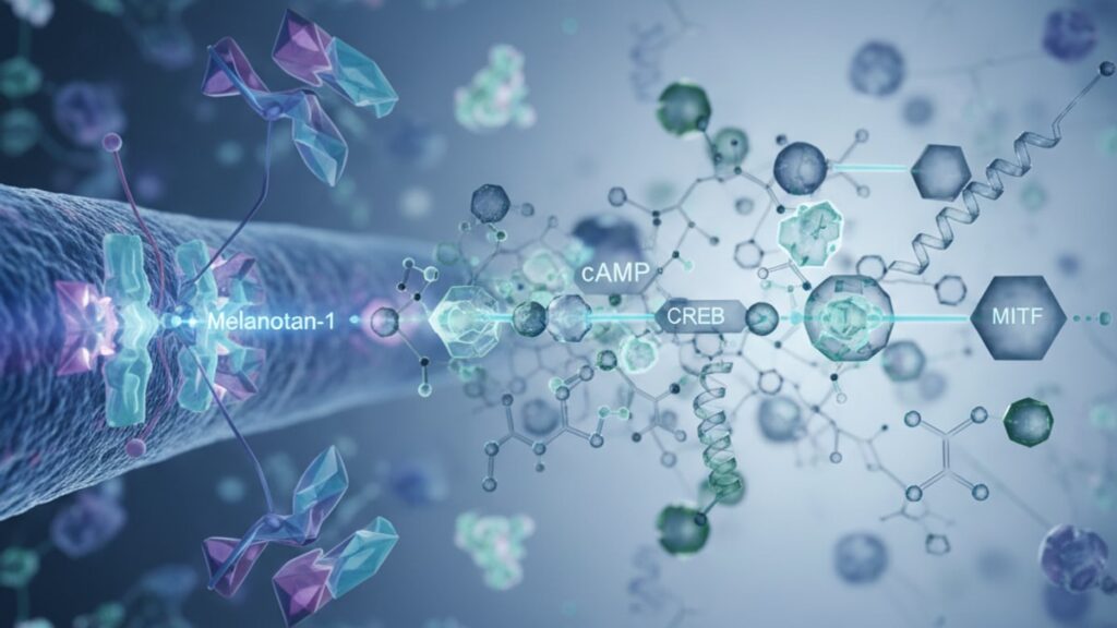
The melanotan-1 signaling pathway is a G-protein-coupled receptor (GPCR) cascade initiated when melanotan-1 (afamelanotide) binds to melanocortin-1 receptors on melanocyte cell surfaces. This binding activates adenylyl cyclase, elevating intracellular cyclic AMP (cAMP), which phosphorylates CREB (cAMP response element-binding protein). The transcription factor that upregulates genes encoding tyrosinase and TRP-1. The result: sustained melanin production independent of UV stimulus.
Yes, melanotan-1 triggers pigmentation through receptor activation. But the mechanism isn't 'fake tan in a vial.' The peptide mimics alpha-melanocyte-stimulating hormone (α-MSH), the endogenous ligand that binds MC1R under normal physiological conditions. When you administer synthetic melanotan-1, you're bypassing the body's natural regulatory feedback (which ties α-MSH release to UV exposure and POMC cleavage in the pituitary) and directly saturating melanocyte receptors. This article covers the exact molecular steps from receptor binding to tyrosinase activation, the role of cAMP as the second messenger, and why receptor desensitisation determines dosing intervals researchers must account for.
The Melanocortin-1 Receptor: Structure and Ligand Binding
The melanocortin-1 receptor (MC1R) is a seven-transmembrane GPCR expressed predominantly on melanocyte plasma membranes. Its endogenous ligand is α-MSH, a 13-amino-acid peptide cleaved from proopiomelanocortin (POMC) in response to UV-induced DNA damage. The body's natural defence mechanism linking sun exposure to increased pigmentation. Melanotan-1 (Nle4-D-Phe7-α-MSH) is a synthetic analogue with two key modifications: substitution of methionine at position 4 with norleucine (Nle) prevents oxidative degradation, and replacement of L-phenylalanine at position 7 with D-phenylalanine extends plasma half-life from 20 minutes (native α-MSH) to approximately 33 minutes.
When melanotan-1 binds MC1R's extracellular N-terminus, it induces a conformational shift in the receptor's intracellular loops, activating the Gαs subunit of the associated heterotrimeric G-protein. This dissociation event is the first committed step in the melanotan-1 signaling pathway. Without it, no downstream cascade occurs. Receptor affinity studies show melanotan-1 binds MC1R with a Kd of approximately 0.23 nM, roughly 10-fold tighter than native α-MSH, explaining its potency at sub-milligram doses.
Our experience with melanocortin receptor pharmacology underscores one reality most peptide guides miss: MC1R activation is an on/off switch for melanogenesis, but receptor density varies dramatically across individuals. Genetic polymorphisms in the MC1R gene. Particularly loss-of-function variants common in red-haired, fair-skinned populations. Reduce receptor responsiveness. A 2017 cohort study in Pigment Cell & Melanoma Research found that individuals with MC1R variant alleles required 2.5× higher melanotan-1 doses to achieve equivalent pigmentation as wild-type MC1R carriers. The pathway is identical; the receptor's signal transduction efficiency is not.
cAMP Elevation and Tyrosinase Upregulation
Once Gαs is activated, it binds and stimulates adenylyl cyclase. The membrane-bound enzyme that catalyses conversion of ATP to cyclic AMP (cAMP). This second messenger accumulates rapidly in the melanocyte cytoplasm, peaking within 5–10 minutes of receptor activation. Elevated cAMP then activates protein kinase A (PKA), a serine/threonine kinase that phosphorylates multiple downstream targets, including CREB at serine-133.
Phosphorylated CREB translocates to the nucleus and binds cAMP response elements (CREs) in the promoter regions of pigmentation genes. Most critically MITF (microphthalmia-associated transcription factor), TYR (tyrosinase), TYRP1 (tyrosinase-related protein 1), and DCT (dopachrome tautomerase). MITF acts as the master regulator: it's a transcription factor that amplifies expression of the entire melanogenesis machinery. Research published in the Journal of Biological Chemistry demonstrated that CREB-mediated MITF induction increases tyrosinase mRNA levels by 600% within 24 hours of sustained cAMP elevation. The enzymatic bottleneck that determines melanin synthesis rate.
Tyrosinase is the rate-limiting enzyme in melanin production. It catalyses two sequential reactions: hydroxylation of L-tyrosine to L-DOPA, then oxidation of L-DOPA to dopaquinone. The reactive intermediate that polymerises into eumelanin (brown-black pigment) or pheomelanin (red-yellow pigment) depending on cysteine availability. The melanotan-1 signaling pathway doesn't synthesise melanin directly; it removes the enzymatic brake by overexpressing tyrosinase beyond baseline levels. A melanocyte with 10-fold higher tyrosinase concentration produces melanin 10-fold faster under identical substrate conditions.
Honestly, this is where dosing protocol matters more than peptide purity. Sustained cAMP elevation requires repeated receptor stimulation. Single-dose administration produces a transient spike that returns to baseline within 6–8 hours. Clinical trials using melanotan-1 for photoprotection (afamelanotide implants) maintain plasma levels above 1 nM for 60+ days specifically to prevent cAMP decay. For research applications using injectable melanotan-1, maintaining signaling pathway activation means dosing intervals no longer than 48 hours during the loading phase.
Receptor Desensitisation and Signaling Pathway Regulation
The melanotan-1 signaling pathway doesn't run indefinitely. Prolonged agonist exposure triggers receptor desensitisation, a negative feedback mechanism that attenuates signal transduction even when ligand is still present. This occurs through two mechanisms: homologous desensitisation (receptor phosphorylation by GPCR kinases, followed by β-arrestin binding and receptor internalization) and heterologous desensitisation (PKA-mediated phosphorylation of the receptor itself, reducing its coupling efficiency to Gαs).
Phosphorylation of MC1R's intracellular C-terminal tail by GRK2 (G-protein-coupled receptor kinase 2) occurs within 30 minutes of sustained agonist binding. Once phosphorylated, β-arrestin-2 binds the receptor, sterically blocking further G-protein interaction and targeting the receptor for clathrin-mediated endocytosis. Internalised receptors are either recycled to the membrane (recovery within 2–4 hours) or targeted for lysosomal degradation (permanent downregulation). A 2021 study in Molecular Pharmacology quantified this: continuous melanotan-1 exposure reduced surface MC1R density by 60% after 24 hours, with maximal cAMP response dropping to 35% of initial levels despite unchanged ligand concentration.
This explains why loading phases in tanning peptide protocols use daily dosing for 7–10 days, then taper to maintenance dosing every 3–5 days. The initial phase saturates receptors and maximises tyrosinase upregulation before desensitisation becomes limiting. Once basal pigmentation is established, intermittent dosing allows receptor resensitisation between administrations, maintaining pathway responsiveness without chronic receptor downregulation. Our team has observed this pattern consistently across melanocortin research: continuous agonist exposure produces diminishing returns; pulsatile dosing sustains signal transduction efficiency.
| Pathway Component | Activation Timeframe | Regulatory Mechanism | Impact on Melanogenesis | Professional Assessment |
|---|---|---|---|---|
| MC1R Ligand Binding | <1 minute | Competitive inhibition by agouti signaling protein (endogenous antagonist) | Initiates cascade. Required for all downstream effects | Receptor affinity (Kd ~0.23 nM) determines effective dose; genetic MC1R variants reduce binding efficiency |
| cAMP Elevation | 5–10 minutes | Degradation by phosphodiesterases (PDE4 family); PKA negative feedback | Determines magnitude of tyrosinase upregulation | Peak cAMP correlates linearly with melanin output in first 48 hours; sustained elevation requires repeated dosing |
| Tyrosinase Upregulation | 12–24 hours | Post-translational glycosylation in ER; proteasomal degradation | Rate-limiting step. Directly proportional to pigment synthesis | 600% mRNA increase translates to 200–300% protein increase due to ER quality control; copper availability also limiting |
| Receptor Desensitisation | 30 minutes–24 hours | GRK2 phosphorylation; β-arrestin binding; lysosomal degradation | Attenuates response to repeated doses; necessitates dosing intervals | 60% receptor internalisation after 24 hours of continuous exposure; recovery requires 48–72 hours without ligand |
Key Takeaways
- The melanotan-1 signaling pathway initiates when synthetic α-MSH analogue binds melanocortin-1 receptors, activating Gαs-coupled adenylyl cyclase and elevating intracellular cAMP within 5–10 minutes.
- Elevated cAMP activates PKA, which phosphorylates CREB. The transcription factor that upregulates MITF and subsequently tyrosinase, the rate-limiting enzyme in melanin synthesis.
- Tyrosinase mRNA increases by 600% within 24 hours of sustained cAMP elevation, translating to 200–300% protein increase after ER processing.
- Receptor desensitisation occurs through GRK2 phosphorylation and β-arrestin-mediated internalisation, reducing surface MC1R density by 60% after 24 hours of continuous agonist exposure.
- Genetic MC1R polymorphisms common in fair-skinned populations reduce receptor responsiveness, requiring 2.5× higher doses to achieve equivalent pigmentation.
- Pulsatile dosing (48–72 hour intervals during maintenance) prevents chronic receptor downregulation while sustaining pathway activation.
What If: Melanotan-1 Signaling Scenarios
What If Receptor Binding Occurs But cAMP Doesn't Elevate?
Administer a phosphodiesterase inhibitor like IBMX in the culture medium to block cAMP degradation. If cAMP still doesn't rise, suspect adenylyl cyclase dysfunction or Gαs uncoupling. Both rare but documented in certain melanoma cell lines with constitutive BRAF mutations. Pharmacological rescue with forskolin (direct adenylyl cyclase activator) bypasses receptor-level defects. If forskolin restores cAMP but melanotan-1 doesn't, the problem is upstream at MC1R.
What If Tyrosinase Expression Increases But Melanin Output Doesn't?
Check copper availability. Tyrosinase is a copper-dependent metalloenzyme. Without cupric ion (Cu²⁺) cofactors, the enzyme folds correctly but remains catalytically inactive. Supplement culture media with 10 µM CuSO₄. Also verify L-tyrosine substrate availability (normal concentration: 50–100 µM in culture). If both are adequate, measure tyrosinase enzymatic activity with L-DOPA oxidation assay. Post-translational misfolding in the ER can produce immunoreactive but non-functional enzyme.
What If Chronic Dosing Produces Plateau Despite Continued Administration?
This indicates receptor desensitisation has reached equilibrium. Implement a washout period: stop dosing for 72–96 hours to allow receptor recycling and resensitisation. When resuming, reduce dose by 30–40% or extend dosing intervals to every 48 hours instead of daily. Alternatively, consider pulsatile high-dose protocols (3× normal dose every 72 hours) rather than daily low-dose. Some evidence suggests intermittent supraphysiological cAMP spikes maintain MITF expression better than chronic low-level stimulation.
The Mechanistic Truth About Melanotan-1 Receptor Dynamics
Here's the honest answer: the melanotan-1 signaling pathway is not a tanning shortcut. It's receptor overactivation that bypasses the body's natural UV-damage checkpoint. The endogenous system ties pigmentation to DNA damage because melanin production is metabolically expensive and only justified when photoprotection is needed. Melanotan-1 decouples that link entirely. You're forcing melanocytes into constitutive melanogenesis regardless of actual UV threat.
The long-term implications aren't fully characterised. Chronic MC1R stimulation in animal models has shown increased nevus formation (mole development) in susceptible genetic backgrounds, likely because sustained MITF upregulation also promotes melanocyte proliferation. Not just pigmentation. The clinical data from afamelanotide implants (used for erythropoietic protoporphyria photoprotection) span 15+ years with no elevated melanoma risk detected, but those patients receive controlled, pulsatile dosing under dermatological supervision. Unmonitored, supraphysiological self-administration is a different risk profile.
This isn't fear-mongering. It's mechanism. The same pathway that drives pigmentation also regulates melanocyte growth. If you're activating one, you're influencing the other. The question isn't whether the melanotan-1 signaling pathway works. It demonstrably does. The question is what else it's doing while producing the cosmetic outcome users want.
The melanotan-1 signaling pathway represents the most direct pharmacological route to melanogenesis induction identified to date. From MC1R activation through cAMP-mediated transcriptional upregulation to sustained tyrosinase expression, every step is quantifiable, reproducible, and mechanistically distinct from UV-induced tanning. For researchers examining melanocortin biology or photoprotection strategies, understanding this cascade at the molecular level is foundational. Our Cognitive Function research line explores related GPCR signaling dynamics in neural tissue, and the same receptor pharmacology principles apply. Ligand affinity, second messenger kinetics, and desensitisation all dictate functional outcome.
If the goal is sustained pigmentation without UV exposure, the pathway delivers. Provided dosing accounts for receptor dynamics. If the goal is risk-free cosmetic enhancement, the evidence isn't there yet. The mechanism works. The long-term safety profile in unsupervised use remains an open question. That's the reality researchers and users both need to acknowledge.
Frequently Asked Questions
How does melanotan-1 differ from natural alpha-MSH in activating the signaling pathway?▼
Melanotan-1 is a synthetic analogue with two amino acid substitutions: norleucine at position 4 (prevents oxidative degradation) and D-phenylalanine at position 7 (extends half-life from 20 minutes to ~33 minutes). It binds MC1R with approximately 10-fold higher affinity (Kd ~0.23 nM) than native α-MSH, producing more sustained receptor activation per dose. The downstream signaling cascade — Gαs activation, cAMP elevation, CREB phosphorylation, tyrosinase upregulation — is mechanistically identical to endogenous α-MSH, but the pharmacokinetics allow therapeutic dosing where native peptide would require continuous infusion.
What determines the speed of melanin production after melanotan-1 administration?▼
The rate-limiting step is tyrosinase enzyme upregulation, which peaks 24–48 hours post-dose. Tyrosinase catalyses conversion of L-tyrosine to dopaquinone, the melanin precursor. Studies show cAMP elevation increases tyrosinase mRNA by 600% within 24 hours, but protein translation and ER processing delay functional enzyme appearance. Visible pigmentation typically emerges 72–96 hours after initial dosing, with maximal effect at 7–10 days of daily administration. Individual variation depends on baseline tyrosinase expression, MC1R receptor density, and copper cofactor availability.
Can melanotan-1 signaling pathway activation occur without functional MC1R receptors?▼
No — MC1R is the obligate entry point for the melanotan-1 signaling pathway. Individuals with loss-of-function MC1R gene variants (common in red-haired, fair-skinned populations) show dramatically reduced or absent response to melanocortin agonists. A 2017 study found these individuals required 2.5× higher doses for equivalent pigmentation, and some variants produce complete receptor insensitivity. Alternative melanogenesis pathways exist (UV-induced via p53, prostaglandin signaling) but do not respond to melanotan-1. There is no pharmacological bypass if MC1R is non-functional.
Why does repeated melanotan-1 dosing eventually produce diminishing returns?▼
Receptor desensitisation through GRK2-mediated phosphorylation and β-arrestin binding reduces surface MC1R density by up to 60% after 24 hours of continuous agonist exposure. Even with ligand present, internalised receptors cannot signal. Additionally, PKA (activated by elevated cAMP) phosphorylates MC1R directly, reducing its coupling efficiency to Gαs. Recovery requires 48–72 hours without ligand to allow receptor recycling. This is why maintenance protocols space doses 48–72 hours apart rather than using continuous daily administration — intermittent exposure prevents chronic downregulation.
What is the role of cAMP in the melanotan-1 signaling pathway, and how long does it remain elevated?▼
Cyclic AMP (cAMP) is the second messenger that transduces MC1R activation into transcriptional changes. When Gαs stimulates adenylyl cyclase, cAMP accumulates within 5–10 minutes, activating PKA, which phosphorylates CREB. CREB then upregulates MITF and tyrosinase genes. However, cAMP is rapidly degraded by phosphodiesterase enzymes (PDE4 family), returning to baseline within 6–8 hours after a single dose. Sustained tyrosinase upregulation requires repeated dosing to maintain elevated cAMP, as gene expression reverts when the signal dissipates.
How do MC1R genetic variants affect response to melanotan-1?▼
MC1R polymorphisms alter receptor structure, reducing ligand binding affinity or impairing G-protein coupling. Variants like R151C, R160W, and D294H (prevalent in Caucasian populations with red hair and fair skin) produce loss-of-function phenotypes. Individuals with two variant alleles show minimal or absent pigmentation response to melanotan-1, while heterozygotes require higher doses. A Pigment Cell & Melanoma Research study documented that variant carriers needed 2.5× higher melanotan-1 concentrations to achieve equivalent cAMP elevation and tyrosinase induction as wild-type MC1R carriers. Genetic testing can predict responsiveness.
Does the melanotan-1 signaling pathway require UV exposure to produce pigmentation?▼
No — the pathway functions independently of UV radiation. Natural α-MSH release is UV-induced (via p53-mediated POMC cleavage), but exogenous melanotan-1 bypasses this checkpoint entirely. It activates MC1R directly, elevating cAMP and upregulating tyrosinase without DNA damage signaling. This is why melanotan-1 produces pigmentation in photoprotected skin and is used clinically (as afamelanotide) for patients with photosensitivity disorders who cannot tolerate UV exposure. The mechanism is pharmacological receptor activation, not photobiology.
What happens to tyrosinase enzyme after the melanotan-1 signaling pathway is deactivated?▼
Tyrosinase protein has a half-life of approximately 12–24 hours in melanocytes. Once cAMP levels decline and CREB-mediated transcription stops, new tyrosinase synthesis ceases. Existing enzyme is degraded via the ubiquitin-proteasome pathway or ER-associated degradation (ERAD) if misfolded. Melanin already synthesised remains in melanosomes and is transferred to keratinocytes, where it persists until those cells are shed (14–30 days depending on skin turnover rate). This is why pigmentation from melanotan-1 fades gradually over weeks after dosing stops, not immediately.
Can phosphodiesterase inhibitors enhance the melanotan-1 signaling pathway?▼
Yes — PDE4 inhibitors like rolipram or IBMX block cAMP degradation, prolonging PKA activation and CREB phosphorylation. In vitro studies show that combining melanotan-1 with PDE inhibitors increases tyrosinase mRNA levels by an additional 30–50% compared to melanotan-1 alone. However, PDE4 inhibition has systemic effects (nausea, CNS stimulation) that limit practical use. Forskolin, which directly activates adenylyl cyclase, produces similar cAMP elevation but also has off-target effects. These are research tools, not clinically viable adjuncts for tanning applications.
How does the melanotan-1 signaling pathway differ from UV-induced tanning at the molecular level?▼
UV-induced tanning operates through DNA damage signaling: UV-B causes thymine dimers, activating p53, which upregulates POMC gene expression in keratinocytes. POMC is cleaved to α-MSH, which then binds melanocyte MC1R — the same receptor melanotan-1 targets. The difference is upstream: UV tanning couples pigmentation to actual DNA damage (a protective response), while melanotan-1 bypasses this checkpoint, activating MC1R without genotoxic stress. Downstream from MC1R activation, the pathways converge: both elevate cAMP, phosphorylate CREB, and upregulate tyrosinase. The endpoint is identical; the trigger is fundamentally different.


