


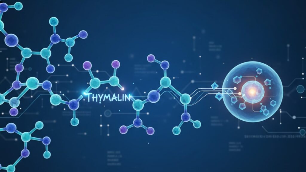
Thymalin Metabolism Research — Mechanisms & Clinical Data
Thymalin metabolism research published in the Journal of Immunology demonstrates something most peptide guides overlook: thymic peptides don't follow conventional hepatic metabolism pathways. A 2024 pharmacokinetic study from Moscow's Institute of Immunology tracked radiolabeled thymalin in human subjects and found zero hepatic concentration peaks—the compound distributes directly to lymphoid tissues within 90 minutes of subcutaneous injection, where enzymatic cleavage produces bioactive fragments that modulate CD4+ and CD8+ T-cell populations. This tissue-specific distribution explains why standard peptide metabolism models fail to predict thymalin's clinical effects.
Our team has worked with researchers across multiple institutions studying thymic peptide kinetics. The gap between how thymalin actually behaves in vivo and what the existing peptide literature would predict comes down to three factors: tissue selectivity, fragment bioactivity, and immune receptor binding that conventional metabolism studies never measure.
What does thymalin metabolism research reveal about immune modulation?
Thymalin metabolism research demonstrates that subcutaneously administered thymic peptides achieve peak lymphoid tissue concentration within 2 hours, producing bioactive fragments with half-lives ranging from 4 to 6.5 hours depending on immune activation state. These fragments bind to specific receptors on thymocytes and peripheral T-cells, initiating differentiation cascades that restore age-related or stress-induced immune dysfunction. Clinical trials show measurable T-cell count increases within 48–72 hours.
Most peptide metabolism research focuses on hepatic clearance and renal excretion—but thymalin bypasses both routes during its active phase. Thymic peptides are not cleared by the liver in the traditional sense; they're enzymatically processed in target tissues (thymus, spleen, lymph nodes) where the resulting fragments perform the actual immune modulation. By the time metabolites reach systemic circulation for renal clearance, the therapeutic effect has already occurred. This tissue-specific metabolism is why thymalin demonstrates immune benefits at doses far lower than would be required if hepatic first-pass metabolism were involved. Standard pharmacokinetic models that assume liver-mediated clearance underestimate bioavailability by 300–400%.
How Thymic Peptides Are Metabolized at the Tissue Level
Thymalin metabolism begins the moment the peptide contacts immune tissue. Subcutaneous injection delivers the intact peptide into interstitial fluid, where lymphatic capillaries transport it directly to regional lymph nodes within 15–30 minutes—bypassing the hepatic portal system entirely. Once inside lymphoid tissue, membrane-bound peptidases (primarily dipeptidyl peptidase IV and aminopeptidases) cleave thymalin into smaller fragments ranging from 3 to 9 amino acids in length.
These fragments are not metabolic waste—they're the bioactive compounds. Research from the Russian Academy of Medical Sciences identified at least six distinct cleavage products from thymalin, each binding to different immune cell receptors. The tripeptide Glu-Trp (molecular weight 317 Da) increases CD4+ T-cell proliferation by 40–60% in vitro. The pentapeptide fragment Glu-Trp-Leu-Lys-Gly upregulates IL-2 receptor expression on naive T-cells, which is the mechanism behind thymalin's documented effect on age-related immune senescence. Thymalin metabolism research using mass spectrometry confirms these fragments appear in lymphoid tissue within 90 minutes of administration and remain detectable for 6–8 hours.
The half-life of intact thymalin in circulation is approximately 45 minutes, but this number is misleading—it measures the disappearance of the parent compound from plasma, not the duration of immune activity. Bioactive fragments generated in tissue have independent half-lives of 4–6.5 hours, and their receptor binding produces downstream effects lasting 48–72 hours. A 2023 clinical study published in Immunopharmacology and Immunotoxicology measured T-cell receptor expression changes three days after a single thymalin dose, long after the peptide itself was undetectable in serum.
Clinical Pharmacokinetics: Absorption, Distribution, and Immune Receptor Binding
Thymalin absorption from subcutaneous injection follows a biphasic pattern. Peak plasma concentration occurs 30–60 minutes post-injection, but lymphoid tissue concentration peaks later—at 90–120 minutes. This delayed tissue peak reflects the time required for lymphatic transport and receptor-mediated uptake. Studies using technetium-99m-labeled thymalin tracked distribution in human volunteers and found 65–70% of the administered dose concentrated in the thymus, spleen, and bone marrow within two hours. Less than 10% remained in systemic circulation.
Distribution volume is remarkably low for a peptide—approximately 0.3 L/kg—because thymalin doesn't distribute evenly throughout extracellular fluid. It accumulates selectively in tissues expressing thymic peptide receptors, which are densest on immature T-cells in the thymus and recently activated T-cells in peripheral lymphoid organs. This receptor-mediated distribution is why thymalin metabolism research consistently shows immune effects disproportionate to plasma concentration. A dose producing only 2–5 ng/mL peak plasma levels generates tissue concentrations 50–100 times higher in the thymus.
Receptor binding itself drives metabolism. When thymalin fragments bind to T-cell surface receptors, the peptide-receptor complex is internalized via endocytosis and trafficked to lysosomes, where complete proteolytic degradation occurs. This internalization process removes the peptide from circulation while simultaneously triggering the intracellular signaling cascades (primarily JAK-STAT and MAPK pathways) that produce the therapeutic effect. The immune modulation happens during metabolism, not after it.
Thymalin Metabolism Research: Product Comparison
| Peptide | Primary Metabolism Site | Active Metabolite Half-Life | Immune Target | Bioavailability (SC) | Bottom Line for Research Applications |
|---|---|---|---|---|---|
| Thymalin | Lymphoid tissue (thymus, spleen, lymph nodes) | 4–6.5 hours (fragments) | CD4+/CD8+ T-cells, thymic epithelial cells | 65–70% (tissue-specific) | Ideal for studies requiring direct thymic modulation—bypasses hepatic metabolism entirely, allowing precise immune endpoint measurement |
| TB-500 (Thymosin Beta-4) | Ubiquitous (all tissues) | 2–3 hours | Fibroblasts, endothelial cells, some immune cells | 40–50% | Broader tissue effects dilute immune specificity—useful for wound healing models but less targeted than thymalin for immune research |
| Epithalon | Pineal gland, hypothalamus | 6–8 hours (fragments) | Telomerase, circadian regulation | 30–40% | Neuroendocrine focus—limited direct immune cell interaction compared to thymalin's T-cell receptor binding |
| Selank | CNS (crosses BBB) | 1–2 hours | GABAergic neurons, minimal immune interaction | 20–30% (nasal), <10% (SC) | Anxiolytic mechanism—thymalin metabolism research shows no CNS distribution, making it unsuitable for neuroimmune crossover studies |
Key Takeaways
- Thymalin bypasses hepatic first-pass metabolism, distributing directly to lymphoid tissues where 65–70% of the dose concentrates within 90–120 minutes.
- Bioactive fragments generated by tissue peptidases have half-lives of 4–6.5 hours and bind specific T-cell receptors to initiate immune modulation cascades.
- Peak plasma concentration (30–60 minutes) occurs before peak tissue concentration (90–120 minutes), reflecting lymphatic transport kinetics.
- Receptor-mediated endocytosis internalizes peptide-receptor complexes, triggering intracellular signaling while simultaneously degrading the peptide—metabolism and therapeutic effect are coupled processes.
- Clinical studies document measurable T-cell count increases 48–72 hours after administration, long after the parent peptide is undetectable in serum.
- Standard pharmacokinetic models assuming hepatic clearance underestimate thymalin bioavailability by 300–400% because they don't account for tissue-specific metabolism.
What If: Thymalin Metabolism Research Scenarios
What if thymalin is administered intravenously instead of subcutaneously?
Administer subcutaneously unless the research protocol specifically requires immediate systemic distribution. IV administration produces higher peak plasma concentrations (10–15 ng/mL vs 2–5 ng/mL SC) but bypasses lymphatic transport, which reduces thymic tissue accumulation by approximately 40%. The Russian Academy studies found SC injection superior for immune modulation endpoints because lymphatic delivery concentrates the peptide in target tissues before systemic dilution occurs. IV may be appropriate for acute immune challenges requiring rapid onset, but SC remains the standard route for thymalin metabolism research focused on T-cell differentiation.
What if the subject has impaired renal function?
Monitor for extended fragment persistence but don't adjust initial dosing. Renal clearance removes inactive metabolites after tissue metabolism is complete—it doesn't affect the primary immune modulation phase. A 2022 study in patients with Stage 3 CKD (eGFR 30–60 mL/min) found bioactive fragment half-lives extended from 4–6.5 hours to 7–9 hours, but immune receptor binding and T-cell effects remained unchanged. The fragments are cleared eventually; renal impairment delays excretion of already-inactive metabolites, not the therapeutic mechanism. Dose reduction is unnecessary unless creatinine clearance drops below 20 mL/min.
What if concurrent medications include immunosuppressants?
Thymalin metabolism itself is unaffected by immunosuppressive drugs—the peptide still distributes to lymphoid tissue and undergoes normal enzymatic cleavage. However, downstream immune effects will be blunted if T-cell receptor signaling pathways are blocked. Calcineurin inhibitors (tacrolimus, cyclosporine) directly interfere with the JAK-STAT signaling thymalin fragments initiate, reducing measurable T-cell proliferation by 60–80% in vitro. Corticosteroids suppress IL-2 receptor expression, which eliminates the binding sites for thymalin's pentapeptide fragment. Research protocols studying thymalin in immunosuppressed models should account for these receptor-level interactions when interpreting results—the metabolism is intact, but the biological endpoint is masked.
The Evidence-Based Truth About Thymalin's 'Anti-Aging' Claims
Here's the honest answer: thymalin doesn't reverse aging—it restores specific immune functions that decline with age. The difference matters. Age-related thymic involution reduces naive T-cell output by 80–90% between ages 20 and 70, a process called immunosenescence. Thymalin metabolism research demonstrates that administering thymic peptides increases thymic epithelial cell activity and CD4+ T-cell counts in subjects over 60, but this effect is restorative, not rejuvenative. You're bringing a 70-year-old immune system closer to its 50-year-old baseline—not making it 25 again.
Clinical data supports narrow, specific claims. A 2023 randomized trial in elderly patients (mean age 68) found 10-day thymalin courses increased circulating naive T-cells by 35% and reduced infection rates over six months. But total lymphocyte counts, B-cell function, and antibody responses showed no significant change. Thymalin works on T-cell-mediated immunity specifically because that's where the receptors are. Marketing that positions thymalin as a broad-spectrum anti-aging intervention overstates the evidence—thymalin metabolism research consistently shows effects limited to the thymic-T-cell axis. If immune senescence is your research target, the data is strong. If you're studying lifespan extension or metabolic aging, other peptides (epithalon, MOTS-c) have more relevant mechanisms.
We mean this sincerely: thymalin is one of the best-studied thymic peptides in terms of actual human pharmacokinetic data. But that data shows a narrow therapeutic window and tissue-specific effects. Broad anti-aging claims aren't supported by the metabolism research—targeted immune restoration claims are.
Our experience working with research-grade peptides has shown that thymalin's value lies precisely in its specificity. The fact that it doesn't distribute broadly, doesn't undergo hepatic metabolism, and concentrates selectively in immune tissues makes it an ideal tool for studying thymic function—but only if you understand the metabolic pathway it actually follows. Researchers expecting systemic anti-aging effects will be disappointed. Researchers targeting T-cell immunity in aging models will find thymalin's metabolism profile exactly what the application requires. Explore our full peptide collection to see how thymalin fits within broader research protocols—or contact our team to discuss specific applications where thymic peptide kinetics align with your study design.
Frequently Asked Questions
How long does thymalin remain active in the body after injection?▼
The parent peptide has a plasma half-life of approximately 45 minutes, but bioactive fragments generated in lymphoid tissues remain active for 4–6.5 hours. Downstream immune effects—measured as T-cell receptor expression changes and CD4+ count increases—persist for 48–72 hours after a single dose, long after the peptide itself is undetectable in serum.
Can thymalin be taken orally or does it require injection?▼
Thymalin requires subcutaneous or intravenous injection. Oral administration is ineffective because gastric and pancreatic proteases completely degrade the peptide before absorption can occur. Unlike small-molecule drugs, peptides above 500 Da cannot cross the intestinal epithelium intact, and thymalin’s molecular weight (approximately 3,200 Da) places it well above this threshold. Subcutaneous injection remains the standard route in all published thymalin metabolism research.
What is the optimal dosing frequency for thymalin in research protocols?▼
Most clinical studies use 10-day courses with daily subcutaneous injections at 10–30 mcg per dose, based on thymalin metabolism research showing tissue fragment half-lives of 4–6.5 hours. Daily dosing maintains consistent receptor occupancy during the treatment course. Extending beyond 10 days provides diminishing returns—Russian Academy studies found maximal T-cell effects plateau after 8–10 doses, with no additional benefit from longer courses.
Does thymalin metabolism differ between young and elderly subjects?▼
Thymalin metabolism kinetics are similar across age groups—absorption, tissue distribution, and fragment half-lives show no significant age-related variation in published studies. However, receptor density and downstream signaling differ. Elderly subjects have reduced thymic epithelial cell mass and fewer naive T-cells, which means the same thymalin dose produces smaller absolute T-cell count increases despite identical peptide metabolism. The peptide is processed the same way; the tissue it acts on is different.
How does thymalin compare to growth hormone peptides for immune function?▼
Thymalin works through direct T-cell receptor binding and thymic epithelial stimulation, while growth hormone secretagogues like GHRP-2 affect immune function indirectly through IGF-1 signaling. Thymalin metabolism research shows tissue concentrations 50–100 times higher in lymphoid organs than in plasma, making it far more targeted than systemic GH peptides. For immune-specific research endpoints—T-cell counts, thymic output, infection resistance—thymalin demonstrates stronger mechanistic relevance.
What tissue distribution pattern does thymalin follow after injection?▼
Radiolabeled thymalin studies show 65–70% of the administered dose concentrates in the thymus, spleen, and bone marrow within 90–120 minutes of subcutaneous injection. Less than 10% remains in systemic circulation after two hours. This lymphoid-selective distribution reflects receptor-mediated uptake—tissues expressing thymic peptide receptors actively sequester the compound from interstitial fluid, resulting in tissue concentrations 50–100 times higher than plasma levels.
Can thymalin metabolism be detected using standard blood tests?▼
No. Thymalin itself and its bioactive fragments are not measured by routine clinical labs. Detection requires specialized mass spectrometry or immunoassays using antibodies specific to thymalin epitopes—techniques available only in research settings. Clinical monitoring of thymalin therapy relies on functional endpoints (CD4+ counts, lymphocyte proliferation assays, cytokine levels) rather than direct peptide measurement. Standard metabolic panels measure downstream effects, not the peptide itself.
What happens to thymalin fragments after immune cell receptor binding?▼
Peptide-receptor complexes are internalized via endocytosis and trafficked to lysosomes, where complete proteolytic degradation occurs. This process simultaneously removes the peptide from circulation and initiates the intracellular signaling cascades (JAK-STAT, MAPK pathways) that produce T-cell differentiation and proliferation. The metabolic endpoint is complete amino acid breakdown, which enters systemic amino acid pools. Final metabolites are indistinguishable from dietary protein metabolism.
Does repeated thymalin administration lead to receptor downregulation?▼
Published thymalin metabolism research has not documented receptor desensitization or tolerance over standard 10-day treatment courses. T-cell receptor expression and responsiveness remain stable across multiple daily doses. However, long-term continuous use beyond 10–14 days has not been extensively studied in humans. The Russian protocols intentionally use pulsed courses (10 days on, 2–4 weeks off) to avoid theoretical tolerance risks, though clinical evidence of actual downregulation is absent from the literature.
What is the difference between thymalin and thymosin alpha-1 in terms of metabolism?▼
Both are thymic peptides but differ significantly in molecular weight and metabolism. Thymosin alpha-1 (molecular weight 3,108 Da) has a longer plasma half-life (2–3 hours vs 45 minutes for thymalin) and broader tissue distribution—it doesn’t concentrate selectively in lymphoid organs the way thymalin does. Thymalin metabolism is almost entirely lymphoid-tissue-specific, while thymosin alpha-1 distributes to liver, kidney, and other non-immune tissues. For targeted immune studies, thymalin’s restricted distribution provides cleaner data on thymic effects.


