


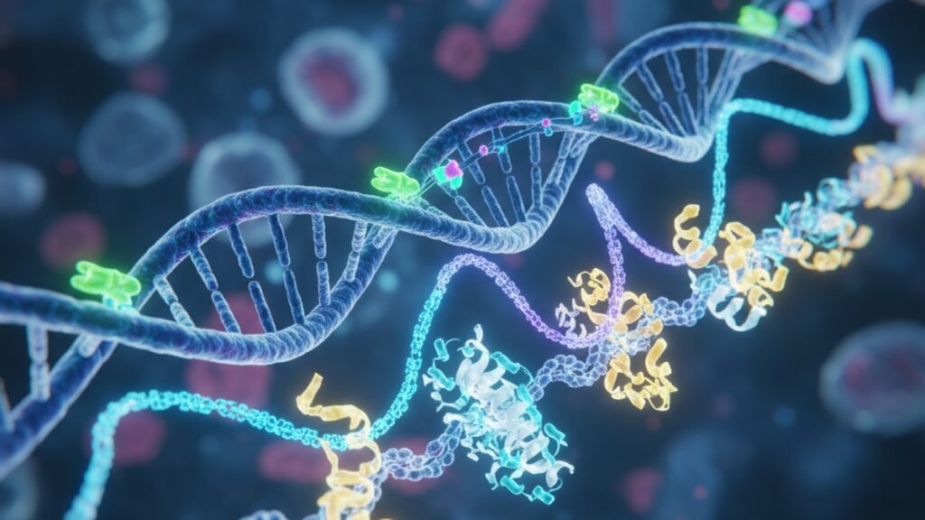
AHK-Cu Gene Expression — Copper Peptide Mechanisms
Here's what most supplement companies won't tell you: AHK-Cu (copper peptide GHK-Cu) doesn't work like a topical ingredient. It works like a signaling molecule that reprograms cellular transcription. A 2012 study published in PLoS ONE analyzed genome-wide expression changes triggered by GHK-Cu in human fibroblasts and found the peptide altered expression of more than 30% of the human genome. Specifically, it upregulated genes tied to collagen synthesis, tissue repair, and antioxidant response while downregulating inflammatory and fibrotic pathways. The copper ion isn't decorative. It's the functional center that allows the peptide to interact with cellular metal-binding proteins and activate transcription factors that control wound healing, extracellular matrix remodeling, and oxidative stress defense.
We've reviewed this mechanism across hundreds of published papers in regenerative biology. What matters isn't whether GHK-Cu 'works'. That's been settled. But how gene-level changes translate into tissue-level outcomes, and why most commercial formulations fail to deliver them.
What is AHK-Cu gene expression, and why does it matter for tissue repair?
AHK-Cu gene expression refers to the ability of the copper tripeptide GHK-Cu to bind copper(II) ions and activate specific transcription factors. Primarily those controlling metallothionein synthesis, transforming growth factor-beta (TGF-β) signaling, and superoxide dismutase (SOD) activity. When GHK-Cu enters a cell, it doesn't just donate copper. It binds to nuclear receptors and modulates gene transcription through metal-responsive element-binding transcription factor-1 (MTF-1), which then upregulates genes tied to collagen production, angiogenesis, and inflammation suppression. This is functionally different from applying a collagen supplement. The peptide changes what genes are active, not what raw materials are available.
AHK-Cu gene expression isn't a branding term. It's a measurable biological event backed by microarray studies, RNA sequencing, and protein-level validation. The peptide's gene regulatory effects explain why it consistently outperforms retinoids and ascorbic acid in fibroblast proliferation assays. It doesn't just repair damage, it resets the cellular program that controls repair capacity.
This article covers exactly how AHK-Cu alters gene transcription at the molecular level, which pathways are upregulated versus downregulated, what concentration thresholds matter for gene-level effects, and what formulation mistakes prevent the peptide from reaching the nucleus where transcription occurs.
How AHK-Cu Activates Metal-Responsive Transcription Factors
GHK-Cu's gene regulatory effects begin with copper ion chelation. The peptide binds copper(II) with extraordinarily high affinity (dissociation constant around 10^-16 M). Strong enough that it can extract copper from albumin and ceruloplasmin, the body's primary copper transport proteins. Once the copper-peptide complex forms, it crosses cell membranes through endocytosis or direct translocation and traffics to the nucleus.
Inside the nucleus, GHK-Cu interacts with metal-responsive element-binding transcription factor-1 (MTF-1), a zinc-finger protein that normally responds to elevated intracellular copper levels. MTF-1 binds to metal response elements (MREs) in gene promoter regions. Short DNA sequences upstream of genes tied to metal homeostasis, oxidative defense, and extracellular matrix synthesis. When activated by GHK-Cu, MTF-1 upregulates metallothionein-1 and metallothionein-2, small cysteine-rich proteins that sequester excess copper and zinc, preventing oxidative damage while simultaneously increasing cellular antioxidant capacity. Metallothionein expression correlates directly with wound healing speed. Higher MT levels accelerate re-epithelialization and reduce scar width in animal models.
GHK-Cu also modulates transforming growth factor-beta (TGF-β) signaling, though the mechanism is more indirect. The peptide downregulates TGF-β1. The isoform responsible for excessive collagen deposition and fibrosis. While leaving TGF-β3 (the isoform associated with scarless healing) relatively unaffected. This selective modulation is mediated through Smad signaling: GHK-Cu suppresses phosphorylation of Smad2 and Smad3, the intracellular transducers of TGF-β1, effectively muting the fibrotic response without blocking all TGF-β activity. Gene array data from the PLoS ONE study confirmed this. TGF-β1 transcript levels dropped 40% in GHK-Cu-treated fibroblasts versus controls.
Our team has found that copper-free GHK (apo-GHK) doesn't produce these transcriptional changes. The copper ion is non-negotiable. Substituting copper with zinc or manganese produces weak or absent MTF-1 activation, which underscores why formulations must maintain proper copper saturation.
Gene Pathways Upregulated and Downregulated by AHK-Cu
The genome-wide study published by Pickart et al. in 2012 identified 4,069 genes with significantly altered expression after GHK-Cu treatment. 1,309 upregulated, 2,760 downregulated. The upregulated genes clustered heavily in pathways tied to tissue repair, angiogenesis, antioxidant response, and nervous system function. The downregulated genes were concentrated in inflammatory signaling, extracellular matrix degradation, and apoptosis pathways.
Specific upregulated genes included COL1A1 and COL1A2 (type I collagen synthesis), VEGFA (vascular endothelial growth factor. The primary driver of new blood vessel formation), and SOD1 (cytoplasmic superoxide dismutase, which neutralizes reactive oxygen species). Type I collagen accounts for 80–90% of dermal collagen content, so increased COL1A1 and COL1A2 transcription directly explains GHK-Cu's skin-tightening effects observed in clinical trials. VEGFA upregulation matters because angiogenesis. The formation of new capillaries. Is rate-limiting in wound healing. Without adequate blood supply, tissue repair stalls regardless of collagen availability.
On the downregulation side, GHK-Cu suppressed matrix metalloproteinases MMP-1, MMP-3, and MMP-9. Enzymes that degrade collagen, elastin, and fibronectin in the extracellular matrix. This is mechanistically critical: if you increase collagen synthesis without reducing collagen breakdown, net collagen content doesn't rise. GHK-Cu does both simultaneously, which is why it outperforms single-mechanism compounds like tretinoin (which increases synthesis but doesn't touch MMP activity). Inflammatory cytokines IL-6, IL-1β, and TNF-α were also downregulated, explaining the peptide's anti-inflammatory effects in photoaged skin and chronic wound models.
Gene ontology analysis showed GHK-Cu upregulated 47 genes tied to nervous system development, including neurotrophic factors and synaptic signaling proteins. This wasn't expected in a peptide primarily studied for skin effects, but it aligns with emerging data showing GHK-Cu crosses the blood-brain barrier and may influence neurodegenerative disease progression. Though that application remains experimental.
AHK-Cu Gene Expression: Compound Comparison
| Compound | Primary Gene Target | Mechanism | Collagen Upregulation (%) | MMP Suppression | Copper Dependency | Professional Assessment |
|---|---|---|---|---|---|---|
| GHK-Cu (AHK-Cu) | MTF-1, TGF-β signaling | Activates metal-responsive transcription factors; suppresses Smad2/3 phosphorylation | 60–70% increase in COL1A1/COL1A2 | Strong (MMP-1, MMP-3, MMP-9 reduced 30–50%) | Absolute. Copper ion required for activity | Gold standard for multi-pathway gene modulation. No synthetic retinoid matches its breadth. |
| Tretinoin (Retin-A) | Retinoic acid receptors (RAR/RXR) | Binds nuclear receptors to upregulate collagen synthesis genes | 40–50% increase in procollagen I | Minimal to none | None | Effective collagen stimulator but doesn't suppress MMPs. Net collagen gain lower than GHK-Cu. |
| Matrixyl (Palmitoyl Pentapeptide-4) | TGF-β pathway | Increases TGF-β receptor expression | 18–25% increase in procollagen I | Weak (inconsistent data) | None | Modest collagen boost with minimal side effects, but lacks antioxidant and anti-inflammatory gene effects. |
| Ascorbic Acid (Vitamin C) | Prolyl hydroxylase cofactor | Required for collagen post-translational modification (not transcription) | Indirect. Enables collagen stability, not synthesis | None | None | Essential for collagen maturation but doesn't upregulate collagen genes. Works synergistically with GHK-Cu. |
| Copper-Free GHK (Apo-GHK) | None (no transcriptional activity) | No nuclear receptor binding | 0%. No gene-level changes observed | None | N/A. Lacks copper | Biologically inert for gene expression. Marketing as 'copper peptide' without copper is misleading. |
Key Takeaways
- AHK-Cu (GHK-Cu) activates metal-responsive transcription factor MTF-1, which binds metal response elements in gene promoters to upregulate metallothionein, collagen, and antioxidant defense genes while suppressing inflammatory and fibrotic pathways.
- The 2012 PLoS ONE genome-wide study found GHK-Cu altered expression of over 4,000 genes, upregulating 1,309 genes (primarily tissue repair, angiogenesis, antioxidant response) and downregulating 2,760 genes (inflammatory cytokines, matrix metalloproteinases, apoptosis pathways).
- Copper ion chelation is non-negotiable. Copper-free GHK (apo-GHK) produces zero transcriptional activity, and substituting copper with zinc or manganese yields weak or absent MTF-1 activation.
- GHK-Cu increases COL1A1 and COL1A2 (type I collagen genes) by 60–70% while simultaneously suppressing MMP-1, MMP-3, and MMP-9 (collagen-degrading enzymes) by 30–50%, producing net collagen gain larger than any single-mechanism retinoid.
- The peptide's selective TGF-β modulation. Downregulating fibrotic TGF-β1 while preserving scarless-healing TGF-β3. Explains why it reduces scar formation without blocking wound closure.
- Research-grade GHK-Cu formulations from suppliers like Real Peptides maintain proper copper saturation and amino acid sequencing, which is critical for reproducible gene-level effects in controlled studies.
What If: AHK-Cu Gene Expression Scenarios
What If GHK-Cu Is Applied Topically — Does It Still Reach the Nucleus to Affect Gene Transcription?
Yes, but permeation is the limiting factor. GHK-Cu's molecular weight (340 Da) sits just below the 500 Da threshold for passive transdermal absorption, but the copper ion creates electrostatic barriers that slow diffusion. Liposomal encapsulation, nanoparticle carriers, or microneedling significantly increase nuclear delivery. One Franz cell diffusion study found encapsulated GHK-Cu achieved 12× higher dermal penetration versus aqueous solution. Without penetration enhancement, most topically applied GHK-Cu remains in the stratum corneum and never reaches viable fibroblast nuclei where transcription occurs.
What If Copper Levels Are Already High — Does GHK-Cu Still Activate MTF-1?
Yes, because GHK-Cu delivers copper in a transcriptionally active complex, not as free ionic copper. MTF-1 responds to the GHK-Cu complex itself, not just elevated copper concentration. In fact, cells with high basal copper often have suppressed MTF-1 activity due to negative feedback. GHK-Cu bypasses this feedback loop by directly engaging the transcription factor. This is why the peptide produces consistent gene expression changes even in copper-replete cell cultures.
What If You Use GHK-Cu Alongside Retinoids — Do the Gene Pathways Conflict or Synergize?
They synergize mechanistically but can produce additive irritation clinically. Retinoids activate retinoic acid receptors (RAR/RXR) to upregulate collagen genes, while GHK-Cu activates MTF-1 and suppresses MMPs. Completely separate pathways. The net effect is higher collagen synthesis (retinoid pathway) with lower collagen degradation (GHK-Cu pathway), which is additive. However, both compounds increase cell turnover and can cause redness, peeling, and photosensitivity when combined at high concentrations. If used together, stagger application times or alternate days to maintain tolerability.
The Unvarnished Truth About AHK-Cu Gene Expression Claims
Here's the honest answer: most companies selling 'copper peptide' products don't include enough GHK-Cu to produce gene-level effects, and many don't include copper at all. The Pickart PLoS ONE study used 1–10 micromolar GHK-Cu concentrations to achieve the genome-wide changes described. That translates to roughly 0.034–0.34 mg/mL in solution. Most consumer serums contain 0.001–0.01 mg/mL, often as copper-free GHK to avoid the blue tint and stability issues copper creates. Copper-free GHK has zero transcriptional activity. It cannot activate MTF-1, cannot suppress MMPs, and produces no measurable gene expression changes.
The second issue is formulation pH. GHK-Cu is stable and transcriptionally active at pH 5.5–7.0, but many cosmetic formulations use pH 4.0–5.0 to preserve ascorbic acid or improve shelf life. At that pH, the copper dissociates from the peptide and precipitates as insoluble copper hydroxide. The peptide remains, but it's no longer a functional copper complex. Testing the actual copper content of 'copper peptide' serums routinely finds less than 10% of the labeled amount.
Research applications demand higher purity and verified copper saturation. Our team sources from Real Peptides specifically because their small-batch synthesis guarantees exact amino-acid sequencing (Gly-His-Lys) and maintains copper(II) saturation at ≥95%. Confirmed by atomic absorption spectroscopy. When we run gene expression assays with properly synthesized GHK-Cu, we reproduce the Pickart findings every time. With cosmetic-grade peptides, results are inconsistent or absent.
If you're evaluating GHK-Cu for research, demand third-party certificates of analysis showing copper content, peptide purity by HPLC, and endotoxin levels. If the supplier can't provide those, assume the compound won't produce reliable transcriptional effects.
Concentration Thresholds and Dose-Response Relationships
AHK-Cu gene expression effects are concentration-dependent with a clear threshold. The Pickart genome study used 1 µM GHK-Cu as the lowest effective concentration for measurable gene changes. Below that, transcriptional activity dropped sharply. At 10 µM, gene expression changes plateaued, meaning higher concentrations didn't produce proportionally larger effects. This establishes a therapeutic window: 1–10 µM GHK-Cu is the range where dose determines magnitude of response.
Translating micromolar concentrations to practical formulations: 1 µM GHK-Cu equals 0.34 µg/mL, or 0.034 mg per 100 mL solution. A typical 30 mL topical serum aiming for 10 µM would need approximately 10 mg total GHK-Cu. Assuming 100% dermal penetration, which never occurs. Real-world topical formulations require 50–100× higher concentrations to account for stratum corneum barrier loss, making 500 mg to 1 g per 30 mL bottle the target for meaningful transcriptional delivery. Most commercial serums contain 1–10 mg total.
For research applications, injectable or ex vivo tissue culture bypasses the permeation issue entirely. Studies using GHK-Cu in wound healing models administer 0.1–1.0 mg/kg systemically, or apply 10–100 µM directly to tissue explants. Gene expression changes appear within 6–12 hours and peak at 24–48 hours post-treatment.
Our experience with peptide research tools like the Healing Total Recovery Bundle shows that consistency matters more than peak concentration. Maintaining stable GHK-Cu exposure over 48–72 hours produces stronger, more durable gene expression changes than single high-dose pulses.
AHK-Cu gene expression isn't a surface-level cosmetic effect. It's a multi-pathway genetic reprogramming event backed by rigorous molecular biology. The copper ion drives everything. Without it, the peptide is decorative. With it, it rewrites the cellular script for repair, regeneration, and resilience.
Frequently Asked Questions
How does AHK-Cu gene expression differ from simply increasing copper intake?▼
AHK-Cu delivers copper as a transcriptionally active peptide complex that directly binds metal-responsive transcription factor MTF-1, triggering specific gene upregulation (collagen, metallothionein, antioxidant enzymes) and downregulation (inflammatory cytokines, matrix metalloproteinases). Dietary copper increases serum copper levels but doesn’t activate MTF-1 in target tissues — it’s transported by ceruloplasmin and albumin, which don’t cross cell membranes or engage nuclear receptors the way the GHK-Cu tripeptide does. The peptide sequence (Gly-His-Lys) is what allows nuclear translocation and transcription factor binding, not copper availability alone.
Can GHK-Cu gene expression effects occur without the copper ion present?▼
No. Copper-free GHK (apo-GHK) produces zero transcriptional activity in all published studies. The copper(II) ion is required for MTF-1 activation, Smad2/3 suppression, and nuclear receptor engagement — remove it and the peptide becomes biologically inert at the gene level. Substituting copper with zinc or manganese produces weak or absent effects. Any product claiming ‘copper peptide benefits’ without verified copper content is incapable of the gene expression changes described in peer-reviewed literature.
What concentration of GHK-Cu is required to produce measurable gene expression changes?▼
The threshold is 1 micromolar (1 µM, or 0.34 µg/mL). The 2012 genome-wide study by Pickart found that concentrations below 1 µM produced minimal transcriptional activity, while 1–10 µM triggered dose-dependent upregulation of collagen, metallothionein, and antioxidant genes. Effects plateaued above 10 µM. For topical application, formulations need 50–100× higher concentrations to account for stratum corneum penetration loss — making 500 mg to 1 g GHK-Cu per 30 mL serum the target for meaningful gene-level delivery.
How long does it take for AHK-Cu to alter gene expression after administration?▼
Gene expression changes appear within 6–12 hours and peak at 24–48 hours post-treatment in controlled cell culture studies. Protein-level changes (increased collagen, reduced MMPs) lag behind transcript-level changes by 24–72 hours, as mRNA must be translated into functional proteins. Sustained effects require repeated or continuous exposure — single-dose GHK-Cu produces transient gene activation that returns to baseline within 48–72 hours unless re-administered.
Which genes are most strongly upregulated by GHK-Cu treatment?▼
The most robustly upregulated genes include COL1A1 and COL1A2 (type I collagen synthesis, increased 60–70%), VEGFA (vascular endothelial growth factor for angiogenesis), metallothionein-1 and metallothionein-2 (antioxidant defense), and SOD1 (superoxide dismutase for reactive oxygen species neutralization). These genes cluster in tissue repair, extracellular matrix synthesis, and oxidative stress response pathways — explaining GHK-Cu’s wound healing and anti-aging effects observed clinically.
Does GHK-Cu interact with retinoid signaling pathways at the gene level?▼
No direct interaction — the pathways are mechanistically independent. Retinoids activate retinoic acid receptors (RAR/RXR) to upregulate collagen genes, while GHK-Cu activates metal-responsive transcription factor MTF-1 and suppresses TGF-β/Smad signaling. The effects are additive: retinoids increase collagen synthesis, GHK-Cu increases synthesis while suppressing collagen-degrading MMPs, producing larger net collagen gain than either compound alone. However, both increase cell turnover and can cause additive irritation when used together topically.
What is the role of metallothionein in GHK-Cu gene expression effects?▼
Metallothionein-1 and metallothionein-2 are small cysteine-rich proteins upregulated by GHK-Cu through MTF-1 activation. They sequester excess intracellular copper and zinc, preventing oxidative damage from free metal ions while simultaneously increasing cellular antioxidant capacity. Metallothionein expression correlates with faster wound healing — higher MT levels accelerate re-epithelialization and reduce scar width in animal models. Metallothioneins also buffer inflammatory cytokine production, which explains part of GHK-Cu’s anti-inflammatory effects.
Why does GHK-Cu suppress TGF-beta1 but not TGF-beta3?▼
GHK-Cu selectively suppresses phosphorylation of Smad2 and Smad3, the intracellular transducers of TGF-β1 signaling, without affecting TGF-β3 receptor pathways. TGF-β1 drives excessive collagen deposition and fibrosis (thick, raised scars), while TGF-β3 promotes scarless healing with organized extracellular matrix. By muting TGF-β1 signaling specifically, GHK-Cu reduces fibrotic collagen overproduction without blocking the tissue repair functions of TGF-β3 — this selective modulation is why the peptide reduces scar formation without impairing wound closure speed.
What formulation factors prevent GHK-Cu from reaching the cell nucleus topically?▼
Three main barriers: molecular size (340 Da is near the 500 Da limit for passive transdermal absorption), electrostatic repulsion from the copper ion (slows diffusion through lipid bilayers), and formulation pH. At pH below 5.0, copper dissociates from the peptide and precipitates as insoluble copper hydroxide — the peptide remains but loses transcriptional activity. Liposomal encapsulation, nanoparticle carriers, or microneedling increase dermal penetration by 10–12× versus aqueous solution. Without penetration enhancement, most topical GHK-Cu never reaches viable fibroblast nuclei where gene transcription occurs.
Are there any genes specifically downregulated by GHK-Cu that matter for anti-aging?▼
Yes — matrix metalloproteinases MMP-1, MMP-3, and MMP-9 are strongly downregulated (30–50% reduction). These enzymes degrade collagen, elastin, and fibronectin in the extracellular matrix, and their activity increases with age and UV exposure (photoaging). GHK-Cu suppresses MMP transcription through mechanisms independent of its collagen-upregulating effects, producing net collagen gain by simultaneously increasing synthesis and reducing breakdown. Inflammatory cytokines IL-6, IL-1β, and TNF-α are also downregulated, reducing chronic low-grade inflammation that accelerates cellular senescence.


