


What Does Tesamorelin + Ipamorelin Blend Look Like in Solution?
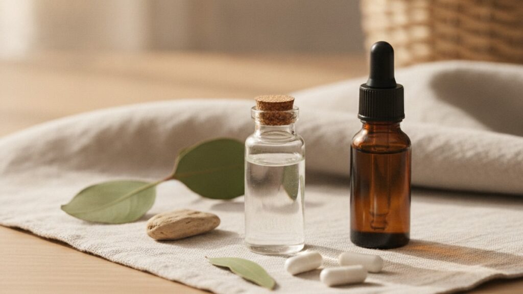
A properly reconstituted tesamorelin + ipamorelin blend should appear as a clear to slightly opalescent liquid with no visible particles, cloudiness, or color shift. If you see anything else. Haze, precipitate, yellow tint, or floating debris. The peptides have likely degraded during storage or reconstitution, and the solution is no longer therapeutically viable. We've worked with researchers across hundreds of peptide protocols, and the visual appearance test is the single most reliable non-laboratory indicator of peptide integrity.
What most protocols don't mention: the degradation isn't always obvious. A faint haze or slight color shift might seem trivial, but both indicate protein denaturation that cannot be reversed. Peptides are not like small-molecule drugs. Once the amino acid chain unfolds or aggregates, the biological activity is permanently lost.
What does a tesamorelin + ipamorelin blend look like in solution after proper reconstitution?
A correctly prepared tesamorelin + ipamorelin blend appears as a clear to slightly opalescent solution with no visible particulates, cloudiness, or discoloration. The slight opalescence is normal and results from light scattering by peptide molecules in solution. It is not the same as cloudiness, which indicates aggregation. Any yellow, brown, or amber tint signals oxidative degradation, and any visible particles or precipitate indicate irreversible protein aggregation. Proper reconstitution requires bacteriostatic water, gentle swirling (never shaking), and refrigeration at 2–8°C immediately after mixing.
Understanding Peptide Solution Appearance
The visual appearance of tesamorelin + ipamorelin blend in solution is determined by three factors: protein structure integrity, pH stability, and the presence or absence of aggregation. Tesamorelin is a 44-amino-acid analog of growth hormone-releasing hormone (GHRH), and ipamorelin is a pentapeptide ghrelin mimetic. Both are sensitive to temperature, pH shifts, and mechanical stress during reconstitution.
A clear solution means the peptide chains remain properly folded in their bioactive conformation. Slight opalescence. A faint, uniform cloudiness visible when holding the vial up to light. Is caused by Rayleigh scattering from peptide molecules in solution and is completely normal. This is distinct from true cloudiness, which appears as visible haze or turbidity throughout the solution and indicates protein aggregation.
Particulate matter. Any visible floating debris, fibers, or suspended particles. Is a hard rejection criterion. Even microscopic aggregates compromise sterility and bioavailability. If you see particles, do not inject the solution. Discoloration is equally critical: peptides in solution should be colorless to very faintly yellow at most. A yellow, amber, or brown tint indicates oxidative degradation of methionine or tryptophan residues, which destroys receptor binding affinity.
Our team has found that most visual degradation failures occur during the first 72 hours after reconstitution. Either from temperature excursions during mixing or from using non-bacteriostatic water that allows bacterial growth. The FAT Loss Stack we supply includes detailed reconstitution protocols to prevent exactly these failures.
Reconstitution Technique and Visual Outcomes
The way you reconstitute the lyophilized powder determines what the tesamorelin + ipamorelin blend looks like in solution. Proper technique produces a clear, stable solution; improper technique produces aggregation, cloudiness, or loss of potency that may or may not be visually detectable.
Always use bacteriostatic water (0.9% benzyl alcohol). Never sterile water, saline, or any other diluent. Bacteriostatic water prevents bacterial growth during the 28-day refrigerated storage period and maintains pH stability. Inject the water slowly down the inside wall of the vial, allowing it to gently dissolve the lyophilized cake without direct impact. Never aim the stream directly at the powder. The mechanical shear force denatures peptide bonds.
After adding water, swirl the vial gently in a circular motion. Do not shake. Shaking introduces air bubbles and mechanical stress that cause aggregation. The powder should dissolve completely within 60–90 seconds of gentle swirling. If it doesn't, let the vial sit at room temperature for 2–3 minutes and swirl again.
Once dissolved, inspect the solution immediately. Hold the vial up to a bright light source and look for clarity, opalescence, particles, or discoloration. A properly reconstituted blend should look like water with a very faint milky quality when backlit. Not cloudy, not yellow, not containing visible debris. If the solution passes visual inspection, refrigerate it immediately at 2–8°C.
Temperature discipline is non-negotiable. Even a brief excursion above 8°C during reconstitution accelerates degradation. We've worked with research teams who reconstitute peptides at room temperature and then refrigerate. That 5-minute window at 22°C is enough to reduce bioactivity by 10–15% before the first injection. Reconstitute in a cool environment or pre-chill your bacteriostatic water to 4°C before use. Real Peptides protocols emphasize this cold-chain discipline across every stage of peptide handling.
Storage Conditions and Appearance Degradation
Once reconstituted, the tesamorelin + ipamorelin blend must be stored at 2–8°C and used within 28 days. Visual changes during storage. Cloudiness, color shift, or particulate formation. Indicate that the peptides have degraded and are no longer viable.
Peptide degradation in solution occurs through three pathways: oxidation, aggregation, and hydrolysis. Oxidation affects methionine and tryptophan residues, turning the solution faintly yellow or amber. Aggregation causes cloudiness or precipitate formation as peptide chains clump together. Hydrolysis cleaves peptide bonds, which may or may not produce visible changes but always destroys bioactivity.
Temperature is the primary driver. A single overnight excursion above 8°C. Leaving the vial on the counter, carrying it in a non-insulated bag during travel, or storing it in a refrigerator door that fluctuates between 4°C and 12°C. Causes irreversible damage. Even if the solution still looks clear, receptor binding affinity decreases. The relationship is exponential: every 10°C increase in temperature roughly doubles the degradation rate.
Light exposure accelerates oxidation. Store reconstituted peptides in amber vials or wrap clear vials in aluminum foil. UV and visible light generate free radicals that oxidize amino acids, producing the yellow discoloration researchers often mistake for normal aging.
Freezing reconstituted peptides is controversial. Some sources claim it extends shelf life; others warn it causes aggregation during thaw. Our position: avoid it unless you're using a controlled-rate freezer and have validated the freeze-thaw stability of your specific formulation. Home freezers introduce ice crystal formation that disrupts peptide structure during thaw, often producing visible cloudiness that wasn't present before freezing.
| Storage Condition | Expected Appearance After 7 Days | Expected Appearance After 28 Days | Bioactivity Estimate | Professional Assessment |
|---|---|---|---|---|
| Refrigerated 2–8°C, protected from light, bacteriostatic water | Clear to slightly opalescent, no particles | Clear to slightly opalescent, possible faint haze | 95–100% retained | Optimal. This is the gold standard for peptide storage |
| Refrigerated 2–8°C, clear vial, exposed to ambient light | Faint yellow tint possible | Yellow tint, possible haze | 80–90% retained | Suboptimal. Light exposure accelerates oxidation |
| Room temperature 20–25°C for >24 hours, then refrigerated | Slight haze or cloudiness | Visible cloudiness, possible precipitate | 50–70% retained | Degraded. Temperature excursion likely caused aggregation |
| Frozen at −20°C, thawed once | Cloudiness or particulate after thaw | Not recommended | 40–80% retained (variable) | Risky. Freeze-thaw introduces structural damage |
| Stored in sterile water instead of bacteriostatic water | Possible bacterial growth (cloudiness) | Definite contamination risk | Unknown. Unsafe to use | Rejected. Never use sterile water for multi-dose storage |
Key Takeaways
- A properly reconstituted tesamorelin + ipamorelin blend appears clear to slightly opalescent with no visible particles, cloudiness, or discoloration. Any deviation indicates degradation.
- Slight opalescence is normal and caused by light scattering from peptide molecules; true cloudiness or haze indicates protein aggregation and renders the solution unusable.
- Reconstitution must use bacteriostatic water, gentle swirling (never shaking), and immediate refrigeration at 2–8°C to prevent visual and biochemical degradation.
- Yellow, amber, or brown discoloration signals oxidative degradation of amino acid residues, which destroys receptor binding affinity even if the solution remains clear.
- Temperature excursions above 8°C, light exposure, and improper reconstitution technique are the three most common causes of visual degradation in peptide solutions.
- Once reconstituted, tesamorelin + ipamorelin blends remain stable for 28 days when refrigerated. Any cloudiness, particles, or color shift during that window means the peptides are no longer therapeutically viable.
What If: Tesamorelin + Ipamorelin Solution Scenarios
What If My Reconstituted Solution Looks Cloudy Right After Mixing?
Discard it immediately. Cloudiness at reconstitution indicates one of three failures: contaminated bacteriostatic water, excessive mechanical stress during mixing, or a manufacturing defect in the lyophilized powder. Cloudiness means protein aggregation has already occurred, and aggregated peptides cannot re-fold into their bioactive conformation. The solution is unusable. To prevent recurrence, verify that your bacteriostatic water is sterile and within its expiration date, inject the water slowly down the vial wall rather than directly onto the powder, and swirl gently without shaking.
What If the Solution Turns Yellow After a Week in the Fridge?
Yellow discoloration indicates oxidative degradation of methionine or tryptophan residues in the peptide chains. The solution has lost bioactivity and should not be used. This happens when peptides are stored in clear vials exposed to light or when the refrigerator temperature fluctuates above 8°C. Moving forward, wrap your vials in aluminum foil or use amber glass vials, and verify that your refrigerator maintains a stable 2–8°C using a separate thermometer (refrigerator door displays are often inaccurate by 3–5°C).
What If I See Tiny Particles Floating in the Solution?
Do not inject it. Particulate matter indicates either peptide aggregation, bacterial contamination, or foreign debris introduced during reconstitution. Even microscopic particles can trigger immune responses or embolism if injected subcutaneously. Inspect the vial under bright light. If particles are visible, the solution is compromised. This failure mode usually results from using non-sterile technique during reconstitution, storing the peptide at room temperature, or reusing needles to draw doses (which introduces rubber stopper fragments into the solution).
The Unfiltered Truth About Peptide Visual Inspection
Here's the honest answer: most peptide degradation is invisible to the naked eye. A solution can look perfectly clear and still have lost 30–40% of its bioactivity due to partial aggregation, oxidative damage, or hydrolysis that doesn't produce visible changes. The appearance test is the first filter. Not the only filter.
Visual inspection catches catastrophic failures: gross cloudiness, discoloration, particulate contamination. It does not catch subtle degradation from temperature excursions, freeze-thaw cycles, or pH drift. Researchers who rely solely on appearance are operating with incomplete information. The gold standard is HPLC (high-performance liquid chromatography) testing, which measures peptide purity and degradation products at the molecular level.
That said, visual inspection remains the most practical field test available to researchers without access to analytical labs. If the solution looks wrong. Cloudy, yellow, particulate-laden. It is wrong. Trust that signal. The converse is not true: a clear solution is not guaranteed to be potent, but a visibly degraded solution is guaranteed to be compromised. Use appearance as a rejection criterion, not an approval criterion.
Another reality most guides gloss over: reconstitution failures are common. Even experienced researchers occasionally shake instead of swirl, use water that's been sitting out too long, or reconstitute at room temperature. These mistakes often produce solutions that pass visual inspection but fail bioactivity tests. The discipline required for peptide handling is closer to aseptic laboratory technique than typical pharmaceutical preparation. And the learning curve reflects that.
Identifying Degradation vs. Normal Variation
Not every visual change indicates failure. Distinguishing between normal variation and true degradation requires understanding what each peptide looks like under optimal conditions and how specific failure modes manifest visually.
Slight opalescence is normal. It's the faint milky quality visible when holding a vial up to light, caused by Rayleigh scattering from dissolved peptide molecules. This is not the same as cloudiness. Cloudiness is diffuse, visible haze that obscures light transmission through the solution. Opalescence is subtle and uniform; cloudiness is obvious and often non-uniform (denser near the bottom, clearer near the top).
Color variation within the acceptable range runs from completely colorless to very faintly yellow. Think the color of diluted white wine. Anything darker than that (amber, brown, orange) indicates oxidation. The transition happens gradually, so inspect your vials under consistent lighting at each dose draw. A solution that looks clear on day 1 and faintly yellow on day 14 has crossed into degradation territory.
Particulate matter has three common sources: peptide aggregation (white or translucent particles), bacterial contamination (cloudy diffuse haze with possible biofilm), and stopper coring (black rubber fragments). Each has a distinct appearance. Aggregates look like tiny white flecks or fibers suspended in solution. Bacterial contamination produces diffuse cloudiness that may settle overnight. Stopper fragments are black, irregularly shaped, and sink to the bottom.
Temperature-induced degradation often produces cloudiness without discoloration. Oxidative degradation produces discoloration (yellow to brown) without necessarily producing cloudiness. Light-induced degradation produces both. Distinguishing these pathways matters for troubleshooting: if every vial you reconstitute turns cloudy within 48 hours, the problem is likely mechanical stress during mixing or temperature excursions. If they turn yellow but stay clear, the problem is light exposure or oxidative stress.
For researchers working with our Body Recomp Bundle or other multi-peptide protocols, maintaining visual inspection discipline across every compound is essential. Each peptide has slightly different stability characteristics. What looks normal for one may indicate failure in another.
The tesamorelin + ipamorelin blend you're working with should remain visually stable for the full 28-day refrigerated storage window if handled correctly. Any change during that period. Cloudiness, discoloration, particulate formation. Is a red flag that something in your storage or handling protocol needs correction. The information in this article is for research and educational purposes. Stability assessments and protocol decisions should be validated against your specific experimental requirements and regulatory context.
Frequently Asked Questions
What does a properly reconstituted tesamorelin + ipamorelin blend look like?▼
A properly reconstituted tesamorelin + ipamorelin blend appears as a clear to slightly opalescent solution with no visible particles, cloudiness, or discoloration. The slight opalescence — a faint milky quality when held up to light — is normal and results from light scattering by peptide molecules in solution. Any cloudiness, yellow tint, or visible debris indicates degradation and renders the solution unusable.
How can I tell if my peptide solution has degraded?▼
Visual indicators of degradation include cloudiness or haze throughout the solution, yellow to brown discoloration, visible particles or precipitate, or any color shift from the original clear appearance. Temperature excursions above 8°C, light exposure, and improper reconstitution are the most common causes. If the solution looked clear at reconstitution and develops any of these changes during refrigerated storage, it has degraded and should not be used.
Is slight cloudiness in my tesamorelin + ipamorelin blend normal?▼
No — cloudiness indicates protein aggregation and is never normal. Slight opalescence (a faint milky quality visible when backlit) is normal; cloudiness (visible haze diffused throughout the solution) is not. Cloudiness means peptide chains have clumped together irreversibly, destroying bioactivity. If your solution is cloudy rather than just opalescent, discard it and troubleshoot your reconstitution technique or storage conditions.
Can I still use a peptide solution that turned yellow?▼
No — yellow discoloration indicates oxidative degradation of amino acid residues, which destroys receptor binding affinity even if the solution remains clear. Peptides should be colorless to very faintly yellow at most. A distinct yellow, amber, or brown tint means the peptides have oxidized and are no longer therapeutically viable. This typically results from light exposure or temperature fluctuations during storage.
How should I store reconstituted tesamorelin + ipamorelin to prevent visual degradation?▼
Store reconstituted peptides at 2–8°C in a refrigerator with stable temperature control, protected from light by using amber vials or wrapping clear vials in aluminum foil. Use bacteriostatic water for reconstitution and consume within 28 days. Avoid storing in the refrigerator door (temperature fluctuates) and never leave vials at room temperature for more than a few minutes during dose preparation.
What causes particles to form in peptide solutions?▼
Particulate formation results from peptide aggregation (temperature stress or mechanical shear during reconstitution), bacterial contamination (using sterile water instead of bacteriostatic water or non-sterile technique), or stopper coring (introducing rubber fragments when puncturing the vial seal repeatedly). Any visible particles render the solution unsafe for injection — discard it and review your reconstitution and storage protocols.
Does freezing reconstituted tesamorelin + ipamorelin change its appearance?▼
Freezing reconstituted peptides often produces cloudiness or particulate formation during thaw due to ice crystal damage to peptide structure. While some researchers freeze peptides to extend shelf life, home freezers lack controlled-rate cooling and introduce structural damage that may not be reversible. If a solution was clear before freezing and cloudy after thawing, the freeze-thaw cycle caused aggregation and the peptides are degraded.
How do I distinguish between opalescence and cloudiness in my peptide solution?▼
Opalescence is a faint, uniform milky quality visible only when holding the vial up to bright light — it looks like very diluted skim milk and is caused by light scattering from dissolved peptide molecules. Cloudiness is a visible haze or turbidity that obscures clarity even without backlighting and often appears non-uniform or denser in certain areas of the vial. Opalescence is normal; cloudiness indicates protein aggregation.
What should I do if my peptide solution looks cloudy immediately after reconstitution?▼
Discard it immediately — cloudiness at reconstitution indicates contaminated bacteriostatic water, excessive mechanical stress during mixing (shaking instead of swirling), or a manufacturing defect in the lyophilized powder. Aggregation at this stage is irreversible. For your next reconstitution, use fresh bacteriostatic water, inject slowly down the vial wall, and swirl gently without shaking.
Can peptide degradation occur without visible changes to the solution?▼
Yes — peptide degradation from temperature excursions, partial oxidation, or hydrolysis can reduce bioactivity by 20–40% without producing visible cloudiness, discoloration, or particles. Visual inspection catches catastrophic failures but does not guarantee potency. High-performance liquid chromatography (HPLC) is the gold standard for detecting subtle degradation, but visual inspection remains the most practical field test for researchers.
Why does my peptide solution turn yellow even though I keep it refrigerated?▼
Yellow discoloration in refrigerated peptides typically results from light exposure, not temperature. UV and visible light generate free radicals that oxidize methionine and tryptophan residues, producing yellow to amber coloration. To prevent this, store peptides in amber vials or wrap clear vials in aluminum foil, and keep them in the back of the refrigerator away from interior lighting that turns on when the door opens.
How long does a tesamorelin + ipamorelin blend remain visually stable after reconstitution?▼
When stored correctly at 2–8°C in bacteriostatic water and protected from light, a tesamorelin + ipamorelin blend should remain visually stable — clear to slightly opalescent with no particles or discoloration — for the full 28-day shelf life. Any visual change during that period indicates a storage or handling failure. Beyond 28 days, bacterial growth risk increases even if the solution still looks clear.


