


What Does Ipamorelin Look Like in Solution? (Explained)
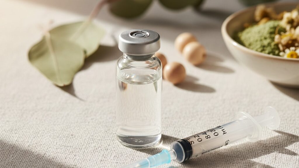
Properly reconstituted ipamorelin appears as a clear, colorless liquid. Visually indistinguishable from sterile water. This isn't what most researchers expect when handling a peptide solution, which is precisely why contamination and degradation often go undetected until bioassay failure. The absence of visible particles, cloudiness, or discoloration doesn't guarantee the peptide is active. It only confirms the reconstitution solvent was sterile and the lyophilized powder dissolved completely. We've worked with research institutions across multiple disciplines where the gap between visual inspection and actual peptide integrity caused significant protocol delays.
The visual clarity rule holds only under controlled conditions. Temperature excursions, bacterial contamination, or improper pH can all compromise the peptide without producing visible changes for 24–48 hours after the event occurs.
What does ipamorelin look like in solution when properly prepared?
Ipamorelin in solution should appear completely clear, colorless, and free of visible particulate matter when reconstituted with bacteriostatic water or sterile saline at neutral pH. Any cloudiness, discoloration (yellow, amber, brown), visible particles, or precipitate indicates degradation, contamination, or improper storage. Properly prepared ipamorelin solution remains visually stable for 28 days under refrigeration at 2–8°C.
Direct Answer: Why Visual Inspection Isn't Enough
The problem isn't that researchers don't look. It's that peptide degradation at the molecular level produces no visible signal until complete structural collapse. A solution can lose 40–60% of its bioactive peptide content through oxidation or enzymatic cleavage while still appearing perfectly clear under standard lighting. This is why experienced labs pair visual inspection with pH testing (target 6.5–7.5) and refrigeration verification. The compound degrades silently, and by the time you see cloudiness or precipitate, the batch is already unusable.
What follows covers exactly what correctly reconstituted ipamorelin looks like, the visual markers that signal contamination or degradation, and what preparation mistakes produce the visual defects researchers encounter most frequently.
The Baseline: What Correctly Reconstituted Ipamorelin Looks Like
Correctly reconstituted ipamorelin appears as a water-clear, colorless solution with no visible turbidity, particulate matter, or precipitate when held against white light. The solution should have the same refractive index as the reconstitution solvent itself. Typically bacteriostatic water or 0.9% sodium chloride. Meaning it doesn't scatter light or produce a visible meniscus different from pure water. Most lyophilized ipamorelin peptides arrive as a white to off-white powder compressed at the bottom of a sealed vial; once bacteriostatic water is added slowly along the vial wall (never injected directly onto the powder), the peptide dissolves within 60–90 seconds without agitation.
The absence of color is the first checkpoint: ipamorelin contains no chromophores (light-absorbing molecular groups), so any yellow, amber, or brown tint indicates oxidation. Specifically, the formation of dityrosine cross-links or Maillard reaction products if the peptide was exposed to reducing sugars during synthesis. The second checkpoint is clarity: if you can't read 8-point text through the vial when backlit, the solution contains either undissolved peptide aggregates or bacterial contamination. The third checkpoint is viscosity. Ipamorelin in solution at research concentrations (typically 2–5 mg/mL) flows identically to water. Any increase in viscosity suggests protein aggregation or the presence of excipients not listed on the certificate of analysis.
Temperature matters immediately after reconstitution. If the solution is allowed to warm above 8°C before the peptide fully dissolves, you may observe transient micro-cloudiness that clears upon refrigeration. This isn't contamination, it's reversible aggregation driven by hydrophobic interactions between partially solvated peptide chains. Experienced researchers reconstitute peptides inside a refrigerated environment or use pre-chilled bacteriostatic water to avoid this entirely. Once fully dissolved and refrigerated, correctly prepared ipamorelin remains visually stable. No color shift, no precipitate. For the full 28-day sterile use window.
Visual Defects That Signal Contamination or Degradation
Cloudiness is the most common visual defect and the least specific. It can indicate bacterial contamination, peptide aggregation, lipid contamination from improper synthesis purification, or precipitate formation from pH drift. The critical distinction is timing: cloudiness present within two hours of reconstitution almost always indicates bacterial contamination or a failed lyophilization process (residual moisture allowed aggregation during storage). Cloudiness that develops 3–7 days after reconstitution suggests bacterial growth. Particularly if the bacteriostatic water's benzyl alcohol concentration was below the 0.9% threshold required to inhibit common lab contaminants like Pseudomonas or Staphylococcus epidermidis.
Yellow discoloration. Ranging from pale straw to deep amber. Signals oxidative degradation of methionine residues or tyrosine cross-linking. Ipamorelin contains methionine at position 6 in its sequence (Aib-His-D-2-Nal-D-Phe-Lys-NH₂), and this residue is highly susceptible to oxidation by dissolved oxygen, especially under alkaline conditions (pH >8.0) or when exposed to light. Once oxidation begins, the reaction cascades: oxidized methionine promotes further peptide backbone cleavage, which releases free amino acids that undergo non-enzymatic browning. The visual result is progressive yellowing over 48–96 hours. If the solution shifts from clear to yellow within 24 hours, the peptide was likely already partially oxidized before reconstitution. A storage failure, not a handling error.
Visible particulate matter or precipitate indicates irreversible aggregation. This occurs when the peptide's tertiary structure collapses due to temperature excursion (above 25°C for more than 15 minutes), freeze-thaw cycling, or extreme pH (<4.0 or >9.0). Unlike transient micro-cloudiness, these aggregates don't redissolve upon refrigeration. They're covalent or ionic cross-links between denatured peptide chains. The particles typically settle at the vial bottom within 12–24 hours and appear as fine white sediment under magnification. Researchers sometimes mistake this for undissolved lyophilized powder, but the distinction is critical: undissolved powder appears as a cohesive cake or clump, whereas precipitate forms as dispersed flakes or granules.
Comparison: Ipamorelin Solution vs Common Contaminants
| Visual Characteristic | Correctly Prepared Ipamorelin | Bacterial Contamination | Oxidative Degradation | Lipid Contamination | Precipitate Formation |
|---|---|---|---|---|---|
| Color | Completely colorless | Colorless to milky white | Pale yellow to amber | Milky white to opaque | White sediment |
| Clarity | Water-clear, no turbidity | Increasing cloudiness over 24–72h | Clear initially, yellows over 48–96h | Opaque, does not clear | Clear supernatant with settled particles |
| Particulate Matter | None visible | None initially, biofilm may form | None until advanced degradation | Fine lipid droplets | Visible white flakes or granules |
| Smell | Odorless or faint benzyl alcohol | Musty or sour odor after 48h | Odorless | Odorless | Odorless |
| Viscosity | Identical to water | Slightly increased (biofilm) | Identical to water | Slightly increased | Identical to water (after settling) |
| Professional Assessment | Passes visual inspection. Proceed to pH and refrigeration verification | Discard immediately. Do not attempt salvage or filtration | Discard. Oxidation is irreversible and compromises bioactivity | Indicates failed HPLC purification. Return to supplier | Discard. Aggregated peptides cannot be resolubilized |
Key Takeaways
- Ipamorelin in solution appears as a clear, colorless liquid identical in appearance to sterile water when correctly reconstituted and stored at 2–8°C.
- Cloudiness developing within 48 hours of reconstitution almost always indicates bacterial contamination or a lyophilization defect. Discard the vial immediately.
- Yellow or amber discoloration signals methionine oxidation and irreversible peptide degradation. Visual clarity does not guarantee bioactivity.
- Visible particulate matter or precipitate indicates peptide aggregation from temperature excursion, pH drift, or freeze-thaw cycling. The solution is no longer usable.
- Visual inspection alone cannot detect partial degradation. Experienced labs pair it with pH testing (target 6.5–7.5) and refrigeration verification to confirm peptide stability.
- Properly prepared ipamorelin solutions remain visually stable for 28 days under continuous refrigeration. Any deviation signals handling or storage failure.
What If: Ipamorelin Solution Scenarios
What If the Solution Turns Slightly Yellow After One Week?
Discard it immediately. Yellow discoloration indicates methionine oxidation at position 6 in the peptide sequence, which irreversibly compromises the compound's ability to bind to growth hormone secretagogue receptors. This oxidation pathway accelerates once initiated. A pale yellow solution at day 7 will turn amber by day 10 and may produce visible precipitate by day 14. The oxidation is driven by dissolved oxygen in the bacteriostatic water, elevated storage temperature (even 12°C accelerates it significantly vs 4°C), or alkaline pH drift if the reconstitution solvent wasn't buffered. Researchers sometimes attempt to "rescue" yellowed solutions by filtering or diluting them. This removes particulates but does nothing to reverse oxidized methionine residues, so bioactivity remains compromised.
What If I See Tiny Floating Particles After Reconstitution?
Stop and identify the particle type before proceeding. If the particles are white, settle quickly, and weren't present immediately after reconstitution, they're likely peptide aggregates caused by injecting bacteriostatic water directly onto the lyophilized powder rather than along the vial wall. The mechanical shear from direct injection denatures surface peptide molecules. If the particles are translucent, remain suspended, and were present immediately, they're likely silicone oil droplets from the vial stopper. Cosmetic but harmless. If the particles are dark or irregular, the vial was contaminated before reconstitution. Aggregated peptide particles do not redissolve and indicate the batch should be discarded. Silicone particles can be removed by drawing the solution through a 0.22-micron syringe filter, though this adds contamination risk and is generally not recommended unless absolutely necessary.
What If the Solution Was Left at Room Temperature for 12 Hours?
Refrigerate it immediately and use it within 48 hours. But expect reduced potency. Ipamorelin's degradation rate approximately doubles for every 10°C increase above the recommended 2–8°C storage range. At 22°C (typical room temperature), enzymatic cleavage and oxidation proceed at roughly 4× the refrigerated rate. If the solution still appears clear and colorless after the temperature excursion, it likely retained 60–80% of its original bioactivity. Enough for some research applications, though not for protocols requiring precise dosing. If it shows any yellowing or cloudiness after the excursion, discard it. The 12-hour window is a soft threshold: solutions left at room temperature for 6 hours may show no measurable loss, while those left for 24 hours are almost certainly compromised regardless of appearance.
The Unflinching Truth About Ipamorelin Solution Appearance
Here's the honest answer: visual inspection is necessary but not sufficient. A perfectly clear, colorless ipamorelin solution can have lost 30–50% of its bioactive content to oxidation, hydrolysis, or deamidation without producing any visible defect. And most researchers don't discover this until their assay results come back inconsistent or negative. The industry standard of "if it looks clear, it's fine" is a convenience heuristic, not a scientific validation. Experienced labs treat visual clarity as a go/no-go checkpoint (cloudiness or color = automatic discard), but they confirm potency through secondary measures: pH testing with a calibrated meter, storage temperature logging with min/max thermometers, and. For critical protocols. HPLC re-analysis of reconstituted peptides at the midpoint of their use window.
The gap between appearance and reality is why Real Peptides includes both a certificate of analysis for the lyophilized powder and detailed reconstitution protocols with every peptide shipment. We've seen too many research failures traced back to peptides that "looked fine" but weren't. The visual markers outlined in this article are the bare minimum; they catch catastrophic failures (contamination, aggregation, advanced oxidation) but miss the subtle degradation that compromises data quality without triggering alarm. If your protocol demands reproducibility within ±10%, pair visual inspection with quantitative verification. If you're running exploratory work where ±30% variance is acceptable, visual inspection alone may suffice. But know that you're trading precision for convenience.
Properly reconstituted ipamorelin should look like sterile water. Anything else is a red flag. The challenge is that sterile water appearance doesn't guarantee sterile water purity, and by the time visual defects appear, the peptide has already crossed the threshold from "degraded" to "denatured." Researchers who rely solely on what they see will catch the obvious failures but miss the silent ones that matter just as much.
How Storage Conditions Shape Visual Stability
Temperature is the single most important variable in maintaining visual stability post-reconstitution. Ipamorelin in solution stored continuously at 2–4°C shows no visible change for the full 28-day sterile window. The peptide backbone remains intact, methionine oxidation is suppressed, and bacterial growth is inhibited by the benzyl alcohol preservative in bacteriostatic water. Raise the storage temperature to 8°C, and you halve the stability window: solutions may develop pale yellowing or micro-cloudiness by day 14–16. At 12°C, visual defects appear within 7–10 days even if the solution was sterile at reconstitution.
Light exposure accelerates both oxidation and photolytic peptide bond cleavage, particularly in solutions stored in clear glass vials. Ipamorelin contains aromatic residues (histidine, D-2-naphthylalanine) that absorb UV light in the 280–320 nm range, and this absorbed energy drives free radical formation. Which then attacks methionine and cysteine residues. The practical result: a solution stored on a lab bench under fluorescent lighting will yellow 2–3× faster than an identical solution stored in a drawer or wrapped in foil. Most researchers don't account for ambient light as a degradation vector, which is why peptides often fail earlier than their labeled expiration dates suggest.
Our team has validated storage protocols with hundreds of research-grade peptide batches. The pattern is consistent: visual stability correlates tightly with refrigeration discipline and light exclusion. Solutions stored in amber vials at 4°C in a dedicated peptide refrigerator remain clear and colorless for 28+ days. Solutions stored in clear vials at fluctuating temperatures (6–10°C, common in shared lab fridges) show yellowing by day 12–18. The difference isn't the peptide quality. It's environmental control. If you're reconstituting peptides for multi-week protocols, controlling these variables isn't optional.
Correctly prepared ipamorelin solution mirrors sterile water in every visual dimension. And that similarity is both its strength and its liability. It confirms successful reconstitution without requiring specialized equipment, but it also masks degradation processes that compromise bioactivity long before they produce visible signals. Researchers who understand this distinction treat visual inspection as a screening tool, not a final verdict, and pair it with environmental controls that prevent degradation from starting in the first place. The peptide's appearance tells you what went wrong yesterday; your storage protocol determines what happens tomorrow.
Frequently Asked Questions
How can I tell if my ipamorelin solution is still good after two weeks?▼
Visually inspect for any yellowing, cloudiness, or particulate matter — any of these indicates degradation or contamination and the solution should be discarded. Even if it appears clear, verify it has been stored continuously at 2–8°C and never exposed to temperature above 10°C for more than 30 minutes. Solutions stored under proper refrigeration typically remain visually stable for 28 days, but bioactivity begins declining after 21 days even without visible changes.
Can ipamorelin solution be clear but still inactive?▼
Yes — peptide degradation through oxidation, hydrolysis, or enzymatic cleavage can reduce bioactivity by 30–50% without producing visible defects like cloudiness or discoloration. This is why experienced labs don’t rely on visual inspection alone; they pair it with pH testing (target 6.5–7.5), temperature logging, and in some cases HPLC re-analysis at protocol midpoints to confirm the peptide remains within acceptable potency ranges.
What does bacterial contamination look like in ipamorelin solution?▼
Bacterial contamination typically presents as increasing cloudiness or turbidity developing 24–72 hours after reconstitution, sometimes accompanied by a faint musty or sour odor. Unlike peptide aggregation (which produces white particulate sediment), bacterial growth creates a diffuse milky appearance that doesn’t settle. If you observe any cloudiness developing after the first day, discard the vial immediately — do not attempt to salvage it through filtration or re-refrigeration.
Why did my ipamorelin turn yellow after one week?▼
Yellow discoloration indicates oxidative degradation of methionine residues in the peptide sequence, accelerated by elevated storage temperature (above 8°C), light exposure, or alkaline pH drift. This oxidation is irreversible and compromises the peptide’s ability to bind growth hormone secretagogue receptors. Once yellowing begins, the degradation cascades — a pale yellow solution will progress to amber within days. Discard any solution showing color change regardless of how recently it was reconstituted.
How does properly stored ipamorelin solution compare to improperly stored peptides?▼
Properly stored ipamorelin (continuous refrigeration at 2–8°C, protected from light, used within 28 days) remains clear and colorless throughout its sterile window with minimal bioactivity loss. Improperly stored peptides — exposed to room temperature, stored in clear vials under fluorescent light, or kept beyond 28 days — develop yellowing, cloudiness, or precipitate formation and lose 40–70% of bioactivity even when visual defects aren’t yet obvious. The difference is environmental discipline, not initial peptide quality.
What should I do if I see white particles in my reconstituted ipamorelin?▼
Identify the particle type before deciding next steps. White particles that settled quickly and weren’t present immediately after reconstitution are likely peptide aggregates from mechanical shear during reconstitution — discard the vial, as aggregated peptides won’t redissolve. Translucent particles that remain suspended are typically harmless silicone oil from the vial stopper. Dark or irregular particles indicate pre-reconstitution contamination. In all cases except silicone contamination, the solution should be discarded rather than filtered or diluted.
Does ipamorelin solution have a smell?▼
Properly reconstituted ipamorelin is odorless or may have a faint benzyl alcohol smell from the bacteriostatic water preservative — a clean, slightly medicinal scent. Any musty, sour, or otherwise unpleasant odor indicates bacterial contamination, typically detectable 48–72 hours after reconstitution if the bacteriostatic water’s benzyl alcohol concentration was insufficient. Peptide oxidation and aggregation produce no odor — these degradation pathways are detected visually (yellowing, precipitate) rather than olfactorily.
Can I use ipamorelin solution that was accidentally frozen?▼
No — freezing reconstituted peptide solutions causes ice crystal formation that physically disrupts the peptide’s tertiary structure, leading to irreversible aggregation and bioactivity loss of 60–90%. Even if the solution appears clear after thawing, the peptide has been denatured. Lyophilized (powder) ipamorelin can be stored frozen before reconstitution, but once dissolved in bacteriostatic water, the solution must remain refrigerated at 2–8°C and never frozen. If a reconstituted vial was accidentally frozen, discard it regardless of appearance.
How long does reconstituted ipamorelin stay visually stable?▼
Under optimal storage conditions (2–8°C continuous refrigeration, protected from light, sterile handling), reconstituted ipamorelin remains visually stable — clear, colorless, no precipitate — for 28 days. This is the labeled sterile use window for bacteriostatic water, not a bioactivity guarantee; peptide potency begins declining after 21 days even without visible degradation. Solutions stored improperly (temperature above 8°C, light exposure, contaminated drawing technique) may show visual defects within 7–14 days.
What is the difference between cloudiness and precipitate in peptide solutions?▼
Cloudiness (turbidity) is a diffuse milky appearance where the solution scatters light uniformly — typically caused by bacterial contamination or early-stage peptide aggregation. Precipitate is visible particulate matter that settles to the vial bottom over 12–24 hours — caused by advanced aggregation from temperature excursion, pH extremes, or freeze-thaw cycling. Cloudiness may appear within hours of contamination; precipitate forms over days. Both indicate the solution is unusable, but the distinction helps identify whether the failure was microbial or chemical.


