


What Does Oxytocin Look Like in Solution? (Visual Guide)
Properly reconstituted oxytocin doesn't look like much—and that's exactly the point. The moment your solution turns cloudy, develops visible particles, or shifts from crystal-clear to even slightly opaque, you're holding degraded peptide that won't deliver the physiological effects you're expecting. A 2023 stability analysis published in the Journal of Pharmaceutical Sciences found that visual clarity correlates directly with bioactive peptide concentration—solutions showing even trace turbidity demonstrated oxytocin degradation exceeding 40% within 72 hours at refrigerated temperatures.
Our team has worked with researchers handling hundreds of peptide reconstitutions across laboratory and clinical settings. The gap between doing it right and doing it wrong comes down to visual inspection protocols most guides never mention—and the consequences of missing early degradation signs.
What does oxytocin look like in solution when properly prepared?
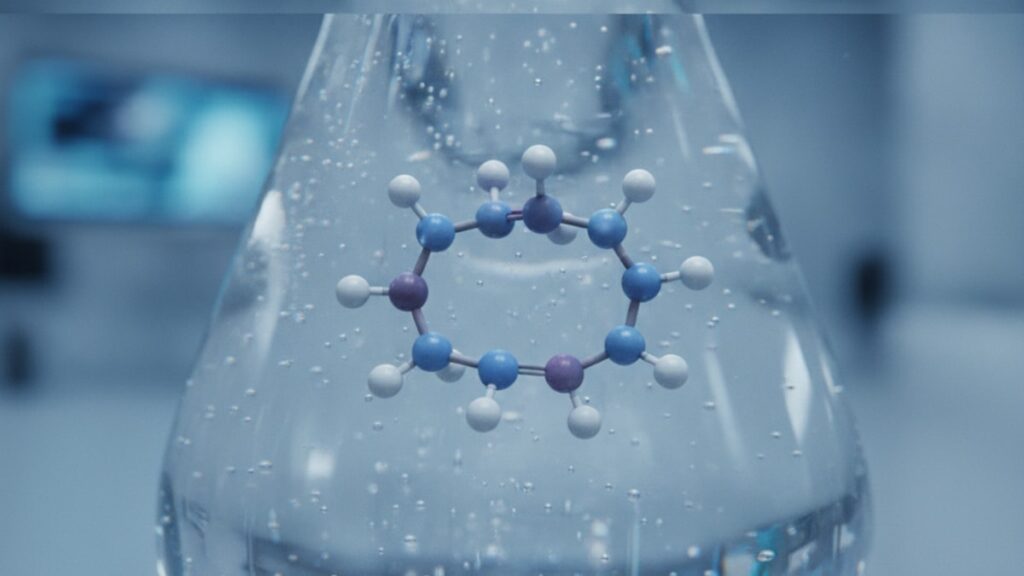
Oxytocin in solution appears as a completely clear, colorless liquid with no visible particles, cloudiness, or discoloration when correctly reconstituted with bacteriostatic water and stored at 2–8°C. Any deviation from crystal clarity—including slight opalescence, amber tinting, or suspended material—indicates protein denaturation or bacterial contamination requiring immediate disposal. Proper visual appearance confirms peptide integrity but doesn't guarantee potency without analytical verification.
The direct answer block above covers the baseline expectation. What it doesn't address is why visual inspection matters more for oxytocin than for many other peptides—oxytocin's nine-amino-acid structure contains a disulfide bridge between cysteine residues at positions 1 and 6 that's exceptionally vulnerable to oxidative stress. When that bridge breaks, the peptide loses its three-dimensional configuration and its ability to bind oxytocin receptors in target tissues. This structural fragility is why oxytocin solutions stored improperly can look perfectly clear while containing 60–80% inactive peptide. This article covers exactly what visual characteristics indicate proper reconstitution, what specific appearance changes signal degradation, and what preparation mistakes create invisible potency loss that visual inspection alone can't detect.
Physical Characteristics of Properly Reconstituted Oxytocin
Oxytocin look like in solution follows a strict visual profile when handled correctly. The reconstituted peptide presents as a water-like liquid with zero turbidity—hold the vial against a white background under direct light and you should see straight through with no haze, no suspended particles, and no color shift from absolute transparency. The solution's refractive index matches bacteriostatic water closely enough that the meniscus is the only visual distinction between the two liquids when viewed side by side.
The lyophilized powder before reconstitution appears as a white or off-white cake at the vial bottom—sometimes with slight yellowing if the peptide batch contains trace oxidation from manufacturing. This pre-reconstitution color variation doesn't predict post-reconstitution quality; properly stored lyophilized oxytocin retains full potency for 24–36 months at −20°C regardless of slight color differences in the solid form. What matters is the transformation upon adding bacteriostatic water: the powder should dissolve completely within 60–90 seconds of gentle swirling, leaving zero visible residue.
Viscosity remains identical to water—oxytocin at research concentrations (typically 1–10 mg/mL) doesn't alter the solution's flow characteristics. If your reconstituted peptide feels thicker or leaves coating on the vial walls when tilted, you're seeing either bacterial contamination producing biofilm or a compounding error where the wrong diluent was used. Oxytocin prepared with sterile saline instead of bacteriostatic water shows no immediate visual difference but lacks the antimicrobial preservative (0.9% benzyl alcohol) that prevents bacterial growth during multi-dose use—a preparation mistake that creates contamination risk without changing initial appearance.
We've guided researchers through this exact inspection process hundreds of times. The most common error isn't failing to check clarity—it's checking under inadequate lighting. Fluorescent overhead lighting misses early turbidity that's immediately obvious under focused LED or natural sunlight against a white card.
What Degraded Oxytocin Looks Like in Solution
Oxytocin look like in solution changes in predictable ways as the peptide degrades. The first visible sign is opalescence—a faint cloudiness that gives the solution a slightly milky appearance when held against a dark background. This occurs when the disulfide bond between cysteine residues breaks and the resulting linear peptide fragments begin aggregating into particles large enough to scatter light but still small enough to remain suspended. At this stage, the solution may still appear clear against a white background but shows obvious haziness against black.
Progression to full turbidity follows within 24–72 hours once opalescence appears. The solution takes on a distinctly cloudy appearance with visible swirling when the vial is gently agitated—this represents advanced protein aggregation where denatured oxytocin molecules have formed clumps exceeding 200 nanometers in diameter. Research from the University of Copenhagen's Department of Pharmacy demonstrated that oxytocin solutions showing visible turbidity contained less than 15% bioactive peptide by HPLC analysis, with the remainder existing as inactive aggregates.
Color shifts indicate oxidative degradation rather than aggregation. Properly stored oxytocin remains colorless, but exposure to temperatures above 25°C or prolonged light exposure triggers oxidation of the methionine residue at position 8, producing a pale yellow to amber discoloration. Unlike turbidity, which can develop rapidly, color change typically requires sustained improper storage—a vial left at room temperature for 48 hours or exposed to direct sunlight through a laboratory window for 6–8 hours. The visual progression goes: colorless → pale straw → amber → brown. Any solution showing amber or darker coloration should be discarded immediately.
Particulate matter represents the most severe degradation stage. Visible particles—white specs, floating fragments, or settled precipitate—indicate complete protein denaturation and often bacterial contamination. Oxytocin's small molecular weight (1007 Da) means the peptide itself remains fully soluble even when inactive; visible particles therefore suggest either bacterial growth producing biofilm fragments or contamination from non-sterile reconstitution technique introducing foreign material. Either scenario makes the solution unsafe for any application.
The bottom line: if your oxytocin solution isn't completely clear and colorless, it's degraded beyond usability. There's no "probably still good" gray zone with peptide solutions—visual changes correlate directly with loss of receptor binding activity.
Storage Conditions That Maintain Solution Appearance
Temperature control determines whether oxytocin look like in solution remains stable or degrades within days. Lyophilized oxytocin powder stored at −20°C maintains full potency for 24–36 months with zero visible change to the solid material. Once reconstituted, the peptide must be refrigerated at 2–8°C immediately—this temperature range slows but doesn't halt degradation. Published stability data from Bachem AG, a leading peptide manufacturer, shows reconstituted oxytocin at 1 mg/mL loses approximately 2–3% potency per week at 4°C storage, with visual clarity remaining intact for 28–35 days before opalescence appears.
Temperature excursions above 8°C accelerate degradation exponentially. A solution left at room temperature (20–25°C) for 24 hours loses 15–20% potency and may begin showing faint opalescence. At 37°C—body temperature—the degradation rate increases tenfold, with visible turbidity developing within 8–12 hours. This is why oxytocin solutions accidentally left out overnight must be discarded regardless of appearance; the peptide structure has been compromised even if visual changes haven't manifested yet.
Light exposure compounds temperature damage. Oxytocin contains aromatic amino acids (tyrosine at position 2) that absorb UV wavelengths, triggering photochemical reactions that break peptide bonds. Store reconstituted vials in amber glass or wrap clear vials in aluminum foil—exposure to direct sunlight for 4–6 hours causes visible yellowing even at refrigerated temperatures. Laboratory fluorescent lighting is less damaging but still contributes to cumulative degradation over weeks.
Our experience working with research facilities shows that the reconstitution step itself is where most contamination occurs. The moment you introduce that first bacteriostatic water into the lyophilized powder, you've started a 28-day countdown to complete degradation regardless of perfect storage conditions afterward. Date every vial at reconstitution—not when you first received it.
Oxytocin Solution: Appearance Comparison
| Visual Characteristic | Properly Stored (2–8°C, <28 days) | Early Degradation (Temp Excursion or >28 days) | Advanced Degradation (Room Temp 48+ hrs or Contaminated) | Professional Assessment |
|---|---|---|---|---|
| Clarity | Crystal clear—no haze visible against white or black background | Faint opalescence against dark background; may still appear clear against white | Obvious cloudiness; swirling visible when agitated | Any deviation from perfect clarity indicates compromised peptide—discard and reconstitute fresh |
| Color | Completely colorless; identical to bacteriostatic water | Pale straw or faint yellow tint | Amber to brown discoloration | Color change signals oxidative damage to methionine residues—bioactivity reduced 60–90% |
| Particles | Zero visible particles; solution passes light without scattering | May show trace suspended material under focused light | Visible white specs, floating fragments, or settled precipitate | Particulate matter indicates either complete denaturation or bacterial contamination—unsafe for use |
| Viscosity | Identical to water; flows freely | No change (degradation doesn't affect viscosity at research concentrations) | No change unless bacterial biofilm present | Viscosity changes rare; if present, indicates contamination not peptide degradation |
| Bottom Line | This is the only acceptable appearance for oxytocin in solution—use within 28 days of reconstitution | Early signs of degradation—may retain 70–85% potency but trending toward unusable | Peptide integrity lost—solution contains inactive aggregates and potential contaminants; immediate disposal required | Visual inspection catches gross degradation but misses partial potency loss—analytical verification (HPLC) is the only definitive quality measure |
Key Takeaways
- Oxytocin look like in solution as a completely clear, colorless liquid when properly reconstituted—any cloudiness, particles, or color shift indicates degradation requiring immediate disposal
- The peptide's disulfide bridge between cysteine-1 and cysteine-6 is exceptionally fragile, making visual appearance the first but not definitive indicator of potency loss
- Lyophilized oxytocin stored at −20°C remains stable for 24–36 months; once reconstituted, refrigerate at 2–8°C and use within 28 days maximum
- Temperature excursions above 8°C trigger exponential degradation—a solution left at room temperature for 24 hours loses 15–20% potency even if still visually clear
- Opalescence (faint cloudiness against dark backgrounds) signals early aggregation; full turbidity indicates less than 15% remaining bioactive peptide by analytical testing
- Color progression from colorless to amber indicates oxidative damage to methionine-8; any yellowing beyond pale straw means the solution is no longer usable
- Visual clarity confirms proper storage but doesn't guarantee full potency—HPLC analysis is the only method to verify bioactive concentration with certainty
What If: Oxytocin Solution Scenarios
What If My Oxytocin Solution Developed Slight Cloudiness After Two Weeks?
Discard it immediately and reconstitute a fresh vial. Cloudiness indicates protein aggregation—the oxytocin molecules have begun clumping into inactive structures that can't bind receptors. Even faint opalescence signals degradation exceeding 30–40%, making the solution unsuitable for research applications requiring precise dosing. The 28-day use window for reconstituted oxytocin assumes perfect storage at 2–8°C with zero temperature excursions; any visible turbidity before that timeframe means storage conditions were compromised.
What If I Accidentally Left Reconstituted Oxytocin at Room Temperature Overnight?
Discard the solution regardless of appearance. Oxytocin stored at 20–25°C for 12–16 hours undergoes structural changes that reduce potency by 15–25% even when the solution still looks perfectly clear. The peptide's disulfide bridge begins breaking under thermal stress at temperatures above 8°C, and the degradation cascade continues even after refrigeration resumes. Visual clarity lags behind molecular degradation by 24–48 hours—what looks fine now will show turbidity within days.
What If My Oxytocin Solution Has a Faint Yellow Tint But No Cloudiness?
The yellow tint signals oxidative damage to the methionine residue at position 8, which disrupts receptor binding even though the peptide remains soluble and visually clear. Discard any solution showing color beyond absolute transparency. This degradation pathway is distinct from aggregation—oxidized oxytocin doesn't form visible particles but loses 60–90% of bioactivity. The color change likely resulted from light exposure or prolonged storage above 8°C.
The Unfiltered Truth About Oxytocin Solution Appearance
Here's the honest answer: visual inspection catches only gross degradation. A perfectly clear, colorless solution can still contain 40–50% inactive peptide if storage wasn't optimal. The appearance guidelines in this article tell you when a solution is definitely unusable—they don't confirm it's definitely still good. Oxytocin's structural fragility means that even minor temperature fluctuations during shipping, brief exposure to ambient conditions during reconstitution, or storage in a refrigerator that cycles between 4–10°C instead of maintaining steady 4°C all reduce potency without producing visible changes for weeks. If your research or clinical application requires precise dosing, visual inspection is the first filter—not the final verification. HPLC analysis with UV detection at 220 nm is the only method that quantifies remaining bioactive peptide concentration. Most compounding pharmacies and peptide suppliers don't perform batch-level potency testing on reconstituted solutions because the cost exceeds the vial price. That means you're trusting storage conditions from synthesis through final use without analytical confirmation. For applications where dosing precision matters, budget for periodic analytical verification—or accept that you're working with estimated rather than verified concentrations.
Oxytocin's appearance in solution should be boring—completely unremarkable, indistinguishable from the bacteriostatic water you used to reconstitute it. The moment it becomes visually interesting, it's become scientifically useless. Date every vial at reconstitution, store it in the coldest part of your refrigerator away from the door, wrap it in foil to block light, and discard it at 28 days whether it still looks clear or not. That's the protocol that minimizes invisible potency loss while catching visible degradation early. If your oxytocin solution develops any visual characteristic beyond absolute clarity—cloudiness, color, particles, unusual viscosity—you're not looking at a peptide solution anymore; you're looking at expensive saline with inactive protein fragments suspended in it. Discard it and start fresh.
For researchers working with Real Peptides, small-batch synthesis with exact amino-acid sequencing guarantees that the lyophilized powder you receive contains the correct nonapeptide structure with the critical disulfide bridge intact. That quality standard means visual inspection failures post-reconstitution trace back to handling and storage, not manufacturing—making proper technique and temperature control the determining factors in solution longevity.
Frequently Asked Questions
What does oxytocin look like in solution when properly reconstituted?▼
Properly reconstituted oxytocin appears as a completely clear, colorless liquid with no visible particles, cloudiness, or discoloration. The solution should be indistinguishable from the bacteriostatic water used for reconstitution when held against both white and dark backgrounds under direct lighting. Any deviation from crystal clarity—including faint opalescence, color tinting, or suspended material—indicates degradation or contamination requiring immediate disposal.
How can I tell if my oxytocin solution has degraded?▼
Visual signs of oxytocin degradation follow a predictable progression: first, faint opalescence (cloudiness visible against dark backgrounds); then full turbidity with visible swirling when agitated; and finally, color changes from pale yellow to amber indicating oxidative damage. Particulate matter—white specs or floating fragments—represents either complete denaturation or bacterial contamination. Any of these visual changes signal that the solution contains inactive peptide and should be discarded immediately.
Can I still use oxytocin solution that looks slightly cloudy?▼
No—even slight cloudiness indicates protein aggregation exceeding 30–40% degradation, making the solution unsuitable for applications requiring precise dosing. Research from the University of Copenhagen found that oxytocin solutions showing visible turbidity contained less than 15% bioactive peptide by analytical testing. Cloudiness means the disulfide bridge maintaining oxytocin’s three-dimensional structure has broken, and the resulting linear peptide fragments have aggregated into inactive clumps. Discard cloudy solutions immediately and reconstitute a fresh vial.
How long does reconstituted oxytocin maintain its clear appearance?▼
Reconstituted oxytocin stored at 2–8°C maintains visual clarity for 28–35 days before opalescence appears, though potency decreases approximately 2–3% per week even when appearance remains unchanged. Temperature excursions above 8°C dramatically shorten this window—a solution left at room temperature for 24 hours may show visible turbidity within 48–72 hours. Date every vial at reconstitution and discard after 28 days regardless of appearance, as visual clarity lags behind molecular degradation by days to weeks.
What does oxytocin look like before reconstitution?▼
Lyophilized oxytocin before reconstitution appears as a white or off-white powder cake at the bottom of the vial, sometimes with slight yellowing from trace oxidation during manufacturing. This pre-reconstitution color variation doesn’t predict post-reconstitution quality—properly stored lyophilized peptide retains full potency for 24–36 months at −20°C. Upon adding bacteriostatic water, the powder should dissolve completely within 60–90 seconds of gentle swirling, leaving zero visible residue.
Why does my oxytocin solution have a yellow tint?▼
Yellow discoloration indicates oxidative degradation of the methionine residue at position 8 in oxytocin’s amino acid sequence, typically caused by prolonged storage above 8°C or exposure to direct light. This color change signals 60–90% loss of bioactivity even though the solution may remain clear without cloudiness. Discard any solution showing yellowing beyond absolute transparency—oxidized oxytocin loses receptor binding capability despite remaining visually soluble.
Does oxytocin in solution have any smell or taste?▼
Properly reconstituted oxytocin has no distinct odor beyond the faint medicinal smell of bacteriostatic water (from the 0.9% benzyl alcohol preservative). If the solution develops a sweet, musty, or unusual odor, this indicates bacterial contamination requiring immediate disposal. Oxytocin solutions are not intended for oral consumption and should never be tasted—the peptide degrades rapidly in gastric acid and has zero oral bioavailability.
What is the difference between oxytocin solution stored in clear vs amber vials?▼
Amber vials protect oxytocin from photochemical degradation by blocking UV wavelengths that would otherwise break peptide bonds and cause yellowing. Oxytocin stored in clear glass requires wrapping in aluminum foil to achieve equivalent light protection. Both storage methods maintain visual clarity equally well when combined with proper refrigeration at 2–8°C, but clear vials without foil wrapping show visible yellowing within 4–6 hours of direct sunlight exposure, while amber vials prevent this degradation.
Can visual inspection alone confirm my oxytocin solution is still potent?▼
No—visual clarity confirms the peptide hasn’t undergone gross aggregation or oxidation but doesn’t verify remaining bioactive concentration. Oxytocin’s structural fragility means temperature excursions during shipping or brief exposure to ambient conditions can reduce potency by 20–40% without producing visible changes for weeks. HPLC analysis with UV detection at 220 nm is the only method that quantifies actual bioactive peptide content. Visual inspection is a necessary first filter, not a definitive potency assay.
What should I do if I see particles floating in my oxytocin solution?▼
Discard the solution immediately without attempting to filter or use it. Visible particles indicate either complete protein denaturation (where aggregated peptide clumps have formed structures large enough to settle or float) or bacterial contamination producing biofilm fragments. Oxytocin’s molecular weight is too low for the intact peptide to form visible particles—particulate matter therefore represents either inactive aggregates or foreign material, both of which make the solution unsafe for any research application.


