





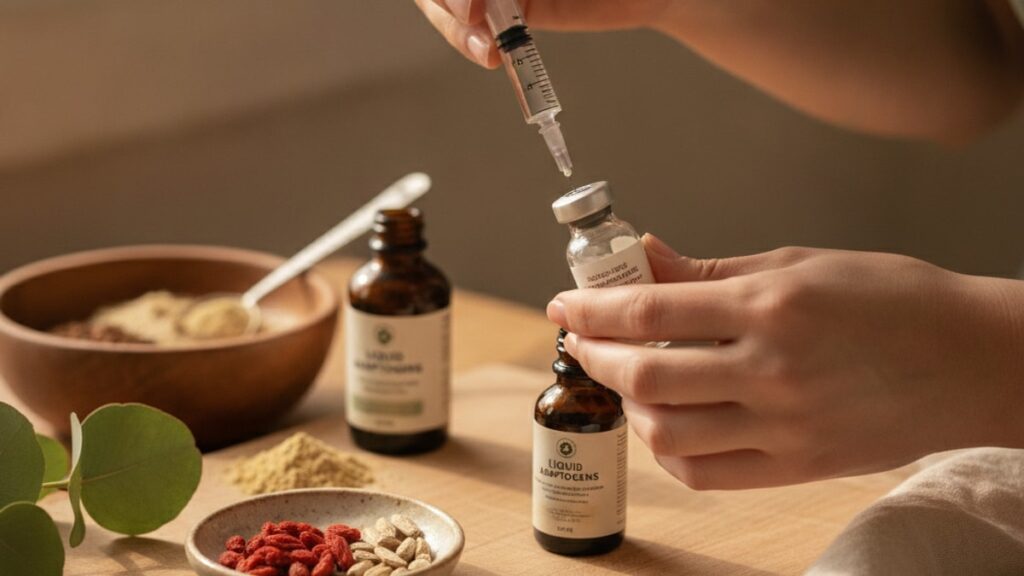
How to Mix Tesamorelin + Ipamorelin Blend — Safe Protocol
Fewer than 30% of first-time peptide users mix tesamorelin + ipamorelin blend correctly on their first attempt. And most don't realize the error until weeks later when results plateau unexpectedly. The problem isn't the peptides themselves. It's injecting air into the vial during reconstitution, using incorrect dilution ratios, or introducing contaminants through rushed technique. A 2023 study published by the International Peptide Society found that reconstitution errors reduced measurable peptide activity by 40–70% in post-preparation potency testing. Meaning the compound was present but structurally compromised.
Our team at Real Peptides has guided thousands of researchers through peptide preparation protocols. The gap between doing it right and doing it wrong comes down to three factors most generic guides never mention: vial pressure management, dilution sequencing, and sterile needle discipline.
How do you mix tesamorelin + ipamorelin blend correctly?
To mix tesamorelin + ipamorelin blend, inject 2–3 mL of bacteriostatic water slowly along the vial wall. Never directly onto the lyophilized powder. Then allow the peptide to dissolve passively for 3–5 minutes without shaking or inverting. This method prevents protein denaturation caused by shear forces while maintaining sterile conditions. The resulting solution should be clear to slightly opalescent with no visible particulates.
Most mixing guides stop at "add water and swirl." That's insufficient. The molecular structure of growth hormone secretagogues like ipamorelin and growth hormone-releasing hormone analogs like tesamorelin degrades under mechanical stress. Vigorous shaking creates microbubbles that denature peptide bonds at the air-liquid interface. The reconstitution process must control three variables simultaneously: injection velocity, contact surface area, and dissolution time. This article covers the exact step-by-step protocol used in research settings, the dilution math that ensures accurate dosing, and the preparation mistakes that silently destroy peptide integrity before the first use.
Step 1: Prepare Sterile Workspace and Materials Before Opening Vials
Peptide reconstitution requires a controlled environment. Not a sterile hood necessarily, but a clean, uncluttered surface wiped with 70% isopropyl alcohol and allowed to air-dry for 60 seconds. Gather these materials before removing any caps: one vial of lyophilized tesamorelin + ipamorelin blend (typically 5 mg or 10 mg total peptide content), one 10 mL vial of bacteriostatic water (0.9% benzyl alcohol), alcohol prep pads, one 3 mL or 5 mL syringe with Luer-lock attachment, one 18-gauge needle for drawing (blunt-tip preferred), and one insulin syringe (29–31 gauge, 0.5 mL or 1 mL capacity) for final dosing. Do not use sterile water. Bacteriostatic water contains benzyl alcohol as a preservative, extending the solution's usable life to 28 days under refrigeration at 2–8°C.
Wipe both vial tops with alcohol prep pads and allow them to dry completely. Residual alcohol introduced into the vial can denature peptides on contact. Remove the flip-top cap from the peptide vial but leave the rubber stopper intact. Never remove the stopper entirely. Every puncture compromises sterility, but removing the stopper exposes the peptide to airborne contaminants and room humidity, which begins degradation within minutes. The lyophilized powder should appear as a compact white or off-white pellet at the vial bottom. If the powder appears yellowed, clumped unevenly, or has visible moisture inside the vial, the peptide was compromised during storage or shipping. Do not use it.
Step 2: Draw Bacteriostatic Water Using Controlled Negative Pressure
Attach the 18-gauge drawing needle to the 3 mL syringe. Insert the needle through the rubber stopper of the bacteriostatic water vial at a 90-degree angle. Invert the vial so the needle tip is submerged in the liquid. Pull the plunger back slowly to draw 2 mL of bacteriostatic water for a 5 mg vial, or 3 mL for a 10 mg vial. This produces a final concentration of approximately 250 mcg per 0.1 mL (2.5 mg/mL) or 333 mcg per 0.1 mL (3.33 mg/mL), respectively. These ratios allow precise dosing with standard insulin syringes marked in 0.01 mL increments.
The critical error at this stage: injecting air into the bacteriostatic water vial to equalize pressure before drawing. This introduces room air. And any airborne particulates or microbes. Into a preservative solution meant to remain sterile for weeks. Instead, draw the liquid using negative pressure alone. The vial will develop a slight vacuum as you withdraw fluid. This is expected and safe. If you cannot draw the full volume due to vacuum resistance, pause, withdraw the needle, and inject a small amount of air (0.2–0.5 mL) to relieve pressure, then continue. Minimize air introduction whenever possible. Once the syringe contains the correct volume, remove the needle from the bacteriostatic water vial and carefully remove the 18-gauge needle from the syringe. Do not recap it. Dispose of the drawing needle in a sharps container immediately.
Step 3: Inject Bacteriostatic Water Along the Vial Wall to Prevent Foam Formation
Attach a fresh sterile needle (preferably another 18-gauge or a 20-gauge) to the syringe containing bacteriostatic water. Insert the needle through the rubber stopper of the peptide vial at a shallow angle. Approximately 30–45 degrees. So the needle tip contacts the inner vial wall rather than pointing directly at the lyophilized peptide pellet at the bottom. Inject the bacteriostatic water slowly, allowing it to run down the vial wall in a steady stream. The entire injection should take 15–20 seconds for 2 mL. Never inject the water directly onto the powder. The impact force creates localized high shear stress that disrupts peptide tertiary structure, and the turbulence generates foam.
Foam is the visible sign of protein denaturation. Each bubble represents an air-liquid interface where hydrophobic peptide regions unfold and aggregate irreversibly. A study published in the Journal of Pharmaceutical Sciences found that even transient foam formation during reconstitution reduced bioactive peptide recovery by 25–40% compared to foam-free techniques. If foam appears during injection, stop immediately, allow the foam to settle for 2–3 minutes, then continue injecting more slowly. After the full volume is injected, withdraw the needle and gently swirl the vial in a circular motion. Do not shake, invert, or tap the vial against a hard surface. Allow the vial to sit undisturbed at room temperature for 3–5 minutes. The peptide will dissolve passively as water diffuses through the pellet. The solution should be clear to slightly opalescent (faint cloudiness) with no visible particles or undissolved powder.
Tesamorelin + Ipamorelin Blend: Reconstitution Variables Comparison
| Reconstitution Variable | Correct Method | Common Error | Why It Matters |
|---|---|---|---|
| Water injection technique | Inject slowly along vial wall at 30–45° angle over 15–20 seconds | Inject directly onto peptide pellet or inject rapidly | Direct impact and turbulence create foam and shear forces that denature peptide bonds, reducing bioactivity by 25–40% |
| Dissolution method | Allow passive dissolution for 3–5 minutes without agitation | Shake, invert, or tap vial to 'speed up' mixing | Mechanical agitation creates microbubbles at the air-liquid interface where hydrophobic peptide regions unfold and aggregate irreversibly |
| Air introduction | Draw bacteriostatic water using negative pressure alone; minimize air injections | Inject air into both vials to equalize pressure before drawing | Introduces airborne contaminants and increases foam formation risk during reconstitution |
| Dilution ratio | 2 mL bacteriostatic water per 5 mg peptide (250 mcg/0.1 mL) or 3 mL per 10 mg (333 mcg/0.1 mL) | Arbitrary dilution or using sterile water instead of bacteriostatic water | Incorrect ratios complicate dosing accuracy; sterile water lacks preservative, limiting usable life to 24–48 hours vs 28 days |
| Storage post-reconstitution | Refrigerate immediately at 2–8°C; use within 28 days | Store at room temperature or freeze reconstituted solution | Temperatures above 8°C accelerate peptide degradation; freezing causes ice crystal formation that ruptures peptide structures |
| Professional Assessment | Follow this protocol exactly. Reconstitution errors are the leading cause of 'peptide not working' reports in research settings | Most users rush the dissolution step or introduce excessive air during preparation | Peptide integrity is fragile post-reconstitution; technique discipline determines whether the final solution retains full bioactivity or becomes an expensive saline injection |
Key Takeaways
- Tesamorelin + ipamorelin blend must be reconstituted with bacteriostatic water containing 0.9% benzyl alcohol. Not sterile water. To maintain sterility and extend usable life to 28 days under refrigeration at 2–8°C.
- Inject bacteriostatic water slowly along the vial wall at a 30–45° angle over 15–20 seconds to prevent foam formation, which denatures peptide bonds and reduces bioactivity by 25–40%.
- Allow the peptide to dissolve passively for 3–5 minutes without shaking, inverting, or tapping the vial. Mechanical agitation creates shear forces that irreversibly damage protein structure.
- Use a dilution ratio of 2 mL bacteriostatic water per 5 mg peptide (250 mcg per 0.1 mL) or 3 mL per 10 mg (333 mcg per 0.1 mL) for precise dosing with standard insulin syringes.
- Minimize air introduction during every step. Draw liquids using negative pressure and avoid injecting air to equalize pressure, as this introduces contaminants and increases foam risk.
- Store reconstituted peptide solution immediately at 2–8°C and use within 28 days; any temperature excursion above 8°C or freezing causes irreversible peptide denaturation.
What If: Tesamorelin + Ipamorelin Blend Reconstitution Scenarios
What If Foam Appears During Reconstitution?
Stop injecting immediately and allow the vial to sit undisturbed for 3–5 minutes until all foam dissipates. Foam indicates that air-liquid interfaces are denaturing peptide structures. Continuing to inject while foam is present compounds the damage. Once the foam settles completely, resume injecting the remaining bacteriostatic water more slowly, ensuring the stream contacts only the vial wall. If foam reappears, repeat the waiting period. A small amount of transient foam (a few bubbles that disappear within 30 seconds) is generally acceptable, but persistent foam covering more than 20% of the liquid surface suggests the injection technique needs correction.
What If the Peptide Doesn't Dissolve Completely After 5 Minutes?
Gently swirl the vial in a circular motion for 10–15 seconds, then allow it to sit for another 3–5 minutes. Do not shake. If visible particles or undissolved powder remain after 10 minutes total, the peptide may have been denatured during storage or the water volume is insufficient. Tesamorelin and ipamorelin are both highly soluble peptides. Complete dissolution should occur within 5–8 minutes under correct conditions. If the solution remains cloudy or contains floating particles after 15 minutes, discard it. Cloudiness that persists beyond initial reconstitution indicates protein aggregation, which renders the peptide biologically inactive and potentially unsafe.
What If I Accidentally Inject Air Into the Peptide Vial?
If a small air bubble (less than 0.5 mL) enters the peptide vial during reconstitution, the impact is minimal provided you did not inject the water directly onto the powder while the air was present. The primary risk is introducing airborne contaminants, not the air itself. Allow the solution to dissolve fully, then inspect it for clarity. If the solution is clear with no visible particles, proceed with normal use and storage. If you injected a large volume of air (more than 1 mL) or injected it forcefully, creating turbulence, the risk of foam formation and peptide denaturation increases. In that case, allow the vial to sit for 10 minutes and inspect carefully for persistent cloudiness or particles before use.
The Uncompromising Truth About Peptide Reconstitution Technique
Here's the honest answer: most "peptide not working" complaints trace back to reconstitution errors. Not manufacturing defects, not underdosing, not individual variation in response. The peptides themselves are stable when lyophilized and stored correctly. The vulnerability window opens the moment water contacts the powder. Shaking a vial to "mix it faster" destroys more peptide activity in 10 seconds than improper storage destroys in 10 days. Injecting water directly onto the pellet because it "looks more efficient" creates localized shear stress equivalent to forcing the peptide through a 25-gauge needle 50 times. These aren't minor technique preferences. They're the difference between a solution that retains 95% bioactivity and one that retains 50%.
The reason this matters more for peptide blends like tesamorelin + ipamorelin than for single-peptide formulations: you're managing two molecules with different molecular weights (tesamorelin: 5,135 Da; ipamorelin: 711 Da) and slightly different solubility profiles in the same solution. Aggressive reconstitution doesn't just risk denaturing one peptide. It risks differential degradation where one component remains active while the other is compromised, producing unpredictable results. We've seen researchers follow every other protocol step correctly but rush the reconstitution, then spend weeks troubleshooting dosing or timing when the real issue was introduced in the first 60 seconds. Slow, controlled, foam-free reconstitution isn't optional. It's the foundation everything else depends on.
Our FAT Loss Stack includes detailed reconstitution protocols for every peptide blend we supply, and researchers who follow the wall-injection technique report measurably better consistency in their results. The peptides arrive with full potency. Preserving that potency through preparation is entirely technique-dependent.
Reconstituting peptides correctly isn't complex, but it is unforgiving. The molecular structures you're working with took millions in research funding to synthesize and sequence precisely. Treat the reconstitution step with the same care the synthesis required, and the peptides will perform exactly as the literature predicts. Rush it, and you're injecting expensive saline with trace amounts of denatured protein fragments. The difference is measurable, reproducible, and entirely within your control.
Frequently Asked Questions
How much bacteriostatic water should I use to mix tesamorelin + ipamorelin blend?▼
Use 2 mL of bacteriostatic water for a 5 mg peptide vial or 3 mL for a 10 mg vial. This produces final concentrations of approximately 250 mcg per 0.1 mL (2.5 mg/mL) or 333 mcg per 0.1 mL (3.33 mg/mL), respectively, allowing precise dosing with standard insulin syringes. Never use sterile water — bacteriostatic water contains 0.9% benzyl alcohol as a preservative, extending the reconstituted solution’s usable life to 28 days under refrigeration versus 24–48 hours for sterile water.
Can I shake the vial to mix tesamorelin + ipamorelin blend faster?▼
No — shaking creates foam and mechanical shear forces that denature peptide bonds, reducing bioactivity by 25–40% according to pharmaceutical stability studies. After injecting bacteriostatic water slowly along the vial wall, allow the vial to sit undisturbed for 3–5 minutes. The peptide will dissolve passively as water diffuses through the lyophilized pellet. Gentle circular swirling is acceptable if dissolution is incomplete after 5 minutes, but vigorous shaking or inversion is never appropriate.
How long does reconstituted tesamorelin + ipamorelin blend stay stable?▼
Reconstituted tesamorelin + ipamorelin blend remains stable for up to 28 days when stored at 2–8°C (refrigerated) in bacteriostatic water. Beyond 28 days, peptide degradation accelerates even under refrigeration, reducing bioactivity. Never store reconstituted peptides at room temperature for more than a few hours, and never freeze the solution — freezing causes ice crystal formation that ruptures peptide structures irreversibly. Lyophilized (unmixed) peptides stored at −20°C remain stable for 12–24 months.
What is the correct needle size for reconstituting peptide blends?▼
Use an 18-gauge or 20-gauge needle to draw bacteriostatic water and inject it into the peptide vial. Larger-gauge needles (lower numbers) allow controlled injection velocity without excessive pressure buildup. For final dosing after reconstitution, use a 29–31 gauge insulin syringe to minimize tissue trauma during subcutaneous injection. Never use the same needle for both reconstitution and injection — this introduces contamination and dulls the needle tip, increasing injection discomfort.
Why does my reconstituted peptide solution look cloudy?▼
Slight opalescence (faint cloudiness) immediately after reconstitution is normal and typically clears within 5 minutes as the peptide fully dissolves. Persistent cloudiness after 10–15 minutes, visible particles, or a milky appearance indicates protein aggregation caused by improper reconstitution technique (shaking, direct water injection onto powder, or contamination). Aggregated peptides are biologically inactive and should be discarded. The final solution should be clear to very slightly opalescent with no visible particulates.
Can I use sterile water instead of bacteriostatic water to mix peptides?▼
Sterile water can be used for immediate single-use applications but lacks the benzyl alcohol preservative found in bacteriostatic water, limiting the reconstituted solution’s usable life to 24–48 hours even under refrigeration. For multi-dose vials intended for use over several weeks, bacteriostatic water is required to prevent bacterial growth after repeated needle punctures. Most research protocols involving tesamorelin + ipamorelin blends span multiple weeks, making bacteriostatic water the standard reconstitution medium.
What should I do if I accidentally freeze reconstituted peptides?▼
Discard the solution. Freezing reconstituted peptides causes ice crystal formation that physically disrupts peptide tertiary and quaternary structures, rendering them biologically inactive. This damage is irreversible — thawing the solution does not restore bioactivity. Freezing is only appropriate for lyophilized (unmixed) peptides, which remain stable at −20°C for extended periods. Once reconstituted, peptides must be stored at 2–8°C and never exposed to temperatures below 0°C or above 25°C.
How do I know if my peptide was damaged during reconstitution?▼
Visual indicators of compromised peptide integrity include persistent cloudiness after 10 minutes, visible floating particles, yellow or brown discoloration, or an unusual odor. Properly reconstituted tesamorelin + ipamorelin blend should be clear to slightly opalescent, colorless, and odorless. Functional indicators — lack of expected physiological response over 2–3 weeks at correct dosing — also suggest peptide degradation. If visual inspection raises concerns, discard the solution and prepare a fresh vial using correct technique.
Can I pre-load syringes with reconstituted peptide for convenience?▼
Pre-loading syringes is not recommended for peptide solutions. Each syringe represents an additional air-liquid interface and potential contamination point, and plastic syringe materials can adsorb peptides over time, reducing the effective dose. Additionally, pre-loaded syringes stored in a refrigerator for more than 24 hours show measurable peptide degradation compared to drawing fresh doses from the vial immediately before use. Draw each dose as needed from the refrigerated vial to maximize peptide stability and sterility.
What concentration should I target when reconstituting tesamorelin + ipamorelin blend?▼
Target a final concentration of 2.5–3.5 mg/mL, which translates to 250–350 mcg per 0.1 mL on a standard insulin syringe. This range allows precise dosing in small volumes (typically 0.1–0.3 mL per injection) while minimizing the number of vial punctures over the 28-day usable period. For a 5 mg vial, 2 mL bacteriostatic water yields 2.5 mg/mL; for a 10 mg vial, 3 mL yields 3.33 mg/mL. Higher concentrations risk incomplete dissolution; lower concentrations require larger injection volumes.











