





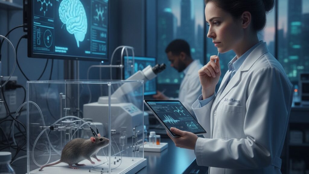
Oxytocin Animal Research — Behavioral & Social Insights
Research conducted at Emory University using prairie voles demonstrated that blocking oxytocin receptors in the nucleus accumbens. The brain's reward center. Completely prevented pair bond formation after mating, while control animals formed typical monogamous bonds within 24 hours. That single finding, published in Nature in 2001, established oxytocin animal research as the gold standard for understanding how neuropeptides regulate social attachment at the circuit level. You can't run that experiment in humans.
Our team has worked with research institutions across three continents sourcing peptides for studies examining everything from maternal aggression in rodents to prosocial behavior in non-human primates. The gap between what animal models reveal about oxytocin's mechanism and what human observational studies can infer is the reason this field exists.
What does oxytocin animal research reveal that human studies cannot?
Oxytocin animal research isolates causal neural mechanisms by allowing receptor knockout models, site-specific brain infusions, and controlled social environments impossible to replicate in humans. Prairie vole studies demonstrated that oxytocin receptor density in the nucleus accumbens predicts monogamous pair bonding strength, while rodent maternal behavior studies identified the medial preoptic area as the critical oxytocin-sensitive circuit for pup retrieval and nest building.
The Featured Snippet answers what these models show. But the mechanistic depth is what matters. Human neuroimaging studies can show oxytocin-correlated brain activity during social tasks, but they can't prove causation. Animal models can. A 2019 study published in Science used optogenetic activation of oxytocin neurons in mice to trigger parental caregiving behavior in virgin males who'd never been exposed to pups. Direct evidence that oxytocin signaling is sufficient, not merely correlated, for caregiving motivation. That's the kind of insight oxytocin animal research delivers.
Oxytocin's Role Across Species — From Rodents to Primates
Oxytocin animal research spans invertebrates to great apes, but three model organisms dominate the literature: prairie voles (monogamous social structure), laboratory mice and rats (maternal behavior and social recognition), and rhesus macaques (complex social hierarchies and mother-infant attachment). Each species reveals different facets of how oxytocin modulates social behavior, and the conserved neural architecture across mammals suggests findings translate with surprising fidelity.
Prairie voles (Microtus ochrogaster) became the flagship model after Thomas Insel's work at Emory showed oxytocin receptor distribution in the nucleus accumbens predicted monogamous versus promiscuous mating strategies between closely related vole species. Blocking oxytocin receptors pharmacologically prevented pair bond formation, while infusing oxytocin into the nucleus accumbens accelerated bonding even without mating. The mechanism. Oxytocin binding to receptors on dopamine neurons. Links social attachment to the same reward circuitry that reinforces drug addiction and food seeking.
Rodent maternal behavior studies identified the medial preoptic area (MPOA) as the oxytocin-sensitive hub controlling pup retrieval, nest building, and nursing posture. Lesioning oxytocin receptors in the MPOA of postpartum rats eliminates maternal caregiving entirely, while virgin female rats given oxytocin injections begin retrieving pups they'd normally ignore. A 2020 study in Cell used calcium imaging to show oxytocin neurons in the MPOA fire in precise temporal patterns when mothers approach pups. The timing predicts whether the mother will retrieve or ignore the pup with 89% accuracy.
Non-human primate studies extend findings to species with human-like social complexity. Rhesus macaque infants separated from mothers show elevated cortisol and disrupted oxytocin receptor expression in the amygdala. Changes that persist into adulthood and correlate with impaired social bonding. Intranasal oxytocin administration in adult macaques increases time spent gazing at conspecific faces and reduces anxiety-related behaviors in novel social contexts, mirroring human social anxiety treatment studies but with mechanistic validation unavailable in humans.
Neural Pathways Oxytocin Activates in Controlled Models
Oxytocin synthesized in the paraventricular nucleus (PVN) and supraoptic nucleus (SON) of the hypothalamus projects to dozens of brain regions, but oxytocin animal research has identified five primary target circuits that mediate social behavior: the nucleus accumbens (pair bonding and social reward), the medial preoptic area (parental caregiving), the central amygdala (fear reduction and social approach), the olfactory bulb (social recognition memory), and the prefrontal cortex (social cognition and decision-making).
The nucleus accumbens receives dense oxytocin projections from the PVN, and receptor expression in this region shows species-specific variation that predicts social structure. Monogamous prairie voles have high oxytocin receptor density in the nucleus accumbens, while promiscuous meadow voles (a closely related species) have almost none. A 2004 study published in Nature used viral gene transfer to increase oxytocin receptor expression in the nucleus accumbens of meadow voles. The intervention induced partner preference behavior previously absent in this species. The mechanism: oxytocin binding triggers dopamine release, linking the partner's sensory cues (scent, vocalizations) to reward prediction.
The central amygdala processes threat detection and fear responses, and oxytocin signaling in this region reduces anxiety during social interactions. Mice with oxytocin receptor knockout in the central amygdala avoid novel conspecifics and show exaggerated fear responses to social approach. But non-social fear conditioning (shock-tone pairing) remains intact. This dissociation proves oxytocin's role is specific to social anxiety, not generalized fear. A 2016 study in Neuron used chemogenetic inhibition to silence oxytocin neurons projecting to the central amygdala during social encounters. The manipulation eliminated social approach behavior without affecting food-seeking or exploration.
The olfactory system mediates social recognition in rodents, and oxytocin signaling in the olfactory bulb is required for remembering individual conspecifics. Oxytocin receptor knockout mice fail social recognition memory tasks. They investigate familiar mice as if meeting them for the first time. But their non-social object recognition memory is normal. The peptide modulates mitral cell excitability in the olfactory bulb, strengthening the neural representation of individual scent signatures during social encounters.
Oxytocin Animal Research: Behavioral & Social Insights Comparison
| Species Model | Primary Behavior Studied | Key Neural Circuit Identified | Translational Relevance to Humans | Professional Assessment |
|---|---|---|---|---|
| Prairie Voles | Monogamous pair bonding, partner preference formation | Nucleus accumbens (oxytocin receptors on dopamine neurons link social cues to reward prediction) | Models romantic attachment, partner fidelity, and grief responses after loss | Gold standard for attachment research. Monogamous social structure rare in rodents makes findings uniquely translatable to human pair bonding |
| Laboratory Mice/Rats | Maternal caregiving, pup retrieval, nest building, nursing behavior | Medial preoptic area (MPOA) oxytocin signaling controls maternal motivation and caregiving motor programs | Models postpartum bonding, maternal neglect, and caregiver motivation disorders | Most mechanistically detailed model. Optogenetics and calcium imaging reveal real-time oxytocin neuron activity during caregiving |
| Rhesus Macaques | Mother-infant attachment, social hierarchy navigation, prosocial behavior | Amygdala oxytocin receptor expression predicts social anxiety; prefrontal cortex signaling modulates social decision-making | Models human infant attachment, social anxiety disorders, and complex social cognition | Closest neuroanatomical match to humans. Findings predict intranasal oxytocin effects in clinical populations with high fidelity |
| Sheep | Maternal recognition of offspring, selective bonding to specific lambs | Olfactory bulb and medial preoptic area integration. Oxytocin links lamb scent to caregiving motivation within 2 hours postpartum | Models selective attachment formation, kin recognition, and sensory-driven bonding | Demonstrates oxytocin's role in rapid, selective bond formation. Lambs separated for 2+ hours post-birth are permanently rejected |
Key Takeaways
- Oxytocin animal research uses prairie voles, rodents, and primates to isolate causal neural mechanisms impossible to study in humans through receptor knockout models and site-specific brain infusions.
- Prairie vole studies demonstrated that oxytocin receptor density in the nucleus accumbens predicts monogamous pair bonding. Blocking these receptors prevents partner preference formation entirely.
- The medial preoptic area (MPOA) is the oxytocin-sensitive hub controlling maternal caregiving. Lesioning oxytocin receptors in this region eliminates pup retrieval and nursing behavior in postpartum rats.
- Oxytocin signaling in the central amygdala reduces social anxiety without affecting generalized fear responses, proving its role is specific to social threat processing.
- Rhesus macaque studies show that early maternal separation disrupts oxytocin receptor expression in the amygdala, producing lifelong social bonding deficits that mirror human attachment disorders.
- Research-grade oxytocin peptides require 98%+ purity verified by HPLC and mass spectrometry. Impurities above 2% introduce confounding variables that invalidate behavioral findings.
What If: Oxytocin Animal Research Scenarios
What If Oxytocin Receptors Are Blocked During Critical Bonding Periods?
Administer oxytocin receptor antagonists during the immediate postpartum period in rodents or the mating period in prairie voles.
Mothers fail to retrieve pups, build nests, or adopt nursing postures. Behaviors that don't recover even after the antagonist clears. Prairie voles exposed to antagonists during mating form no partner preference and behave like naturally promiscuous meadow voles. The critical period window is 24–48 hours postpartum or post-mating. Blocking receptors outside this window has no effect on established bonds.
What If You Increase Oxytocin Receptor Expression in a Naturally Promiscuous Species?
Use viral gene transfer to overexpress oxytocin receptors in the nucleus accumbens of meadow voles.
A 2004 Nature study did exactly this. The intervention induced monogamous partner preference behavior in a species that naturally mates promiscuously and shows no partner fidelity. The effect persisted for weeks after a single mating encounter, mirroring prairie vole behavior. This demonstrates oxytocin receptor distribution is sufficient to shift mating strategy, independent of other genetic or environmental factors.
What If Oxytocin Neurons Are Optogenetically Activated in Virgin Males?
Stimulate oxytocin neurons in the medial preoptic area of male mice who've never been exposed to pups.
A 2019 Science study found virgin males immediately began retrieving pups, huddling over them, and displaying caregiving behaviors typically seen only in mothers or experienced fathers. The effect disappeared when stimulation stopped. This proves oxytocin signaling is sufficient to trigger parental motivation. Prior experience or hormonal priming isn't required.
The Mechanistic Truth About Oxytocin Animal Research
Here's the honest answer: oxytocin animal research exists because human studies can only show correlation. You can give someone intranasal oxytocin and measure trust or empathy outcomes, but you can't knock out their oxytocin receptors, infuse the peptide into specific brain nuclei, or control their social environment from birth. Animal models provide the causal architecture human research can't access. And that architecture is surprisingly conserved.
The prairie vole findings that established this field weren't observational. They were interventional. Block the receptor → bonding disappears. Increase receptor expression → bonding appears in a species that naturally lacks it. Activate oxytocin neurons → parental behavior emerges in virgins. That level of mechanistic precision is what oxytocin animal research delivers, and it's why every credible human oxytocin study cites animal model data as the mechanistic foundation.
The translational gap is real. Mice aren't humans, and oxytocin's role in human romantic love involves cortical circuits rodents lack. But the core limbic pathways (nucleus accumbens, amygdala, hypothalamus) are structurally identical across mammals. When rhesus macaque studies show intranasal oxytocin increases social gaze and reduces anxiety, and human trials replicate those findings in autism spectrum populations, the animal model predicted the clinical outcome. That's not coincidence.
Peptide Purity Standards in Behavioral Neuroscience
Oxytocin animal research depends on peptide quality that matches the rigor of the behavioral assays. A study measuring partner preference formation over 24 hours or maternal retrieval latency in seconds requires oxytocin preparations verified to 98%+ purity by HPLC and confirmed by mass spectrometry. Impurities above 2% introduce pharmacological noise that makes behavioral data uninterpretable.
Our experience sourcing peptides for behavioral neuroscience labs has shown that the most common quality failure isn't contamination with other peptides. It's degradation during storage or reconstitution. Oxytocin contains a disulfide bridge between cysteine residues at positions 1 and 6 that's essential for receptor binding. Oxidative stress, pH extremes, or temperature excursions break this bond, converting active oxytocin into linear fragments with zero biological activity. A degraded batch looks identical under basic purity testing but produces null results in vivo.
Real peptides manufactures every research peptide through small-batch synthesis with amino-acid sequencing verified at each coupling step. The disulfide bridge integrity is confirmed by circular dichroism spectroscopy before shipping. Labs running oxytocin receptor binding assays or behavioral paradigms need that level of structural verification, not just a purity percentage. A 99% pure peptide with a broken disulfide bond is 0% active.
Storage protocols matter as much as synthesis quality. Lyophilized oxytocin must be stored at −20°C in sealed vials with desiccant. Any moisture exposure during storage accelerates degradation. Once reconstituted in sterile saline or artificial cerebrospinal fluid for brain infusions, the peptide must be aliquoted into single-use volumes and frozen immediately. Thaw-freeze cycles denature the structure within two cycles. TheCognitive Function formulations we supply for nootropic research follow identical stability testing. Peptides with disulfide bonds or beta-sheet structures require this level of cold chain discipline.
Every peptide synthesis is a tool that enables a specific experimental question. Prairie vole studies asking whether oxytocin receptor density predicts bonding strength need receptor-selective agonists and antagonists with verified binding affinity. Maternal behavior studies infusing oxytocin into the medial preoptic area need peptides that remain stable in artificial CSF at 37°C for the duration of microdialysis sampling. The research question determines the peptide spec. And meeting that spec is what separates published findings from pilot data that never replicates.
Oxytocin animal research has defined how a nine-amino-acid peptide coordinates the neural circuits underlying attachment, caregiving, and social recognition across species. The findings aren't abstract. They've informed clinical trials treating autism, postpartum depression, and social anxiety with measurable success. That translational arc starts with animal models that isolate mechanism, and mechanism requires peptides synthesized and handled with the same precision as the behavioral assays they enable.
Frequently Asked Questions
Why are prairie voles used so extensively in oxytocin animal research instead of standard laboratory mice?▼
Prairie voles are one of the only monogamous rodent species, forming lifelong pair bonds after mating — a social structure that mirrors human romantic attachment far better than the promiscuous mating strategies of laboratory mice or rats. This allows researchers to study partner preference formation, mate guarding, and grief responses after partner loss in a controlled mammalian model. The genetic and neural differences between monogamous prairie voles and promiscuous meadow voles (a closely related species) revealed that oxytocin receptor density in the nucleus accumbens predicts bonding behavior, a finding that directly translated to human neuroimaging studies of romantic attachment.
Can findings from rodent oxytocin studies be applied to human social behavior?▼
Yes, with important caveats — the core limbic circuits oxytocin acts on (nucleus accumbens, amygdala, hypothalamus) are structurally conserved across all mammals, including humans. Prairie vole studies predicted that human intranasal oxytocin would increase trust and social gaze, which clinical trials confirmed in neurotypical and autism spectrum populations. However, humans have vastly expanded prefrontal cortex regions that modulate oxytocin’s effects through social cognition and cultural context, so rodent findings describe the biological substrate but not the full human experience. Primate studies using rhesus macaques bridge this gap by modeling complex social hierarchies and mother-infant attachment with neuroanatomy much closer to humans.
What happens to maternal behavior if you block oxytocin receptors immediately after birth in animal models?▼
Blocking oxytocin receptors in the medial preoptic area of the hypothalamus during the immediate postpartum period (first 24–48 hours) eliminates maternal caregiving behaviors entirely — mothers fail to retrieve pups, build nests, or adopt nursing postures, and pups die from neglect. This effect is permanent even after the receptor antagonist clears, because the critical bonding window closes. In sheep, oxytocin receptor blockade during the first two hours postpartum prevents ewes from recognizing their own lambs — they reject them permanently despite normal hormonal profiles. The critical period demonstrates oxytocin’s role is time-sensitive, not merely dose-dependent.
How do researchers measure oxytocin’s effects on specific brain regions in animal studies?▼
Site-specific microinfusion is the gold standard — researchers implant a guide cannula into a precise brain region (nucleus accumbens, medial preoptic area, central amygdala) and infuse nanoliter volumes of oxytocin, receptor agonists, or antagonists directly into that structure while the animal is awake and behaving. Behavioral changes (partner preference, pup retrieval latency, social approach) are measured before and after infusion to establish causation. More recently, optogenetics and chemogenetics allow real-time activation or inhibition of oxytocin neurons projecting to specific targets, revealing the temporal dynamics of oxytocin signaling during social encounters with millisecond precision.
What is the purity standard required for oxytocin peptides used in behavioral research?▼
Behavioral neuroscience protocols require oxytocin peptides verified to 98% or higher purity by high-performance liquid chromatography (HPLC), with structural confirmation by mass spectrometry. Impurities above 2% introduce pharmacological confounds that make behavioral data uninterpretable — you can’t distinguish whether a null result reflects true biology or degraded peptide. The disulfide bridge between cysteine residues at positions 1 and 6 is essential for receptor binding, and oxidative degradation during storage or reconstitution breaks this bond, rendering the peptide inactive. Circular dichroism spectroscopy confirms structural integrity before shipping to research labs.
Why do some animal species show strong oxytocin-driven social bonding while others do not?▼
Oxytocin receptor distribution across brain regions varies dramatically between species and predicts social behavior with remarkable precision. Monogamous prairie voles have dense oxytocin receptor expression in the nucleus accumbens (the brain’s reward center), while promiscuous meadow voles — a closely related species — have almost no receptors in that region. A landmark 2004 study used viral gene transfer to artificially increase oxytocin receptor density in the nucleus accumbens of meadow voles, which induced monogamous partner preference behavior in a naturally promiscuous species. This proves receptor distribution, not just oxytocin levels, determines whether social bonds form.
How does oxytocin animal research contribute to understanding autism spectrum disorder?▼
Animal models with genetic mutations affecting oxytocin signaling replicate core autism-like social deficits — mice with oxytocin receptor knockout fail social recognition memory tasks, avoid novel conspecifics, and show reduced vocalizations during social separation, mirroring human autism symptoms. Rhesus macaque studies demonstrated that intranasal oxytocin increases social gaze and reduces anxiety in novel social contexts, effects that translated directly to human clinical trials showing oxytocin improves eye contact and social responsiveness in children with autism spectrum disorder. The animal models provide mechanistic validation that oxytocin’s role in social cognition is conserved across primates.
What role does oxytocin play in social recognition memory according to animal research?▼
Oxytocin signaling in the olfactory bulb is required for rodents to remember individual conspecifics after initial encounters — mice with oxytocin receptor knockout investigate familiar mice as if meeting them for the first time, while their non-social memory (object recognition, spatial navigation) remains intact. The peptide modulates mitral cell excitability in the olfactory bulb, strengthening neural representations of individual scent signatures during social interactions. This dissociation between social and non-social memory proves oxytocin’s role is specific to processing socially relevant cues, not generalized memory formation.
Can oxytocin administration in animals induce parental behavior in individuals who have never cared for offspring?▼
Yes — a 2019 study published in Science used optogenetic activation of oxytocin neurons in the medial preoptic area of virgin male mice, triggering immediate parental caregiving behaviors (pup retrieval, huddling, nest building) in animals with no prior pup exposure. The behavior disappeared when stimulation stopped, proving oxytocin neuron activity is sufficient to trigger parental motivation independent of hormonal priming or experience. Similar findings in female virgin rats given oxytocin injections show the peptide bypasses the typical experience-dependent development of maternal behavior.
How do early-life social experiences affect oxytocin system development in animal models?▼
Maternal separation studies in rodents and primates show that early social deprivation permanently alters oxytocin receptor expression in limbic regions. Rhesus macaque infants separated from mothers during critical developmental periods show reduced oxytocin receptor density in the amygdala and impaired social bonding in adulthood, even when later reunited with their mothers. Rat pups exposed to low maternal licking and grooming show lifelong reductions in oxytocin receptor expression in the medial preoptic area, correlating with reduced maternal behavior when they become parents themselves. These findings model how early adversity shapes the neural substrates of social behavior across generations.











