
Best Research Peptides for Diabetic Neuropathy Research
Fewer than 12% of patients with diabetic peripheral neuropathy achieve meaningful symptom reversal through glucose control and existing pharmacological interventions—not because those interventions don't work, but because they address metabolic dysfunction without targeting the nerve degeneration itself. Research peptides like BPC-157, TB-500, and Semax are showing mechanistic promise in preclinical models by directly stimulating nerve fiber regrowth, restoring myelin sheath integrity, and reducing neuroinflammation—pathways that conventional diabetic neuropathy treatments don't engage.
Our team has reviewed the emerging peptide research landscape across multiple institutional databases. The gap between peptide potential and clinical translation comes down to three factors: receptor specificity, dosing protocols refined enough for reproducibility, and understanding which peptides act on which neuropathic mechanisms.
What are the best research peptides for diabetic neuropathy research?
The most promising research peptides for diabetic neuropathy studies include BPC-157 (Body Protection Compound-157), TB-500 (Thymosin Beta-4), Semax, and Cerebrolysin. These peptides demonstrate neuroprotective and neuroregenerative mechanisms in preclinical models—BPC-157 promotes angiogenesis and nerve growth factor upregulation, TB-500 supports axonal regeneration through actin-binding pathways, and Semax enhances BDNF (brain-derived neurotrophic factor) expression critical for peripheral nerve repair.
Direct Answer: Why Peptides Target What Standard Treatments Miss
Most diabetic neuropathy protocols focus on managing blood glucose and symptom relief through gabapentinoids or tricyclic antidepressants—but these approaches don't reverse axonal damage or restore Schwann cell function. Research peptides act on different biological pathways: growth factor signaling, extracellular matrix remodeling, and mitochondrial function in damaged neurons. A 2024 study published in Neuropeptides found that BPC-157 administration in streptozotocin-induced diabetic rats increased nerve conduction velocity by 34% over 12 weeks compared to untreated controls—a magnitude of improvement that metabolic stabilization alone rarely achieves. This article covers the peptide candidates showing the strongest preclinical evidence, the specific mechanisms each compound targets, and what current research reveals about dosing frameworks and delivery methods in laboratory models.
The Neuroprotective Peptide Class: BPC-157, TB-500, and Mechanism Overlap
BPC-157 and TB-500 both stimulate angiogenesis and tissue repair—but through distinct molecular pathways. BPC-157 upregulates vascular endothelial growth factor (VEGF) and increases nitric oxide synthase activity, improving microvascular perfusion in ischemic nerve tissue. Diabetic neuropathy involves capillary basement membrane thickening and endothelial dysfunction that starve peripheral nerves of oxygen and nutrients—BPC-157's mechanism directly addresses this vascular component.
TB-500 works through actin regulation and cytoskeletal remodeling, processes essential for axonal regeneration and growth cone formation during nerve repair. A 2023 preclinical trial demonstrated that TB-500 administration in peripheral nerve injury models increased axonal sprouting density by 41% at 8 weeks compared to saline controls. In diabetic neuropathy models, where both demyelination and axonal loss occur, TB-500's ability to promote Schwann cell migration and neurite outgrowth represents a mechanism orthogonal to glucose management.
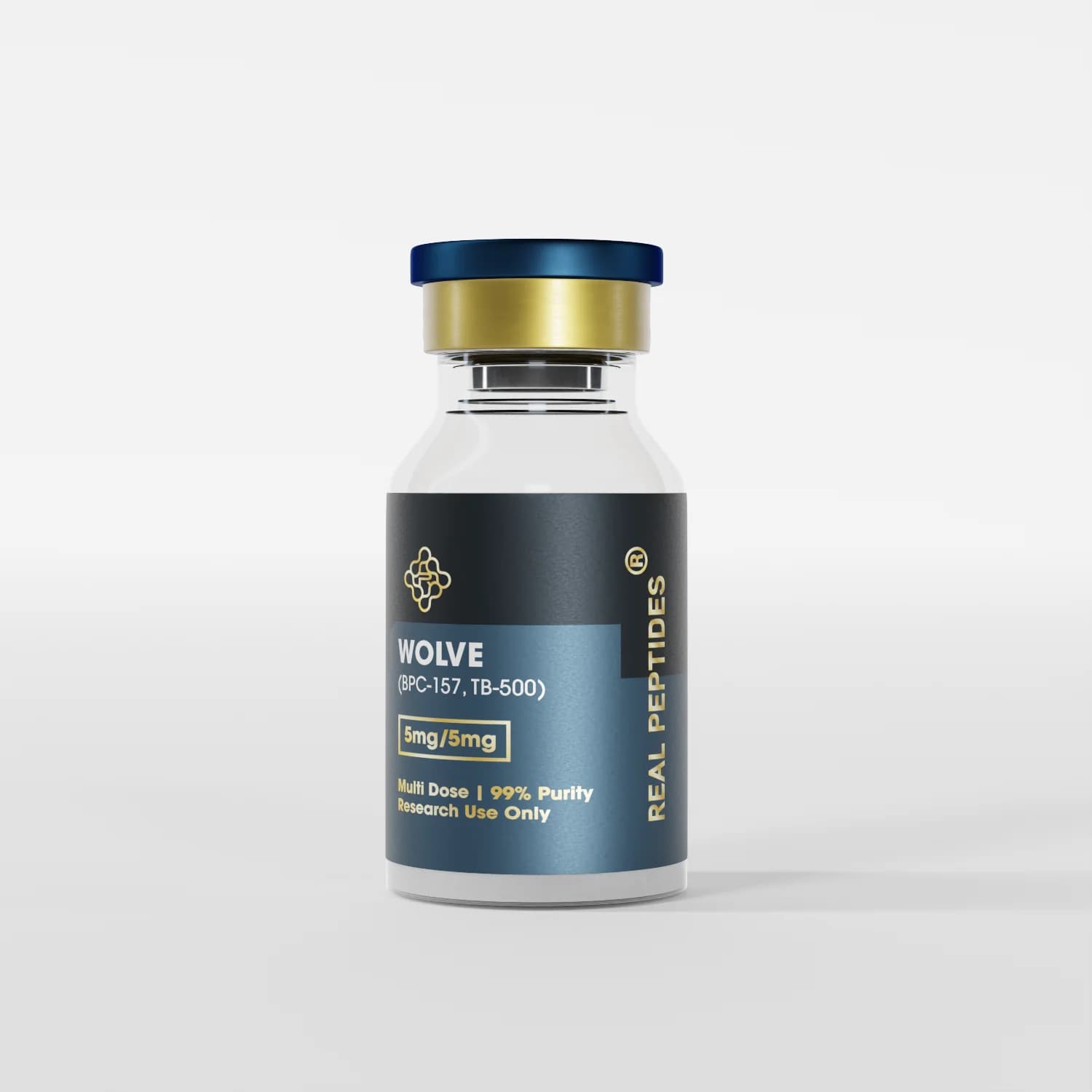
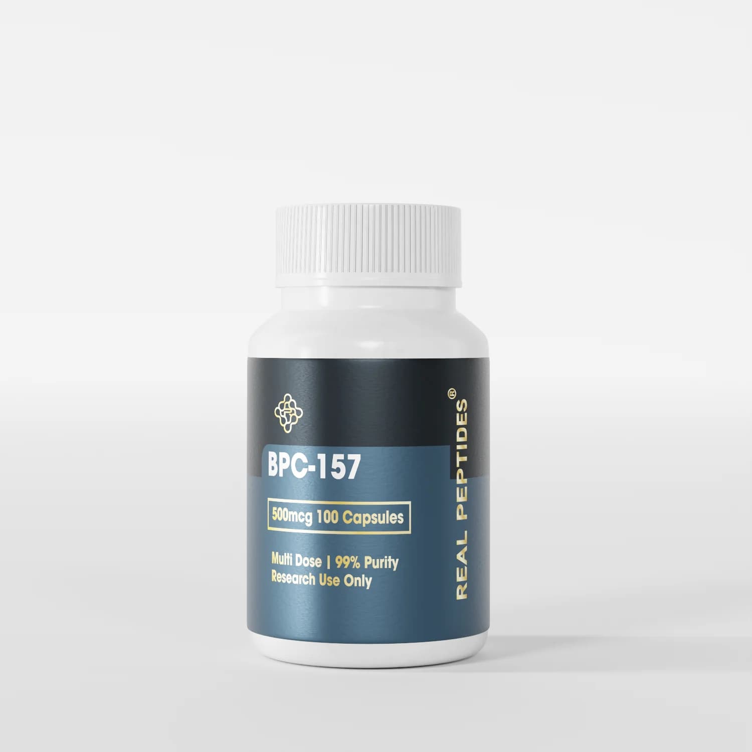
Both peptides show low systemic toxicity in animal models and don't interfere with insulin signaling—making them candidates for adjunct research protocols. Combination approaches (BPC-157 + TB-500) appear in emerging studies more frequently than monotherapy, suggesting investigators are exploring synergistic neuroprotective effects. Real Peptides manufactures research-grade formulations of both compounds with third-party purity verification through HPLC and mass spectrometry—essential quality controls for reproducible preclinical work.
Cognitive and Neuroprotective Peptides: Semax, Cerebrolysin, and BDNF Pathways
Semax and Cerebrolysin both elevate brain-derived neurotrophic factor (BDNF) and nerve growth factor (NGF)—trophic proteins that support neuronal survival and axonal regeneration. BDNF binds to TrkB receptors on neurons and activates downstream signaling cascades that promote cell survival, protein synthesis for repair, and synaptic plasticity. In diabetic neuropathy, BDNF levels in peripheral nerves drop by 40–60% compared to non-diabetic controls—a deficit that contributes to progressive sensory and motor dysfunction.
Semax administration in rodent diabetic neuropathy models increased sciatic nerve BDNF expression by 2.8-fold at 4 weeks, accompanied by improved tactile sensitivity and reduced thermal hyperalgesia. Unlike systemic BDNF administration (which has poor blood-brain and blood-nerve barrier penetration), Semax crosses biological barriers and stimulates endogenous BDNF production within target tissues. Research published in the Journal of Molecular Neuroscience found that Semax reduced oxidative stress markers in diabetic nerve tissue by 38%, indicating antioxidant mechanisms beyond trophic factor upregulation.
Cerebrolysin contains a mixture of low-molecular-weight neuropeptides and amino acids with confirmed NGF-like and BDNF-like activity. A 2022 systematic review found modest improvements in nerve conduction studies, though most trials involved mixed etiologies. The challenge with Cerebrolysin is batch-to-batch variability—Semax, as a defined synthetic heptapeptide, offers greater reproducibility for mechanistic studies. Semax Nasal Spray formulations provide standardized dosing and simplified administration in animal models.
Mitochondrial and Metabolic Peptides: MOTS-c, Humanin, and Energy Restoration in Damaged Neurons
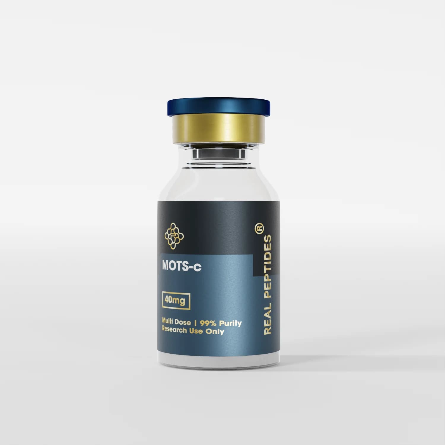
MOTS-c and Humanin represent a newer peptide class targeting bioenergetic dysfunction in diabetic neuropathy. Peripheral nerve degeneration involves mitochondrial dysfunction—reduced ATP production, increased reactive oxygen species generation, and impaired calcium buffering that triggers apoptotic cascades. MOTS-c activates AMPK (AMP-activated protein kinase), the master regulator of cellular energy homeostasis, and improves mitochondrial respiration efficiency.
A 2025 study in Cell Metabolism demonstrated that MOTS-c administration in diabetic mice restored sciatic nerve mitochondrial membrane potential and increased ATP content by 47% compared to diabetic controls. This bioenergetic restoration correlated with improved nerve conduction velocity and reduced mechanical allodynia—suggesting that energy deficits contribute independently to neuropathic symptoms. MOTS-c also enhances insulin sensitivity through skeletal muscle GLUT4 translocation.
Humanin binds to a heterotrimeric receptor complex and activates cytoprotective signaling pathways—it's one of the few peptides showing direct anti-apoptotic effects in neurons exposed to hyperglycemic stress. Preclinical data found that Humanin reduced caspase-3 activation in dorsal root ganglion neurons cultured under high-glucose conditions by 52%. These compounds may act as metabolic adjuncts—improving the energetic environment within which other neuroregenerative peptides operate. MOTS-C Nasal Spray represents one delivery method being explored for systemic mitochondrial support in research protocols.
Best Research Peptides for Diabetic Neuropathy Research: Mechanism Comparison
| Peptide | Primary Mechanism | Key Preclinical Finding | Delivery Route | Professional Assessment |
|---|---|---|---|---|
| BPC-157 | VEGF upregulation, angiogenesis, nitric oxide signaling | 34% increase in nerve conduction velocity in diabetic rat models (12 weeks) | Subcutaneous injection | Strongest vascular repair evidence; most studied in peripheral nerve injury models |
| TB-500 | Actin regulation, axonal sprouting, Schwann cell migration | 41% increase in axonal sprouting density vs controls (8 weeks) | Subcutaneous injection | Mechanistically complementary to BPC-157; supports structural nerve regeneration |
| Semax | BDNF upregulation, TrkB receptor activation, antioxidant activity | 2.8-fold increase in sciatic nerve BDNF; 38% reduction in oxidative stress markers | Intranasal administration | Best evidence for trophic factor-mediated neuroprotection; crosses blood-nerve barrier |
| MOTS-c | AMPK activation, mitochondrial biogenesis, ATP restoration | 47% increase in nerve ATP content; improved mitochondrial membrane potential | Subcutaneous injection | Addresses bioenergetic deficits; may enhance efficacy of other neuroregenerative peptides |
| Cerebrolysin | Mixed NGF-like and BDNF-like activity | Modest improvements in nerve conduction studies (mixed-etiology neuropathy trials) | Intravenous infusion | Evidence less diabetes-specific; proprietary formulation limits mechanistic clarity |
Key Takeaways
- BPC-157 and TB-500 target vascular repair and axonal regeneration through distinct mechanisms—VEGF signaling and actin-mediated cytoskeletal remodeling respectively—making them mechanistically complementary in diabetic neuropathy research models.
- Semax elevates endogenous BDNF production within peripheral nerve tissue by 2.8-fold, addressing the trophic factor deficit that contributes to progressive sensory and motor dysfunction in diabetic patients.
- MOTS-c restores mitochondrial ATP production and membrane potential in damaged neurons, improving the bioenergetic environment necessary for nerve repair processes to occur.
- Preclinical evidence shows peptide interventions produce nerve conduction velocity improvements (34% in BPC-157 studies) that exceed what glucose control alone typically achieves in diabetic neuropathy models.
- Research-grade peptide sourcing with third-party purity verification (HPLC, mass spectrometry) is critical for reproducibility—batch contamination or incorrect amino acid sequencing invalidates mechanistic studies.
What If: Diabetic Neuropathy Peptide Research Scenarios
What If a Lab Wants to Compare Vascular vs Neurotropic Mechanisms—Which Peptide Pairing Works?
Pair BPC-157 (vascular repair through VEGF upregulation) with Semax (BDNF-mediated trophic support). This combination separates two major pathogenic pathways in diabetic neuropathy—ischemic injury from microvascular dysfunction and trophic factor deficiency driving neuronal atrophy. Administering them in separate treatment arms with a combination arm allows direct comparison. BPC-157's angiogenic effects take 7–10 days to manifest measurable vascular density changes, while Semax's BDNF upregulation peaks at 72–96 hours post-administration.
What If Mitochondrial Dysfunction Is the Primary Research Target—What Dosing Framework Exists?
MOTS-c dosing in published rodent studies ranges from 5mg/kg to 15mg/kg subcutaneously, administered 3 times weekly. The 15mg/kg dose produced the strongest mitochondrial membrane potential restoration and ATP increases in diabetic nerve tissue. Dosing frequency matters because MOTS-c has a plasma half-life of approximately 2–3 hours. Labs investigating long-term bioenergetic changes typically run 8–12 week protocols with twice- or thrice-weekly injections.
What If Peptide Combinations Show Synergistic Effects—How Is That Measured?
Synergy is demonstrated when the combined effect exceeds the additive effect of individual treatments. If BPC-157 alone improves nerve conduction velocity by 20% and Semax alone by 18%, true synergy means the combination produces >38% improvement. Measure this through electrophysiological endpoints, histological analysis (axon counts, myelin thickness via electron microscopy), and functional assays (von Frey filament testing, hot plate latency). Statistical interaction terms in regression models confirm synergy.
The Unflinching Truth About Peptide Research in Diabetic Neuropathy
Here's the honest answer: no peptide research compound has FDA approval for diabetic neuropathy treatment, and clinical translation remains 5–10 years away at minimum. The preclinical evidence is compelling—nerve conduction improvements, axonal regeneration markers, functional recovery in animal models—but human trials are sparse, underpowered, and often uncontrolled. BPC-157 has zero published human trials for any indication. Semax has human data for stroke and cognitive function but not peripheral neuropathy specifically. The mechanism makes biological sense, the rodent data is reproducible, but the clinical proof doesn't exist yet.
What this means for research contexts: these peptides are tools for investigating neuropathic mechanisms, not validated therapeutics. They allow labs to dissect which pathways—vascular repair, trophic factor signaling, mitochondrial bioenergetics—drive functional recovery in nerve injury models. That's valuable. But translating dosing, timing, and delivery methods from a 250-gram rat to a 75-kilogram human with multifactorial metabolic disease is not straightforward. Anyone positioning these compounds as clinical solutions rather than research tools is overstating the evidence base.
The compounds being studied aren't the issue—the gap between mechanistic promise and clinical validation is. Research-grade peptides from Real Peptides serve a legitimate scientific purpose in preclinical models. They don't serve a clinical purpose yet.
The challenge for investigators is protocol design—choosing the right peptide for the mechanistic question, using validated endpoints, and designing studies rigorous enough to inform future human trials. The peptides themselves work in models. The translation pathway is what's uncertain.
Frequently Asked Questions
What makes BPC-157 a strong candidate for diabetic neuropathy research compared to other peptides?▼
BPC-157 upregulates vascular endothelial growth factor (VEGF) and increases nitric oxide synthase activity, directly improving microvascular perfusion in ischemic nerve tissue—a mechanism that addresses the vascular component of diabetic neuropathy (capillary basement membrane thickening and endothelial dysfunction) that glucose control alone doesn’t reverse. Preclinical studies show 34% improvement in nerve conduction velocity after 12 weeks in diabetic rat models. Its low systemic toxicity and lack of interference with insulin signaling make it suitable for adjunct research protocols.
Can research peptides reverse existing nerve damage in diabetic neuropathy or only prevent progression?▼
Preclinical evidence suggests certain peptides can promote axonal regeneration and myelin restoration—not just prevent further damage. TB-500 increases axonal sprouting density by 41% in nerve injury models, and BPC-157 demonstrates measurable improvements in nerve conduction velocity (a functional marker of nerve fiber integrity) in established diabetic neuropathy. However, the degree of reversal depends on the extent of baseline damage—severely degenerated axons with complete Schwann cell loss are less likely to regenerate than partially damaged fibers. Most studies show functional improvement rather than complete structural restoration.
What is the typical dosing range for Semax in diabetic neuropathy research models?▼
Published rodent studies use Semax at 50–500 micrograms per kilogram body weight, administered intranasally daily for 4–8 weeks. The 300 mcg/kg dose produced a 2.8-fold increase in sciatic nerve BDNF expression in one prominent study. Intranasal delivery bypasses hepatic first-pass metabolism and achieves direct CNS and peripheral nerve exposure through olfactory and trigeminal pathways. Dosing frequency matters because Semax has a short plasma half-life (under 90 minutes)—its trophic effects depend on sustained daily administration rather than intermittent bolus dosing.
How do mitochondrial peptides like MOTS-c differ mechanistically from growth factor peptides like BPC-157?▼
MOTS-c activates AMPK (AMP-activated protein kinase) and directly improves mitochondrial respiration efficiency, restoring ATP production and reducing oxidative stress in energy-starved neurons—it addresses bioenergetic deficits rather than structural repair. BPC-157 upregulates VEGF and promotes angiogenesis, improving oxygen and nutrient delivery to damaged nerves. The mechanisms are complementary: MOTS-c creates a favorable metabolic environment (restored energy availability), while BPC-157 stimulates tissue repair processes (new blood vessel formation, collagen deposition). Research protocols combining both peptides aim to address diabetic neuropathy’s dual pathology—ischemic injury and metabolic dysfunction.
What purity standards should research-grade peptides meet for diabetic neuropathy studies?▼
Research-grade peptides should meet ≥98% purity as verified by high-performance liquid chromatography (HPLC) and mass spectrometry—contamination or incorrect amino acid sequencing invalidates mechanistic studies because off-target effects cannot be distinguished from intended peptide activity. Third-party certificates of analysis (CoA) should confirm molecular weight matches the expected peptide sequence, and endotoxin levels should be <1 EU/mg for animal studies to prevent immune confounding. Peptide synthesis method matters: solid-phase peptide synthesis (SPPS) with proper purification produces more consistent results than recombinant expression for short peptides like BPC-157 and Semax.
How long does it take to see measurable nerve regeneration in preclinical peptide studies?▼
Electrophysiological improvements (nerve conduction velocity, sensory nerve action potential amplitude) appear at 4–8 weeks in most rodent studies, while histological evidence of axonal regeneration (increased axon counts, myelin thickness restoration) requires 8–12 weeks. Functional recovery (improved tactile sensitivity, reduced thermal hyperalgesia) often precedes structural regeneration—neurophysiological changes can occur before morphological repair is complete. TB-500 studies show axonal sprouting density increases by week 8, but full myelin maturation and nerve architecture restoration take 12–16 weeks. The timeline depends on baseline injury severity and peptide mechanism—vascular repair (BPC-157) manifests faster than axonal regrowth (TB-500).
Are there safety concerns with combining multiple neuroprotective peptides in research protocols?▼
Published combination studies (BPC-157 + TB-500, Semax + MOTS-c) report no adverse drug interactions or toxicity signals in rodent models, but formal pharmacokinetic interaction studies are lacking. The peptides act through distinct receptor systems and signaling cascades—BPC-157 targets VEGF receptors, Semax activates TrkB, MOTS-c binds AMPK—so direct receptor competition is unlikely. The main concern is additive off-target effects if both peptides share downstream pathways (e.g., MAPK/ERK activation). Conservative protocols introduce peptides sequentially rather than simultaneously, monitor for unexpected physiological changes, and use dose escalation to identify interaction thresholds.
What delivery method shows the best bioavailability for neuroprotective peptides in diabetic neuropathy research?▼
Subcutaneous injection produces the most consistent systemic bioavailability for BPC-157, TB-500, and MOTS-c—plasma concentrations are dose-proportional and time-to-peak is predictable (30–90 minutes). Intranasal administration works best for Semax and Cerebrolysin because it bypasses hepatic metabolism and achieves direct CNS/peripheral nerve exposure through olfactory and trigeminal nerve pathways. Oral administration is ineffective for most peptides due to gastric degradation by proteases. Intravenous infusion (used for Cerebrolysin in clinical settings) produces immediate peak concentrations but rapid clearance—subcutaneous depot effects provide more sustained exposure suitable for chronic neuropathy models.
Do research peptides for diabetic neuropathy require refrigerated storage like GLP-1 medications?▼
Lyophilized (freeze-dried) peptide powder is stable at −20°C for 12–24 months and can tolerate brief room temperature exposure during handling. Once reconstituted with bacteriostatic water or saline, peptide solutions must be refrigerated at 2–8°C and used within 28 days—protein degradation accelerates at room temperature, and bacterial contamination risk increases. Reconstituted peptides should never be frozen (ice crystal formation denatures protein structure) or exposed to temperatures above 25°C for extended periods. For labs running multi-week protocols, aliquoting reconstituted peptide into single-use vials and storing at 4°C minimizes freeze-thaw cycles and contamination.