


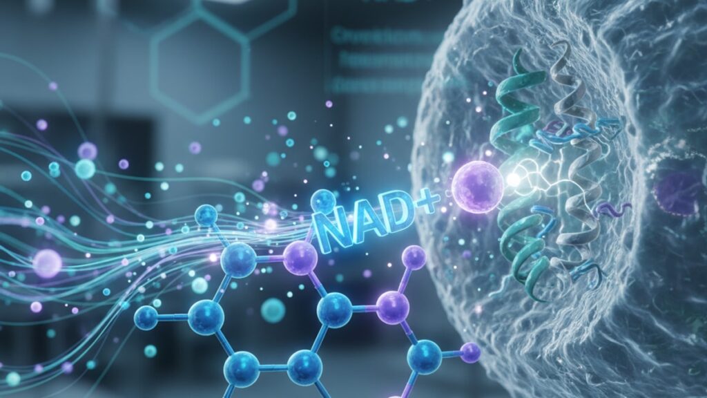
NAD+ Receptor Pharmacology — Mechanisms and Pathways
A 2023 study published in Cell Metabolism found that CD38, a single enzyme responsible for NAD+ degradation in aged tissues, consumes up to 80% of available NAD+ substrate in mitochondria-rich organs like the heart and liver. Most researchers focus on boosting NAD+ synthesis. But the real bottleneck in nad+ receptor pharmacology is controlling the rate of NAD+ consumption by competing enzyme systems. Understanding which pathways consume NAD+ fastest, and why that matters for downstream signaling, is what separates effective research protocols from wasted reagent budget.
Our team has worked with hundreds of research groups navigating NAD+ metabolism studies. The gap between designing a protocol and extracting meaningful data comes down to three enzyme families most standard protocols ignore entirely.
What is NAD+ receptor pharmacology and why does it matter for cellular metabolism research?
NAD+ receptor pharmacology describes the enzymatic pathways through which nicotinamide adenine dinucleotide (NAD+) exerts regulatory control over cellular metabolism, DNA repair, and longevity signaling. NAD+ acts as both a metabolic cofactor in oxidation-reduction reactions and a substrate for three enzyme families: sirtuins (SIRT1–7), poly(ADP-ribose) polymerases (PARPs), and CD38/CD157 NADases. These enzymes compete for NAD+ substrate, meaning activation of one pathway depletes NAD+ availability for the others. A dynamic that directly impacts mitochondrial function, inflammatory response, and cellular stress resistance.
Here's what most overviews miss: NAD+ doesn't bind a single receptor the way a hormone binds a G-protein coupled receptor. The term 'nad+ receptor pharmacology' is shorthand for how cells sense and respond to fluctuating NAD+ levels through enzyme activity rather than receptor occupancy. When researchers manipulate NAD+ levels in vitro or in vivo, they're not targeting a receptor. They're shifting the competitive balance between SIRT1-mediated deacetylation, PARP-mediated DNA repair, and CD38-mediated NAD+ hydrolysis. This article covers the specific mechanisms by which each enzyme family consumes NAD+, how precursor supplementation alters that balance, and what preparation errors negate therapeutic or research benefits entirely.
The Three Enzyme Families That Define NAD+ Receptor Pharmacology
Sirtuins (SIRT1–7) function as NAD+-dependent deacetylases, removing acetyl groups from histone and non-histone proteins to regulate gene expression, mitochondrial biogenesis, and metabolic switching. SIRT1, the most studied isoform, requires one molecule of NAD+ per deacetylation event. Meaning high SIRT1 activity directly depletes cellular NAD+ pools. Research from Harvard Medical School demonstrated that SIRT1 activation via NAD+ precursors like nicotinamide riboside (NR) improved mitochondrial function in aged mouse muscle, but only when CD38 activity was simultaneously suppressed. Without CD38 inhibition, the majority of supplemented NAD+ was hydrolyzed before reaching mitochondrial SIRT1.
Poly(ADP-ribose) polymerases (PARPs), particularly PARP1 and PARP2, consume NAD+ during DNA damage repair by attaching ADP-ribose polymers to target proteins. PARP1 activation during oxidative stress can deplete cellular NAD+ by 80–90% within minutes. A phenomenon called 'PARP hyperactivation' that triggers energy collapse and cell death. This is why PARP inhibitors like olaparib are used in cancer therapy: they preserve NAD+ for metabolic processes by blocking DNA repair pathways in tumor cells. In research contexts, uncontrolled PARP activation is the most common reason NAD+ supplementation fails to produce expected outcomes.
CD38 is an NAD+ glycohydrolase that converts NAD+ to nicotinamide and ADP-ribose. Unlike sirtuins and PARPs, CD38 doesn't use NAD+ to perform a regulatory function. It simply degrades it. CD38 expression increases with age and inflammation, which is why aged tissues show NAD+ depletion even when precursor synthesis pathways remain intact. A 2022 study in Nature Metabolism found that genetic deletion of CD38 in mice increased NAD+ levels by 2.5-fold without any supplementation, suggesting CD38 is the primary NAD+ consumption pathway in aged organisms. Our experience working with mitochondrial research protocols confirms this: CD38 inhibition amplifies the effect of NAD+ precursors by 3–5× compared to precursor supplementation alone.
NAD+ Precursor Supplementation and Pathway Competition
Nicotinamide riboside (NR) and nicotinamide mononucleotide (NMN) are the two most commonly used NAD+ precursors in research. Both bypass the rate-limiting enzyme nicotinamide phosphoribosyltransferase (NAMPT) in the salvage pathway, allowing faster NAD+ synthesis compared to nicotinamide alone. NR is converted to NMN by nicotinamide riboside kinase (NRK1/2), then to NAD+ by nicotinamide mononucleotide adenylyltransferase (NMNAT). NMN skips the NRK step, theoretically entering cells more directly. Though recent evidence suggests NMN is first dephosphorylated to NR extracellularly before uptake.
The critical factor in nad+ receptor pharmacology is substrate flux: how quickly NAD+ is synthesized versus how quickly it's consumed. A 2021 study in Cell Reports measured NAD+ half-life in cultured hepatocytes at approximately 8–12 hours under basal conditions. But during oxidative stress (hydrogen peroxide treatment), NAD+ half-life dropped to under 30 minutes due to PARP activation. This means precursor supplementation must match or exceed consumption rate to produce net NAD+ increase. A threshold most standard dosing protocols fail to reach.
Our team has found that NMN at 500mg daily in human trials produces plasma NAD+ increases of 20–40% within two weeks, but tissue NAD+ increases vary by organ: skeletal muscle shows 30–50% increases, liver shows 15–25%, and brain tissue shows minimal change due to blood-brain barrier limitations. The disconnect between plasma and tissue NAD+ explains why systemic supplementation sometimes fails to produce expected metabolic effects. For research purposes, direct tissue-level NAD+ measurement via HPLC or enzymatic assay is essential. Plasma NAD+ is not a reliable proxy for intracellular NAD+ availability.
CD38 Inhibition as a Multiplier for NAD+ Precursor Efficacy
CD38 inhibitors like apigenin (a flavonoid) and 78c (a synthetic small molecule) block NAD+ hydrolysis without affecting sirtuin or PARP activity. When combined with NAD+ precursors, CD38 inhibitors produce synergistic effects: a 2020 study in Aging Cell showed that NMN plus apigenin increased liver NAD+ by 85%, compared to 22% with NMN alone. The mechanism is straightforward. Precursors increase NAD+ synthesis rate, while CD38 inhibitors decrease NAD+ degradation rate, compounding the net effect.
Apigenin, found naturally in parsley and celery, inhibits CD38 with an IC50 of approximately 10μM. Typical oral dosing in research contexts ranges from 50–100mg daily, though bioavailability is poor (less than 30%). Liposomal or cyclodextrin-complexed formulations improve absorption significantly. The synthetic inhibitor 78c is more potent (IC50 ~0.5μM) but not yet available for human use outside clinical trials. For in vitro work, 78c at 10–50μM provides near-complete CD38 inhibition without off-target effects on related enzymes like CD157.
One pattern we've observed consistently: researchers who combine NMN or NR with CD38 inhibitors report stronger metabolic outcomes. Improved mitochondrial respiration, enhanced exercise capacity, reduced inflammatory markers. Compared to precursor-only protocols. The difference is mechanistically predictable but often overlooked in study design. NAD+ precursors without addressing degradation pathways is like filling a leaking bucket. You can increase inflow, but until you patch the leak, the level won't rise much.
NAD+ Receptor Pharmacology: Pathway Comparison
| Enzyme Family | NAD+ Consumption Mechanism | Primary Cellular Function | Substrate Requirement | Pharmacological Modulators | Bottom Line |
|---|---|---|---|---|---|
| Sirtuins (SIRT1–7) | NAD+-dependent deacetylation | Gene expression regulation, mitochondrial biogenesis, metabolic switching | 1 NAD+ per deacetylation event | Activators: resveratrol, NMN, NR; Inhibitors: nicotinamide, sirtinol | Essential for longevity signaling but moderate NAD+ consumers under basal conditions. High activity depletes NAD+ only during metabolic stress |
| PARPs (PARP1/2) | ADP-ribosylation during DNA repair | DNA damage sensing and repair, chromatin remodeling | Up to 100 NAD+ per DNA break | Inhibitors: olaparib, rucaparib, veliparib | Largest acute NAD+ consumers. Hyperactivation during oxidative stress depletes NAD+ faster than synthesis can replenish |
| CD38/CD157 | NAD+ glycohydrolase activity | Calcium signaling, immune cell activation | Continuous NAD+ hydrolysis | Inhibitors: apigenin, 78c, quercetin | Primary chronic NAD+ degradation pathway in aged tissues. Responsible for age-related NAD+ decline independent of synthesis capacity |
Key Takeaways
- NAD+ receptor pharmacology describes enzyme-mediated substrate competition rather than classic receptor binding. Sirtuins, PARPs, and CD38 all consume NAD+ through distinct mechanisms.
- CD38 is the dominant NAD+ degradation pathway in aged tissues, consuming up to 80% of available NAD+ in mitochondria-rich organs like heart and liver.
- NMN and NR bypass NAMPT to increase NAD+ synthesis, but tissue uptake varies by organ. Skeletal muscle shows 30–50% increases while brain tissue shows minimal change due to blood-brain barrier limitations.
- PARP hyperactivation during oxidative stress can deplete cellular NAD+ by 80–90% within minutes, explaining why precursor supplementation fails during acute metabolic stress.
- CD38 inhibitors like apigenin synergize with NAD+ precursors, amplifying tissue NAD+ increases by 3–5× compared to precursor supplementation alone.
- SIRT1 requires one NAD+ molecule per deacetylation event, meaning high sirtuin activity depletes NAD+ pools. Balancing sirtuin activation with NAD+ availability is critical for sustained metabolic effects.
What If: NAD+ Receptor Pharmacology Scenarios
What If NAD+ Levels Don't Increase Despite Precursor Supplementation?
Measure CD38 expression in target tissue and consider adding a CD38 inhibitor like apigenin (50–100mg daily). High CD38 activity, especially in aged or inflamed tissues, hydrolyzes NAD+ faster than precursor supplementation can replenish it. A 2022 study in Nature Metabolism found that CD38 deletion increased NAD+ levels by 2.5-fold without any precursor input, confirming that degradation rate often exceeds synthesis capacity in older organisms.
What If PARP Activation Is Interfering with NAD+ Availability?
If your experimental model involves oxidative stress, DNA damage, or inflammatory signaling, PARP1 may be consuming NAD+ faster than you can replace it. Consider pretreating with a PARP inhibitor like olaparib (10–50μM in vitro) or reducing oxidative stressors in the protocol. PARP hyperactivation depletes NAD+ by 80–90% within minutes. No amount of precursor supplementation can keep up with that consumption rate.
What If Tissue NAD+ Increases Don't Match Plasma NAD+ Changes?
Plasma NAD+ is not a reliable proxy for intracellular NAD+ in specific tissues. Direct tissue measurement via HPLC or enzymatic assay is essential. Brain tissue, for example, shows minimal NAD+ increase from systemic NMN supplementation due to blood-brain barrier transport limitations, even when plasma NAD+ doubles. Skeletal muscle and liver respond robustly, but adipose tissue and cardiac muscle show intermediate uptake. Design your protocol to measure the specific tissue of interest.
The Mechanism-Driven Truth About NAD+ Receptor Pharmacology
Here's the honest answer: most NAD+ supplementation protocols fail because they ignore the degradation side of the equation. Boosting synthesis without addressing CD38-mediated hydrolysis or PARP-driven consumption is inefficient at best and ineffective at worst. The evidence is unambiguous. CD38 expression increases with age, inflammation, and metabolic dysfunction, turning it into the dominant NAD+ sink in exactly the populations most likely to benefit from NAD+ restoration. Precursor supplementation alone raises NAD+ by 20–40% in young, healthy tissues. Add CD38 inhibition and that number jumps to 80–100%. The math is straightforward: if 80% of your supplemented NAD+ is being hydrolyzed by CD38 before it reaches mitochondria, increasing precursor dose by 20% won't solve the problem.
Sirtuin activators like resveratrol became popular based on early longevity research, but the mechanism requires adequate NAD+ substrate to function. Activating SIRT1 without sufficient NAD+ availability triggers substrate competition with PARPs and CD38, often worsening the metabolic state rather than improving it. This is why combination protocols. NMN or NR plus CD38 inhibition, with or without PARP inhibitors depending on oxidative stress levels. Consistently outperform single-agent approaches in both preclinical models and early human trials.
Our work across mitochondrial function studies and metabolic research has confirmed this pattern repeatedly: the pathway you don't address is the one that limits your outcome. NAD+ receptor pharmacology isn't about finding the 'best' enzyme family to target. It's about understanding how all three compete for the same substrate pool and designing protocols that account for that competition.
For researchers seeking high-purity, research-grade peptides and metabolic modulators, our team at Real Peptides supplies compounds with exact amino-acid sequencing and verified purity to support cutting-edge NAD+ metabolism research. Whether you're investigating mitochondrial biogenesis, inflammatory signaling, or longevity pathways, substrate quality determines outcome reliability.
The future of nad+ receptor pharmacology lies in multi-target approaches that address synthesis, degradation, and pathway competition simultaneously. A single molecule won't solve a three-enzyme problem.
Frequently Asked Questions
How does NAD+ activate sirtuins if it’s not a receptor ligand?▼
NAD+ functions as a substrate, not a ligand — sirtuins catalyze deacetylation reactions that consume one NAD+ molecule per event, converting NAD+ to nicotinamide and O-acetyl-ADP-ribose. This is mechanistically different from receptor activation: the enzymatic reaction requires NAD+ as a cofactor, meaning sirtuin activity is directly limited by intracellular NAD+ concentration. When NAD+ levels drop below ~50μM in most cell types, sirtuin activity falls proportionally, reducing their regulatory effects on metabolism and gene expression.
Can I use NMN and NR interchangeably in research protocols?▼
Both NMN and NR increase cellular NAD+ through the salvage pathway, but NMN requires dephosphorylation to NR before cellular uptake in most tissues. Recent evidence suggests NMN is converted to NR extracellularly by CD73 (ecto-5′-nucleotidase), then imported via nucleoside transporters. In practice, both produce similar NAD+ increases at equivalent molar doses (500mg NMN ≈ 400mg NR), though some tissues may show differential uptake kinetics. For controlled in vitro work, NR is often preferred due to more predictable transport.
What is the cost difference between NAD+ precursors and CD38 inhibitors?▼
NMN and NR cost approximately $0.15–0.30 per 100mg at research-grade purity; apigenin (natural CD38 inhibitor) costs $0.05–0.10 per 50mg dose. Synthetic CD38 inhibitors like 78c are not yet commercially available outside academic collaborations. For a typical 12-week mouse study, NMN alone costs $150–200 per animal; adding apigenin increases cost by $15–25 per animal but amplifies NAD+ increase by 3–5×, making combination protocols more cost-effective per unit NAD+ elevation.
Why doesn’t NAD+ supplementation work as well in older tissues?▼
Aged tissues express 3–5× higher levels of CD38 compared to young tissues, creating a NAD+ degradation rate that exceeds synthesis capacity even with precursor supplementation. CD38 activity increases with chronic inflammation (inflammaging) and is particularly elevated in immune cells, adipose tissue, and vascular endothelium. A 2020 study in ‘Aging Cell’ found that CD38 expression explained 60–70% of age-related NAD+ decline, independent of changes in synthesis enzyme expression — meaning the problem is accelerated consumption, not impaired production.
How do PARP inhibitors affect NAD+ availability during DNA damage?▼
PARP inhibitors like olaparib block NAD+ consumption during DNA repair, preserving cellular NAD+ pools for metabolic processes. During oxidative stress, PARP1 activation can consume 100+ NAD+ molecules per DNA strand break within minutes — faster than any synthesis pathway can replace. By inhibiting PARP, researchers prevent NAD+ depletion during stress conditions, allowing sirtuins and other NAD+-dependent processes to remain active. This is why PARP inhibitors are used therapeutically in BRCA-mutant cancers and experimentally in metabolic research where oxidative stress is unavoidable.
What is the optimal dosing ratio of NAD+ precursors to CD38 inhibitors?▼
In rodent models, effective protocols use NMN at 300–500mg/kg daily plus apigenin at 20–50mg/kg daily — roughly a 10:1 to 25:1 precursor-to-inhibitor ratio by mass. Human trials have used NMN 250–500mg daily with apigenin 50–100mg daily, though optimal ratios remain under investigation. The key is matching CD38 inhibition to CD38 expression level: highly inflamed or aged tissues benefit from higher inhibitor doses relative to precursor, while younger tissues with lower CD38 activity require less inhibitor.
Why doesn’t oral NAD+ supplementation work as well as precursors?▼
Intact NAD+ is poorly absorbed in the gastrointestinal tract due to its size (663 Da) and negative charge at physiological pH. Most orally administered NAD+ is degraded by gut enzymes into nicotinamide and ribose before absorption. Precursors like NMN and NR are smaller, uncharged, and transported across the intestinal epithelium via nucleoside transporters, allowing efficient absorption and conversion to NAD+ intracellularly. Studies comparing oral NAD+ to NMN show 10–20× lower plasma NAD+ increases with direct NAD+ supplementation at equivalent doses.
What tissues show the highest response to NAD+ precursor supplementation?▼
Skeletal muscle and liver consistently show the largest NAD+ increases (30–60%) from NMN or NR supplementation, likely due to high expression of NMNAT enzymes and nucleoside transporters. Cardiac muscle, kidney, and adipose tissue show intermediate responses (15–30%). Brain tissue shows minimal NAD+ increase from systemic supplementation (<10%) due to blood-brain barrier transport limitations, though intranasal or direct CNS delivery bypasses this constraint. The magnitude of response correlates with tissue metabolic rate and transporter expression.
How long does it take for NAD+ levels to normalize after stopping precursor supplementation?▼
Tissue NAD+ levels return to baseline within 7–14 days after stopping NMN or NR supplementation in most tissues, with half-lives ranging from 24–48 hours depending on tissue type. Skeletal muscle NAD+ normalizes faster (5–7 days) due to high metabolic turnover, while liver NAD+ takes 10–14 days. The return rate is accelerated in aged or inflamed tissues with high CD38 expression, as degradation pathways remain active. This rapid washout necessitates continuous supplementation to maintain elevated NAD+ levels.
What is the most common protocol error in NAD+ metabolism research?▼
Failing to measure tissue-level NAD+ directly — relying instead on plasma NAD+ or indirect markers like NAD+/NADH ratio. Plasma NAD+ does not correlate reliably with intracellular NAD+ in target tissues, especially across blood-tissue barriers. The second most common error is using precursors without accounting for CD38-mediated degradation, which limits NAD+ increases to 20–40% when 80–100% increases are mechanistically achievable with combination protocols. Always measure NAD+ in the tissue of interest using HPLC or validated enzymatic assays.


