


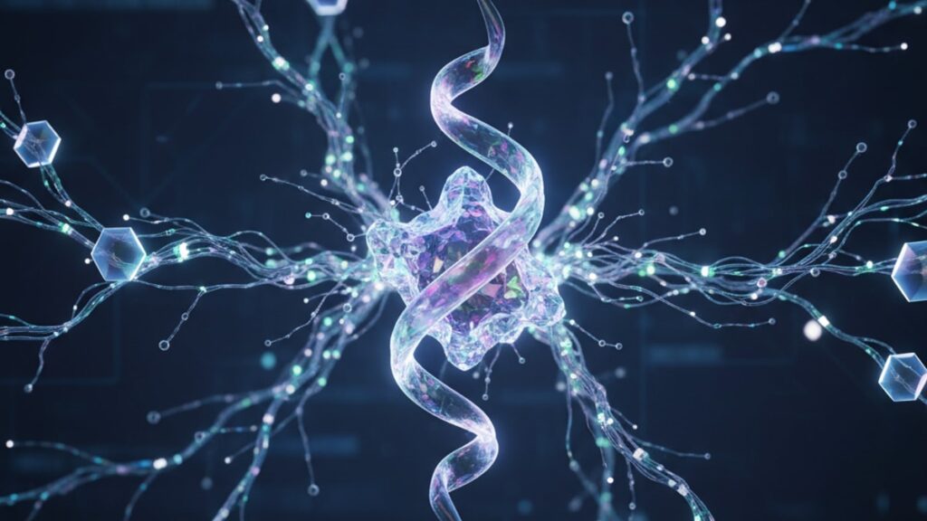
Oxytocin Oxytocin Receptor Mechanism — How It Works
Research from the University of North Carolina School of Medicine found that oxytocin receptor density in the hypothalamus increases by 300–500% during late pregnancy. A biological amplification that transforms the same circulating hormone concentration into drastically different physiological effects. The receptor mechanism isn't passive signal reception. It's active signal transformation, where the same ligand produces uterine contraction in one tissue and anxiolytic effects in another based entirely on which intracellular pathway dominates.
Our team has worked with researchers studying peptide signaling pathways across reproductive endocrinology and social neuroscience applications. The gap between understanding oxytocin as "the bonding hormone" and understanding how oxytocin receptors actually transduce that signal into cellular action is where most explanations stop. And where mechanism-based insights begin.
How does the oxytocin oxytocin receptor mechanism produce such diverse effects across different tissues?
The oxytocin receptor (OXTR) is a G-protein coupled receptor that activates three distinct intracellular signaling cascades simultaneously: Gq/11 pathway triggering calcium release and smooth muscle contraction, Gi/o pathway modulating cAMP levels and neuronal excitability, and β-arrestin pathway regulating receptor internalization and MAP kinase activation. Each tissue type expresses different ratios of these downstream effectors, which is why the same hormone concentration produces uterine contraction in myometrium, milk ejection in mammary tissue, and anxiolytic effects in amygdala neurons.
The critical insight most basic explanations miss: oxytocin binding to its receptor doesn't create one universal "oxytocin effect." The receptor's conformational change upon ligand binding exposes multiple intracellular domains that recruit different signaling proteins depending on what's available in that specific cell type. In uterine smooth muscle, Gq dominance drives calcium mobilization and contraction. In limbic neurons, Gi modulation of potassium channels reduces neuronal firing and anxiety. In lactotrophs, both pathways coordinate to produce pulsatile prolactin release. This article covers the three major signaling pathways activated by OXTR, how tissue-specific expression patterns determine which effects dominate, and what happens when receptor density or downstream signaling components are disrupted.
The Three Signaling Pathways Activated by Oxytocin Receptor Binding
Oxytocin receptor activation isn't a binary on-off switch. It's a simultaneous activation of three parallel intracellular signaling cascades, each coupled to different G-protein subunits. The Gq/11 pathway is the most studied and clinically relevant for reproductive physiology. When oxytocin binds OXTR in uterine myometrium, the receptor undergoes conformational change that activates Gq proteins, which in turn stimulate phospholipase C (PLC). PLC cleaves phosphatidylinositol 4,5-bisphosphate (PIP2) into two second messengers: inositol 1,4,5-trisphosphate (IP3) and diacylglycerol (DAG). IP3 binds to receptors on the endoplasmic reticulum, triggering massive calcium release into the cytoplasm. Elevated intracellular calcium binds calmodulin, activating myosin light-chain kinase (MLCK), which phosphorylates myosin and initiates smooth muscle contraction. This entire cascade. From oxytocin binding to forceful uterine contraction. Completes in under 30 seconds at physiological receptor density.
The Gi/o pathway operates in parallel but produces opposing effects in many tissues. OXTR coupling to Gi proteins inhibits adenylyl cyclase, reducing cyclic AMP (cAMP) production and decreasing protein kinase A (PKA) activity. In hypothalamic neurons, this pathway modulates potassium channel conductance, hyperpolarizing the membrane and reducing neuronal excitability. The molecular basis for oxytocin's anxiolytic effects. The β-arrestin pathway, discovered more recently, doesn't directly produce second messengers but instead regulates receptor internalization and activates mitogen-activated protein kinase (MAPK) cascades. β-arrestin binding to activated OXTR triggers clathrin-mediated endocytosis, removing receptors from the membrane surface and terminating signaling. A desensitization mechanism that prevents overstimulation during sustained oxytocin exposure. In central neurons, β-arrestin-mediated MAPK activation influences gene transcription and long-term synaptic plasticity, linking acute oxytocin signaling to persistent changes in social behavior circuits.
Tissue-Specific Receptor Expression Determines Physiological Outcomes
The same oxytocin molecule circulating in plasma produces radically different effects depending on where its receptors are located and what signaling machinery those cells express. In uterine smooth muscle, OXTR density increases 200-fold during late pregnancy under the influence of estrogen, transforming the uterus from oxytocin-insensitive to exquisitely sensitive. Myometrial cells express high levels of Gq-coupled effectors. Particularly the IP3 receptor subtype that produces rapid calcium transients. Which is why oxytocin administration during labor produces powerful, coordinated contractions. Mammary myoepithelial cells surrounding alveoli express similar receptor density and Gq dominance, producing milk ejection through smooth muscle contraction around the milk-producing acini. This explains why the same hormone triggers both uterine contractions and breastfeeding. Mechanistically identical processes in different anatomical locations.
Central nervous system OXTR distribution follows a distinct pattern concentrated in limbic structures: amygdala, nucleus accumbens, bed nucleus of the stria terminalis, and ventromedial hypothalamus. These regions regulate social recognition, anxiety modulation, and pair-bonding behaviors. Neurons in these areas express different G-protein coupling profiles. Gi/o pathways dominate over Gq, producing net inhibitory effects that reduce amygdala activity during social interaction. A 2021 study published in Nature Neuroscience used optogenetic activation of OXTR-expressing neurons in the medial amygdala and found that selective Gi pathway activation (while blocking Gq signaling pharmacologically) was sufficient to produce social approach behavior in previously isolated mice. The Gq pathway wasn't required for behavioral effects. Pituitary lactotrophs express yet another receptor configuration, where both Gq and Gi pathways coordinate to produce pulsatile prolactin secretion that maintains milk production between nursing bouts.
Receptor Desensitization and the Role of Sustained Exposure
Oxytocin receptor signaling doesn't maintain constant output during sustained hormone exposure. The system actively desensitizes through multiple mechanisms that reduce cellular response over time. The β-arrestin pathway drives receptor internalization within 5–10 minutes of continuous oxytocin exposure. β-arrestin proteins bind to phosphorylated serine residues on the OXTR intracellular tail (phosphorylated by G-protein receptor kinases following receptor activation), sterically blocking further G-protein coupling while simultaneously recruiting clathrin coat proteins that pull the receptor into endocytic vesicles. Once internalized, receptors can be recycled back to the membrane or targeted for lysosomal degradation depending on the duration and intensity of agonist exposure. In myometrial tissue during prolonged oxytocin infusion for labor augmentation, this mechanism reduces contraction amplitude by 30–40% after 4–6 hours despite constant hormone concentration. A clinical phenomenon called tachyphylaxis.
Downstream signaling components also undergo adaptation. Prolonged Gq activation depletes endoplasmic reticulum calcium stores, reducing the magnitude of subsequent IP3-triggered calcium transients. Phosphodiesterases upregulate in response to sustained cAMP suppression via the Gi pathway, creating compensatory mechanisms that restore baseline signaling even with the receptor still occupied. At the gene transcription level, chronic oxytocin exposure in central neurons triggers expression of regulator of G-protein signaling (RGS) proteins that accelerate GTPase activity on G-proteins, shortening the duration of each signaling event. Research from Stanford University demonstrated that mice lacking functional β-arrestin showed persistent social behavior responses to oxytocin that didn't habituate. They continued approaching novel conspecifics even after 60 minutes of continuous central oxytocin infusion, while wild-type mice showed habituation after 15–20 minutes. The desensitization machinery isn't a system failure. It's adaptive regulation preventing overstimulation and allowing the system to detect changes in oxytocin concentration rather than absolute levels.
Oxytocin Receptor Mechanism Comparison
| Signaling Pathway | G-Protein Subtype | Primary Second Messenger | Tissue Where Dominant | Physiological Effect | Activation Timescale |
|---|---|---|---|---|---|
| Gq/11 pathway | Gq, G11 | IP3 → intracellular Ca²⁺ release | Uterine myometrium, mammary myoepithelium | Smooth muscle contraction (labor, milk ejection) | 10–30 seconds |
| Gi/o pathway | Gi, Go | Reduced cAMP, altered K⁺ conductance | Limbic neurons (amygdala, NAc, BNST) | Neuronal inhibition, anxiolysis, social approach | 30–90 seconds |
| β-arrestin pathway | (Not G-protein mediated) | MAPK activation, receptor internalization | All OXTR-expressing tissues | Receptor desensitization, gene transcription changes | 5–15 minutes |
Key Takeaways
- Oxytocin receptor activation simultaneously triggers three distinct intracellular pathways (Gq, Gi, β-arrestin), each producing different cellular outcomes depending on tissue-specific expression of downstream effectors.
- The Gq/11 pathway generates IP3 and releases intracellular calcium within 10–30 seconds, producing the smooth muscle contractions responsible for labor and milk ejection.
- Central nervous system OXTR expression is concentrated in limbic structures where Gi-mediated neuronal inhibition reduces amygdala activity and produces anxiolytic and prosocial behavioral effects.
- Receptor density in uterine tissue increases 200-fold during late pregnancy under estrogen influence, amplifying cellular response to the same circulating oxytocin concentration.
- β-arrestin-mediated receptor internalization begins within 5–10 minutes of sustained oxytocin exposure, creating tachyphylaxis that reduces contraction amplitude during prolonged clinical infusions by 30–40%.
- Tissue-specific G-protein coupling ratios determine whether oxytocin produces contraction (Gq-dominant), inhibition (Gi-dominant), or coordinated pulsatile secretion (mixed coupling) in target cells.
What If: Oxytocin Receptor Mechanism Scenarios
What If Oxytocin Receptor Density Is Abnormally Low at Term?
Administer synthetic oxytocin (Pitocin) at standard doses and expect reduced contractile response requiring dose escalation. Insufficient upregulation of OXTR expression in late pregnancy. Often due to inadequate estrogen priming or genetic polymorphisms in the OXTR promoter region. Means fewer binding sites per cell despite normal circulating hormone levels. The Gq signaling cascade per receptor remains intact, but total calcium mobilization across the tissue is inadequate to generate coordinated forceful contractions. Clinically this presents as prolonged labor or failure to progress despite adequate oxytocin administration, and often requires dose titration to 2–3 times standard protocols or consideration of cesarean delivery when even maximum oxytocin fails to generate effective contractions.
What If Both Gq and Gi Pathways Are Blocked Simultaneously?
The oxytocin signal is effectively silenced at the cellular level despite receptor occupancy. Pharmacological or genetic blockade of both primary G-protein coupling pathways leaves only β-arrestin signaling intact, which cannot produce acute physiological effects like contraction or neuronal modulation. Animal studies using dual Gq/Gi inhibitors showed that oxytocin infusion produced normal receptor binding but zero contractile response in myometrium and no behavioral effects in social approach paradigms. The β-arrestin pathway alone can internalize receptors and activate MAPK but cannot generate the immediate functional responses that define oxytocin's physiological roles. This scenario demonstrates that the receptor's effects are entirely dependent on G-protein coupling, not merely on hormone binding.
What If Continuous Oxytocin Exposure Depletes Calcium Stores?
Expect progressive reduction in contraction amplitude despite maintained receptor activation. The desensitization phenomenon seen clinically during prolonged labor augmentation. The endoplasmic reticulum cannot replenish calcium stores as fast as IP3-triggered release depletes them during sustained Gq pathway activation. After 4–6 hours of continuous oxytocin infusion at constant dose, myometrial cells show normal receptor surface expression and Gq activation but blunted calcium transients because the releasable pool is partially depleted. Compensatory mechanisms upregulate plasma membrane calcium channels to restore some contractility, but peak contraction force remains 30–40% below initial response. Clinical management often involves temporarily discontinuing oxytocin for 30–60 minutes to allow store refilling, then resuming at lower dose with better response than continuing high-dose infusion through depleted reserves.
The Mechanistic Truth About Oxytocin Receptor Signaling
Here's the mechanistic truth: oxytocin's diverse effects aren't magic. They're the result of one receptor activating three parallel intracellular pathways whose relative dominance depends entirely on what signaling proteins the target cell expresses. The same hormone binding event produces uterine contraction in one cell, neuronal inhibition in another, and receptor internalization in both. Not because oxytocin "does different things," but because the cellular context determines which of its activated pathways produce observable effects. The receptor itself is a signal transducer, not a signal specifier. Understanding this distinction is what separates surface-level "oxytocin causes bonding" explanations from mechanism-based insight into why disrupting β-arrestin prevents desensitization, why estrogen priming is required for labor induction to work, and why central versus peripheral oxytocin administration produce entirely different behavioral outcomes despite using the same receptor protein.
The complexity isn't in the hormone. It's in the 50+ proteins downstream of receptor activation that determine whether that signal becomes a contraction, a behavior change, or a gene transcription event. Single-nucleototide polymorphisms in the OXTR gene that alter receptor expression or coupling efficiency have been associated with variations in social behavior, autism spectrum disorder traits, and labor dystocia. Not because the variants change what oxytocin does, but because they change how much signal gets transduced per unit of hormone present. Researchers working with Real Peptides high-purity synthetic oxytocin can control ligand concentration and purity to eliminate one variable while studying how receptor density, G-protein expression, or arrestin function modulate downstream outcomes. That level of experimental control. Knowing the ligand isn't the problem. Is how mechanism gets dissected from correlation in signaling research.
Receptor Polymorphisms and Individual Variation in Oxytocin Sensitivity
Genetic variation in the OXTR gene doesn't change the receptor's structure or signaling capability in most cases. It changes how much receptor gets expressed and where. The most studied single-nucleotide polymorphism (SNP), rs53576, lies in the third intron of OXTR and influences gene transcription efficiency. Individuals homozygous for the G allele show higher OXTR mRNA expression in limbic brain regions and report greater social sensitivity, empathy, and stress resilience in behavioral studies compared to A-allele carriers. A 2019 meta-analysis of 17,000 subjects published in Molecular Psychiatry confirmed that rs53576 A-allele carriers show reduced amygdala OXTR density and higher baseline anxiety scores, despite identical circulating oxytocin levels. The difference is how much signal gets transduced, not how much hormone is present.
Other polymorphisms affect receptor function rather than expression. The rs2254298 SNP alters a regulatory region that influences receptor desensitization rate. Individuals with the A-allele variant show faster β-arrestin recruitment and more rapid tachyphylaxis during sustained oxytocin exposure. This has clinical relevance in obstetrics: women carrying this variant require higher oxytocin doses for labor augmentation and show earlier plateau in contraction response during continuous infusion. Pharmacogenomic studies are beginning to incorporate OXTR genotyping into labor management protocols, though this remains investigational. The broader insight is that "oxytocin resistance" in any physiological context. Whether behavioral, reproductive, or lactational. May reflect receptor genetics rather than hormone deficiency. Measuring serum oxytocin without knowing receptor genotype and expression provides incomplete information about signaling capacity.
Oxytocin receptor mechanisms represent the intersection of endocrinology, neuroscience, and cellular signaling. Three intracellular pathways, infinite tissue-specific combinations, and one hormone whose effects are determined entirely by the machinery waiting to receive its signal. The receptor doesn't create effects. It transduces binding energy into pathway activation, and the cell does the rest. Understanding this distinction is what transforms oxytocin from a folk-biology "bonding molecule" into a mechanistically tractable signaling system where interventions can be designed with pathway specificity rather than hoping one ligand produces one outcome. For researchers working with high-purity peptides like those from Real Peptides, controlling the ligand variable means every observed effect can be attributed to receptor and downstream pathway function. The starting point for mechanism-based discoveries in reproductive biology, social neuroscience, and beyond.
Frequently Asked Questions
How does oxytocin binding to its receptor produce different effects in different tissues?▼
Oxytocin receptor activation triggers three parallel intracellular signaling pathways simultaneously — Gq/11, Gi/o, and β-arrestin — but different tissues express different ratios of the downstream effector proteins these pathways activate. In uterine smooth muscle, high expression of Gq-coupled phospholipase C and IP3 receptors means the Gq pathway dominates, producing calcium release and contraction. In limbic neurons, higher Gi expression and potassium channel density mean the Gi pathway dominates, producing neuronal inhibition and anxiolysis. The receptor mechanism is identical — the cellular context determines which activated pathway produces observable effects.
What is the role of β-arrestin in oxytocin receptor signaling?▼
β-arrestin binds to activated oxytocin receptors and serves two functions: it terminates G-protein signaling by sterically blocking further Gq or Gi coupling, and it triggers receptor internalization through clathrin-mediated endocytosis. This desensitization mechanism prevents overstimulation during sustained oxytocin exposure — in clinical labor augmentation, β-arrestin activity is why contraction amplitude decreases 30–40% after 4–6 hours of continuous infusion despite constant hormone concentration. β-arrestin also activates MAPK pathways that influence long-term gene transcription, linking acute signaling to persistent cellular changes.
Why does prolonged oxytocin infusion cause reduced contraction strength?▼
Sustained oxytocin receptor activation triggers multiple desensitization mechanisms: β-arrestin internalizes receptors from the cell surface within 5–10 minutes, reducing available binding sites; repeated IP3-triggered calcium release depletes endoplasmic reticulum stores faster than they refill; and downstream signaling components upregulate negative regulators like RGS proteins that shorten G-protein activation duration. After 4–6 hours of continuous infusion, myometrial cells show 30–40% reduction in contraction amplitude despite maintained hormone concentration — a phenomenon called tachyphylaxis that often requires temporarily stopping oxytocin to allow receptor recycling and calcium store replenishment before resuming at lower dose.
Can genetic differences affect oxytocin receptor function?▼
Yes — single-nucleotide polymorphisms in the OXTR gene alter receptor expression levels and desensitization rates without changing the receptor protein structure. The rs53576 SNP influences transcription efficiency, with G-allele homozygotes showing higher limbic OXTR density and greater social sensitivity than A-allele carriers. The rs2254298 variant affects β-arrestin recruitment speed, causing faster tachyphylaxis during sustained exposure — women with this variant require higher oxytocin doses during labor augmentation. These polymorphisms explain why individuals with identical circulating oxytocin levels show different behavioral and physiological responses; the difference is signal transduction capacity, not hormone concentration.
What is the difference between central and peripheral oxytocin receptor activation?▼
Peripheral oxytocin receptors (uterus, breast) couple predominantly to Gq pathways producing smooth muscle contraction, while central receptors (amygdala, hypothalamus) couple predominantly to Gi pathways producing neuronal inhibition and behavioral modulation. Peripherally administered oxytocin does not cross the blood-brain barrier in significant amounts, so intravenous Pitocin during labor produces uterine contractions without direct central nervous system effects. Central oxytocin release — triggered by sensory stimuli like nipple stimulation or social interaction — activates both peripheral and central receptors simultaneously through distinct anatomical pathways (magnocellular neurons projecting to posterior pituitary for peripheral release, parvocellular neurons projecting within the brain for central effects).
How quickly does oxytocin receptor signaling produce cellular effects?▼
The Gq/11 pathway produces measurable calcium transients within 10–30 seconds of receptor activation — this is the timescale of uterine contraction initiation and milk ejection following oxytocin exposure. The Gi/o pathway modulates neuronal excitability on a 30–90 second timescale through changes in potassium channel conductance and cAMP levels. β-arrestin-mediated receptor internalization begins 5–10 minutes after sustained activation. MAPK activation and gene transcription changes occur over hours to days. The immediate physiological effects of oxytocin — contraction, secretion, behavior — are mediated by the fast G-protein pathways, while long-term adaptations involve the slower β-arrestin-MAPK pathway.
What happens if Gq signaling is blocked but Gi signaling remains intact?▼
Oxytocin would lose its contractile effects in smooth muscle (no labor contractions, no milk ejection) but retain its central nervous system effects on behavior and anxiety. Animal studies using selective Gq pathway inhibitors showed that oxytocin still produced social approach behavior and amygdala inhibition through intact Gi signaling, but uterine tissue showed zero contractile response despite normal receptor binding. This demonstrates that behavioral and reproductive effects of oxytocin are mechanistically separable — they use the same receptor but different downstream pathways, which is why drugs targeting one pathway can selectively enhance or block specific oxytocin effects without affecting others.
Why does estrogen increase oxytocin receptor density before labor?▼
Estrogen directly activates transcription of the OXTR gene through estrogen response elements in the promoter region, increasing receptor mRNA production and protein expression by 200-fold in uterine tissue during late pregnancy. This receptor upregulation transforms the uterus from oxytocin-insensitive early in pregnancy (when high progesterone suppresses OXTR expression) to exquisitely oxytocin-sensitive at term. The rise in estrogen relative to progesterone in the final weeks before labor is the primary trigger for this receptor amplification — without adequate estrogen priming, oxytocin administration produces weak contractions regardless of dose because there are insufficient receptors to transduce the signal into calcium release and contraction.
Can oxytocin receptor signaling be selectively enhanced in specific tissues?▼
Theoretically yes, through pathway-selective agonists or tissue-selective delivery, though no clinically approved drugs currently do this. Research compounds exist that preferentially activate Gq over Gi pathways (potentially enhancing contractile effects while minimizing central effects) or that stabilize β-arrestin interactions while blocking G-protein coupling (producing MAPK activation without acute signaling). Tissue selectivity could also be achieved through localized delivery — intranasal oxytocin reaches central receptors with minimal peripheral exposure, while intravenous administration targets peripheral receptors. The mechanistic understanding that one receptor activates multiple pathways opens therapeutic possibilities for drugs that enhance or block specific oxytocin effects without affecting others, though this remains investigational.
What role does receptor recycling play in sustained oxytocin response?▼
After β-arrestin-mediated internalization, oxytocin receptors can be recycled back to the plasma membrane or targeted for lysosomal degradation depending on the duration and intensity of agonist exposure. Short-term oxytocin pulses (like physiological release during nursing) favor receptor recycling, allowing rapid resensitization within 20–30 minutes as internalized receptors return to the membrane surface. Prolonged high-dose exposure (like clinical labor augmentation) shifts the balance toward degradation, progressively reducing total receptor number and causing the tachyphylaxis clinicians observe after hours of continuous infusion. This is why intermittent dosing or temporary discontinuation can restore responsiveness — it allows depleted receptor pools to be replenished through new protein synthesis and recycling of internalized receptors rather than fighting against ongoing degradation.


