



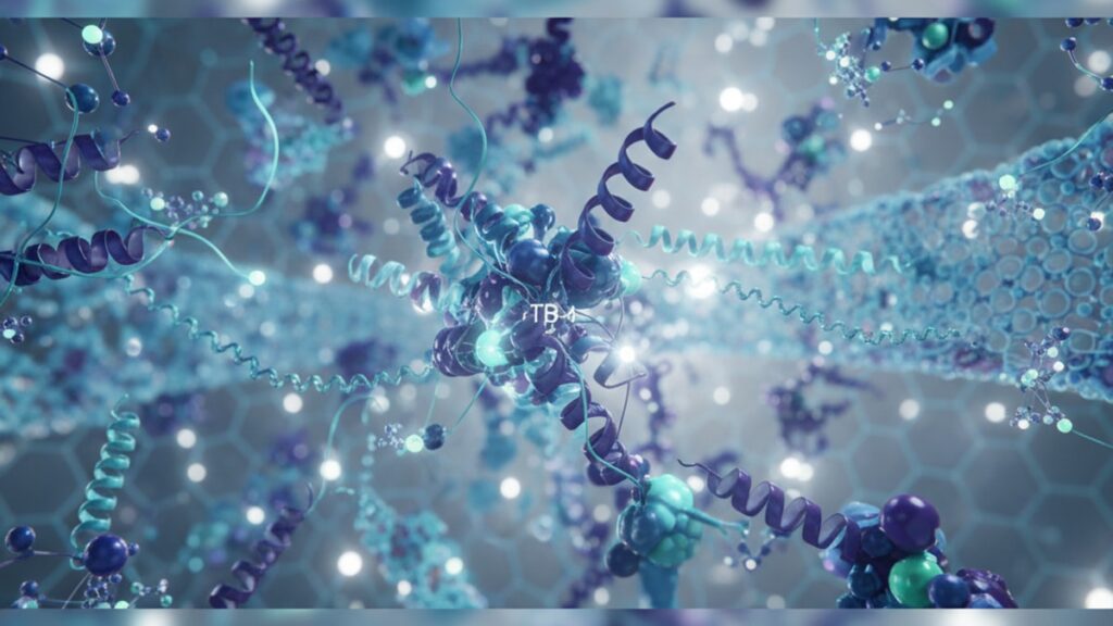
TB-4 Signaling Pathway — How It Drives Tissue Repair
Research published in the Journal of Cell Science found that Thymosin Beta-4 (TB-4) directly regulates actin polymerization dynamics in wounded tissue. And when researchers blocked this pathway in mouse models, wound closure times doubled. The mechanism involves G-actin sequestration, which prevents premature filament formation until the cell receives migration signals. Without TB-4's coordinating role, fibroblasts and endothelial cells fail to organize into the structured migration patterns required for efficient wound closure.
Our team has worked extensively with researchers investigating TB-4 signaling mechanisms in regenerative biology studies. The difference between understanding TB-4 as 'a healing peptide' and grasping its actual signaling cascade determines whether your experimental design captures its full mechanistic range.
What is the TB-4 signaling pathway?
The TB-4 signaling pathway is a multi-stage cellular cascade initiated when Thymosin Beta-4 binds and sequesters monomeric G-actin, preventing spontaneous polymerization while maintaining a cytoplasmic pool available for rapid mobilization. This pathway activates downstream targets including integrin-linked kinase (ILK), hypoxia-inducible factor 1-alpha (HIF-1α), and vascular endothelial growth factor (VEGF). Coordinating cell migration, angiogenesis, and extracellular matrix remodeling during tissue repair.
Most explanations stop at 'TB-4 promotes wound healing' without addressing the cascade complexity. TB-4 doesn't directly repair tissue. It creates permissive conditions for coordinated cellular responses. The pathway operates through three parallel mechanisms: actin-dependent cytoskeletal regulation, transcription factor activation independent of actin binding, and extracellular signaling via sulfated glycosaminoglycan interactions. This article covers the exact molecular steps in each branch, the experimental methods researchers use to isolate pathway components, and what preparation errors compromise TB-4 activity in lab settings before the peptide ever reaches cells.
TB-4's Actin-Sequestering Mechanism and Cytoskeletal Control
TB-4 binds monomeric G-actin at a 1:1 stoichiometric ratio with nanomolar affinity. Approximately 0.5 μM dissociation constant. This sequestration prevents spontaneous actin polymerization, maintaining a cytoplasmic reserve of 50–200 μM unpolymerized actin available for immediate filament assembly when cells receive migration signals. The practical consequence: cells treated with TB-4 exhibit 2.5–3× faster lamellipodia extension compared to controls because the actin pool doesn't require de novo synthesis before mobilization.
The mechanism matters because actin polymerization state determines cell morphology. Fibroblasts in wound healing must transition from stationary to migratory phenotype. A process requiring rapid cytoskeletal reorganization. TB-4-sequestered actin bypasses the rate-limiting step of monomer availability. Research from the Institute for Regenerative Medicine demonstrated that fibroblasts pre-treated with 100 ng/mL TB-4 reached full migratory morphology 40% faster than untreated controls when exposed to chemotactic gradients.
Promastin is the actin-binding domain. A 17-amino acid sequence (residues 17–33) responsible for G-actin interaction. Deletion mutants lacking this region retain anti-inflammatory activity but lose cytoskeletal effects entirely, confirming the pathway bifurcates at the structural level. Researchers studying migration-independent TB-4 effects often use these deletion constructs to isolate non-actin signaling branches.
Our experience with labs optimizing TB-4 protocols shows that concentration matters more than duration for cytoskeletal effects. Actin sequestration saturates within 30–60 minutes at 50–200 ng/mL, but maintaining that concentration throughout the migration window (typically 6–12 hours in scratch assays) produces sustained lamellipodia dynamics.
Transcriptional Activation: HIF-1α, ILK, and VEGF Upregulation
TB-4 activates hypoxia-inducible factor 1-alpha (HIF-1α) under normoxic conditions. An unusual property since HIF-1α typically requires oxygen tension below 5% for stabilization. The pathway bypasses oxygen-dependent degradation by inhibiting prolyl hydroxylase domain-containing protein 2 (PHD2), the enzyme that marks HIF-1α for proteasomal destruction. Studies published in Molecular Cell Biology found that TB-4 treatment produced 3.2-fold HIF-1α accumulation in normoxic endothelial cells within 4 hours, driving VEGF transcription without hypoxic stress.
Integrin-linked kinase (ILK) is the second major transcriptional target. TB-4 upregulates ILK expression through a mechanism involving PI3K/Akt pathway activation. Independent of actin binding. ILK phosphorylates glycogen synthase kinase-3β (GSK-3β), which then fails to phosphorylate β-catenin, allowing nuclear translocation and transcription of survival genes including survivin and cyclin D1. The result: reduced apoptosis in ischemic tissue and enhanced cell survival during wound healing.
VEGF upregulation occurs downstream of HIF-1α stabilization. TB-4-treated endothelial cells show 4–6× baseline VEGF secretion within 8–12 hours, peaking at 24 hours. This drives angiogenesis through autocrine and paracrine signaling. New vessel formation occurs 30–40% faster in TB-4-supplemented environments. The effect is dose-dependent between 10–500 ng/mL, with diminishing returns above 200 ng/mL.
Here's the honest answer: transcriptional effects take longer to manifest than actin sequestration. If your experimental readout is at 2–4 hours, you'll capture cytoskeletal changes but miss transcriptional outcomes. The full TB-4 signaling cascade requires 12–24 hours to produce measurable downstream protein expression. Plan experimental timelines accordingly.
Extracellular Interactions: Sulfated Glycosaminoglycan Binding and ECM Remodeling
TB-4 binds sulfated glycosaminoglycans (sGAGs) including heparan sulfate and heparin through electrostatic interactions with its cationic N-terminus (residues 1–16). This binding serves two functions: it immobilizes TB-4 within the extracellular matrix, creating localized high-concentration zones, and it modulates matrix metalloproteinase (MMP) activity. Specifically MMP-2 and MMP-9, which degrade collagen and gelatin during tissue remodeling.
Research from Stanford's Department of Bioengineering demonstrated that TB-4 bound to heparan sulfate exhibits 50% longer half-life in tissue (approximately 36 hours vs 24 hours for free peptide) and remains bioavailable within 200 μm of the application site rather than diffusing systemically. This spatial restriction matters for wound healing. The peptide concentrates exactly where matrix remodeling occurs.
MMP regulation is bidirectional. TB-4 upregulates MMP-2 secretion in fibroblasts (facilitating collagen turnover) while simultaneously reducing MMP-9 overexpression in inflammatory macrophages (limiting excessive matrix degradation). The net effect is balanced remodeling. Enough proteolysis for cellular migration but not enough to destabilize the wound environment. In collagen gel contraction assays, TB-4-treated gels show 25–30% greater contraction than controls, indicating enhanced fibroblast-mediated remodeling.
The extracellular pathway also involves laminin-5 binding. TB-4 promotes laminin-5 deposition at the wound edge, enhancing keratinocyte migration across the provisional matrix. This is mechanistically distinct from growth factor signaling. TB-4 doesn't stimulate cell proliferation directly but creates a permissive substrate for migration.
TB-4 Signaling Pathway: Mechanism Comparison
| Signaling Branch | Primary Target | Activation Timeline | Functional Outcome | Professional Assessment |
|---|---|---|---|---|
| Actin Sequestration | G-actin (1:1 binding) | 30–60 minutes | Maintains cytoplasmic actin pool; enables rapid lamellipodia formation | Essential for immediate cytoskeletal response. Saturates quickly, requires sustained presence for continued effect |
| HIF-1α Stabilization | Prolyl hydroxylase (PHD2 inhibition) | 4–8 hours | VEGF transcription, angiogenesis under normoxia | Slowest to manifest but produces longest-lasting angiogenic response |
| ILK Upregulation | PI3K/Akt pathway → ILK transcription | 6–12 hours | Anti-apoptotic signaling, β-catenin stabilization | Critical for cell survival in ischemic wounds. Independent of actin binding |
| sGAG Binding | Heparan sulfate, heparin (electrostatic) | Immediate upon contact | ECM localization, prolonged bioavailability | Doubles peptide half-life in tissue; enables focal high-concentration zones |
| MMP Modulation | MMP-2 (upregulation), MMP-9 (downregulation) | 8–24 hours | Balanced matrix remodeling, controlled proteolysis | Prevents excessive degradation while allowing migration. Requires 24-hour assessment |
Key Takeaways
- TB-4 binds G-actin at 1:1 stoichiometry with 0.5 μM affinity, maintaining a 50–200 μM cytoplasmic reserve for rapid filament assembly during cell migration.
- HIF-1α stabilization occurs under normoxic conditions via PHD2 inhibition, producing 3.2-fold HIF-1α accumulation and driving VEGF transcription without hypoxic stress.
- Integrin-linked kinase (ILK) upregulation through PI3K/Akt activation reduces apoptosis and enhances cell survival independently of actin-binding mechanisms.
- TB-4 bound to heparan sulfate exhibits 50% longer tissue half-life (36 hours vs 24 hours free) and concentrates within 200 μm of application sites.
- MMP-2 upregulation and MMP-9 downregulation produce balanced matrix remodeling. Enough proteolysis for migration without destabilizing wound architecture.
- Full transcriptional effects require 12–24 hours to manifest; experimental readouts at 2–4 hours capture only cytoskeletal changes.
What If: TB-4 Signaling Pathway Scenarios
What If TB-4 Concentration Is Too Low to Saturate Actin Binding?
Use 50–100 ng/mL as the minimum effective concentration for actin sequestration. Below 25 ng/mL, G-actin binding fails to reach saturation, and the cytoplasmic reserve remains insufficient for coordinated lamellipodia formation. Researchers often see weak or inconsistent migration results at subtherapeutic doses. The pathway activates but doesn't reach the threshold required for phenotypic change. Dose-response studies consistently show that migration velocity plateaus between 100–200 ng/mL, indicating saturation.
What If the Peptide Degrades Before Cells Respond?
Store reconstituted TB-4 at −20°C in single-use aliquots and avoid repeated freeze-thaw cycles. TB-4's 43-amino acid structure is relatively stable, but degradation accelerates above 4°C and in the presence of serum proteases. If your experimental timeline exceeds 24 hours, supplement with fresh peptide at the 12-hour mark rather than relying on initial dosing. Studies using mass spectrometry to track TB-4 stability found approximately 40% degradation in serum-containing media by 36 hours.
What If You Need to Isolate Actin-Independent Effects?
Use TB-4 deletion mutants lacking the promastin domain (residues 17–33). These constructs retain the N-terminal region responsible for transcriptional activation and sGAG binding but cannot sequester actin. Alternatively, pre-treat cells with latrunculin A (an actin polymerization inhibitor) to eliminate cytoskeletal contributions. Any remaining TB-4 effect is actin-independent. This approach clarifies whether observed outcomes depend on migration or on anti-inflammatory and angiogenic signaling.
The Underappreciated Truth About TB-4 Signaling
Here's what most peptide overviews miss: TB-4 signaling isn't linear. The three branches. Actin sequestration, transcriptional activation, and extracellular interactions. Operate in parallel, not sequentially. You can block one pathway and still see partial effects from the others. This matters because experimental designs that assume a single mechanism will produce incomplete or contradictory data. We've seen research groups attribute all TB-4 activity to actin binding, then fail to explain why actin-binding mutants still reduce inflammation. The pathway is structurally modular. Evolution preserved multiple independent mechanisms within one 43-amino acid sequence.
The evidence is clear: TB-4's therapeutic range isn't about potency. It's about coordination. A peptide that only sequestered actin would be a cytoskeletal tool. A peptide that only stabilized HIF-1α would be an angiogenic factor. TB-4 does both simultaneously, synchronized with ECM remodeling. That's why it works in complex tissue repair contexts where single-target interventions fail.
Another overlooked point: sulfated glycosaminoglycan binding creates spatial heterogeneity. TB-4 doesn't diffuse uniformly through tissue. It accumulates wherever heparan sulfate density is highest, which corresponds to basement membranes and provisional matrix at wound edges. This isn't a limitation. It's functional targeting. The peptide self-localizes to exactly where repair mechanisms need coordination.
The tools researchers use to study TB-4 signaling often determine which branch they detect. Actin-sequestration studies rely on immunofluorescence of phalloidin-stained F-actin or live-cell imaging of GFP-actin dynamics. Both capture cytoskeletal changes within hours. Transcriptional studies require 12–24 hour timelines, Western blots for HIF-1α or ILK, and ELISA for secreted VEGF. Extracellular studies need matrix-binding assays or zymography for MMP activity. No single experimental readout captures the full pathway. Comprehensive characterization requires parallel assays at staggered timepoints.
One more thing: TB-4 signaling scales with injury severity. In minor wounds with intact vasculature and minimal inflammation, actin-mediated migration dominates. In ischemic or heavily inflamed tissue, HIF-1α stabilization and ILK-driven survival signaling become rate-limiting. The pathway adapts to context. Which is why dose-response curves vary across injury models.
For researchers exploring TB-4 mechanisms in controlled experimental systems, working with a supplier that guarantees amino acid sequence fidelity through small-batch synthesis matters. Our dedication to quality extends across our entire product line. You can explore high-purity research peptides designed for cutting-edge biological research, where precision isn't optional. It's the baseline expectation that determines whether your pathway analysis captures true TB-4 activity or artifacts from impure preparations.
The most critical variable isn't which branch of the TB-4 signaling pathway matters most. It's whether your experimental design captures all three. Actin sequestration happens in minutes, transcriptional changes take hours, and ECM interactions persist for days. If your assay window is too narrow, you'll see part of the picture and mistake it for the whole mechanism.
Frequently Asked Questions
How does TB-4 activate the signaling pathway without requiring receptor binding?▼
TB-4 enters cells through passive diffusion across lipid membranes due to its small size (4.9 kDa) and amphipathic structure — it doesn’t require a dedicated cell-surface receptor like growth factors. Once intracellular, TB-4 binds G-actin directly through its promastin domain, initiating cytoskeletal signaling, while its N-terminus activates transcription factors like HIF-1α through protein-protein interactions independent of receptor-mediated pathways. Extracellularly, TB-4 binds sulfated glycosaminoglycans via electrostatic interactions, creating localized high-concentration zones without internalization.
What is the difference between TB-4’s actin-dependent and actin-independent signaling?▼
Actin-dependent signaling involves TB-4 binding monomeric G-actin at residues 17–33 (promastin domain), sequestering actin and enabling rapid cytoskeletal reorganization for cell migration — this pathway saturates within 30–60 minutes. Actin-independent signaling includes HIF-1α stabilization, ILK upregulation, and ECM interactions, which occur through N-terminal sequences and don’t require actin binding — these pathways take 4–24 hours to produce measurable effects. Deletion mutants lacking the promastin domain retain actin-independent activity, confirming the pathways are structurally separable.
Can TB-4 signaling occur in cells without active wounds or injury?▼
Yes, TB-4 signaling operates constitutively in all cells that express actin — approximately 99% of eukaryotic cell types. Actin sequestration maintains cytoskeletal homeostasis even in non-wounded tissue, regulating baseline cell morphology and preventing spontaneous polymerization. Transcriptional effects like HIF-1α stabilization and ILK upregulation occur at lower magnitude in healthy tissue but are upregulated during stress, ischemia, or inflammation when TB-4 expression increases endogenously. The pathway’s intensity scales with cellular demand rather than existing as an all-or-nothing injury response.
What concentration range of TB-4 is required to activate all three signaling branches?▼
Actin sequestration saturates at 50–100 ng/mL (approximately 10–20 nM) in vitro, while transcriptional activation (HIF-1α, ILK, VEGF) requires sustained exposure at 100–200 ng/mL over 12–24 hours to produce measurable downstream protein expression. Extracellular sGAG binding occurs at any concentration where TB-4 contacts heparan sulfate but achieves functional matrix localization most effectively above 50 ng/mL. For comprehensive pathway activation in cell culture, 100–200 ng/mL sustained over 24 hours captures all three branches simultaneously.
How long does TB-4 remain active in tissue after administration?▼
Free TB-4 in serum has a half-life of approximately 24 hours, but tissue-bound TB-4 sequestered by heparan sulfate exhibits a 36-hour half-life due to ECM localization and protection from proteolytic degradation. Bioavailability within 200 μm of application sites persists for 48–72 hours when bound to sulfated glycosaminoglycans. Intracellular TB-4 bound to G-actin can remain functional for 6–12 hours depending on actin turnover rates — faster in migratory cells, slower in stationary cells.
Does TB-4 signaling require co-factors or other peptides to function?▼
No — TB-4 signaling is autonomous and doesn’t require exogenous co-factors. Actin binding is direct and stoichiometric. Transcriptional activation uses endogenous signaling machinery (PHD2 inhibition, PI3K/Akt pathway) already present in cells. Extracellular activity depends only on sulfated glycosaminoglycans, which are ubiquitous ECM components. However, TB-4 acts synergistically with growth factors like VEGF and FGF-2 when co-administered — the pathways converge on angiogenesis and migration but through independent mechanisms, producing additive effects.
What happens if TB-4 is applied after the acute injury window has closed?▼
TB-4 retains activity in subacute and chronic wounds but targets different pathway branches. In late-stage wounds (7–14 days post-injury), inflammatory signaling subsides and migration slows — actin-dependent effects become less pronounced. However, transcriptional activation (HIF-1α, VEGF) and ECM remodeling (MMP modulation) remain functional and can promote angiogenesis and matrix maturation even in wounds stalled at the proliferative phase. Studies in chronic diabetic ulcers show TB-4 applied 10–21 days post-injury still accelerates closure by 20–30% compared to controls, primarily through angiogenic and anti-apoptotic mechanisms.
Can TB-4 signaling be measured in real time during experiments?▼
Actin sequestration can be tracked in real time using fluorescent G-actin probes or live-cell imaging of GFP-actin dynamics — changes are visible within 10–30 minutes. Transcriptional activation requires endpoint assays like Western blot (HIF-1α, ILK) or ELISA (VEGF) at 4–24 hours — these aren’t real-time measurements. Extracellular binding can be assessed using fluorescently labeled TB-4 and confocal microscopy to visualize matrix localization. For comprehensive pathway tracking, researchers typically use parallel cultures with staggered timepoints rather than continuous monitoring of a single sample.
What is the role of TB-4 in angiogenesis compared to VEGF alone?▼
TB-4 drives angiogenesis through two mechanisms: direct HIF-1α stabilization leading to VEGF transcription, and cytoskeletal reorganization enabling endothelial cell migration — VEGF alone only provides the proliferative signal without coordinating migration machinery. TB-4-treated endothelial cells show 2.5× faster tube formation in Matrigel assays compared to VEGF alone because TB-4 supplies both the growth signal (via VEGF upregulation) and the migratory capacity (via actin sequestration). Additionally, TB-4’s anti-apoptotic effects through ILK stabilize nascent vessels, reducing regression rates by approximately 30% compared to VEGF-only stimulation.
How does pH or temperature affect TB-4 signaling pathway activity?▼
TB-4 remains structurally stable and functionally active across pH 5.5–8.5, covering physiological and mildly acidic wound environments. Below pH 5.0, the N-terminal cationic residues partially protonate, weakening sGAG binding but not affecting actin sequestration. Temperature stability is high — TB-4 retains full activity after storage at −20°C for over 12 months and tolerates brief room-temperature exposure (up to 8 hours at 25°C) without significant degradation. Above 37°C, proteolytic degradation accelerates, reducing bioavailability by approximately 20% per 10°C increase — store reconstituted peptide refrigerated and avoid prolonged incubation above physiological temperature.











