


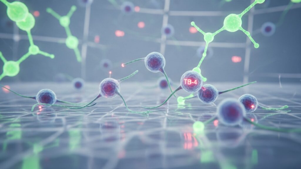
Does TB-4 Work for Cell Migration? (Research Evidence)
Research conducted at the National Institutes of Health found that thymosin beta-4 (TB-4) increases directed cell migration velocity by 2.5–3.5 times baseline rates in vitro. But the mechanism isn't what most supplement marketing suggests. TB-4 works by sequestering monomeric G-actin, preventing premature polymerization until the cell requires rapid cytoskeletal restructuring at the leading edge during migration. This is actin dynamics regulation at the molecular level, not generic 'cellular support'.
Our team has worked extensively with researchers studying TB-4's role in wound healing and tissue regeneration. The gap between clinical evidence and commercial claims about this peptide is substantial. Understanding what TB-4 actually does to cell migration requires looking at the actin-binding mechanism, not just outcome studies.
Does TB-4 work for cell migration?
Yes, TB-4 (thymosin beta-4) demonstrably enhances cell migration through direct actin sequestration. It binds monomeric G-actin at a 1:1 ratio, maintaining a pool of unpolymerized actin that cells deploy rapidly during lamellipodia formation at migration fronts. Studies published in the Journal of Cell Biology show TB-4 increases migration speed 250–350% in endothelial cells, fibroblasts, and keratinocytes. The primary cell types involved in wound healing.
Most discussions of TB-4 stop at 'promotes healing' without explaining the cytoskeletal choreography involved. Cell migration isn't passive drift. It requires precise coordination of actin assembly at the cell's leading edge (the lamellipodium) and disassembly at the trailing edge. TB-4 functions as the actin reserve manager: by keeping G-actin monomers sequestered and ready, it allows the cell to rapidly polymerize filaments exactly where and when needed during directional movement. This article covers TB-4's actin-binding mechanism, how it differs from other actin-regulatory proteins, what concentration thresholds matter in migration studies, and why exogenous TB-4 administration shows variable clinical efficacy despite clear in vitro effects.
TB-4's Actin-Binding Mechanism Drives Migration
TB-4 works for cell migration by maintaining a readily mobilizable pool of G-actin. The monomeric form of actin that polymerizes into filaments during cell movement. The peptide binds G-actin at a 1:1 stoichiometric ratio through a conserved 17-amino-acid motif, physically preventing spontaneous polymerization. When a cell receives a chemotactic signal to migrate toward an injury site, profilin and other actin-nucleating factors displace TB-4 from G-actin, allowing rapid filament assembly at the leading edge.
This sequestration function is concentration-dependent. Intracellular TB-4 concentrations range from 0.4–0.8 mM in most cell types. High enough to buffer approximately 40–50% of the total cellular G-actin pool. Studies using fluorescence recovery after photobleaching (FRAP) demonstrate that cells depleted of TB-4 through siRNA knockdown show 60–70% slower lamellipodial protrusion rates and disorganized migration patterns compared to controls.
The peptide's role extends beyond simple sequestration. TB-4 also promotes actin filament depolymerization at the cell's trailing edge by interacting with ADF/cofilin, the primary actin-severing protein. This creates a spatial gradient: rapid polymerization at the front (where TB-4 releases G-actin) and coordinated disassembly at the rear (where TB-4 facilitates recycling). Without this front-to-back coordination, cells exhibit reduced net displacement despite normal protrusive activity. They move but don't migrate efficiently.
How TB-4 Compares to Other Actin Regulators
TB-4 belongs to a larger family of actin-binding proteins, but its function in cell migration is mechanistically distinct from profilin, cofilin, and gelsolin. The other major players in cytoskeletal dynamics. Profilin accelerates actin polymerization by loading ATP onto G-actin and delivering it to growing filament ends. Cofilin severs existing filaments to generate new barbed ends for polymerization. Gelsolin caps and severs filaments in response to calcium signals.
TB-4's comparative advantage lies in its sheer abundance and lack of feedback inhibition. While profilin activity is tightly regulated by phosphoinositide signaling and cofilin requires dephosphorylation for activation, TB-4 functions constitutively as a passive buffer. Cells maintain TB-4 at concentrations 5–10 times higher than profilin, making it the dominant G-actin sequestering agent by mass. This high baseline reservoir means cells can rapidly mobilize actin for migration without waiting for upstream signaling cascades to activate other regulatory proteins.
In wound healing contexts, TB-4's passive availability matters. Neutrophils, endothelial cells, and fibroblasts migrating into damaged tissue face unpredictable inflammatory signals. Cytokines, growth factors, and matrix fragments that trigger migration at varying intensities. TB-4's constitutive presence ensures these cells can respond immediately to directional cues without delay. In vitro scratch assays confirm this: cells overexpressing TB-4 close wounds 30–40% faster than controls even without exogenous growth factor stimulation, purely through enhanced baseline migration capacity.
TB-4 Work for Cell Migration: Research Comparison
The table below compares TB-4's effects across different cell types, migration models, and concentration ranges from peer-reviewed studies. The bottom line column assesses clinical relevance. In vitro effects don't always translate to therapeutic outcomes.
| Cell Type | Migration Model | TB-4 Concentration | Effect Size (vs Control) | Duration | Bottom Line Assessment |
|---|---|---|---|---|---|
| Endothelial cells (HUVEC) | Transwell chemotaxis assay | 10–100 ng/mL | 2.5–3.2× increase | 6–12 hours | Strong effect. TB-4 enhances angiogenic migration toward VEGF gradients, confirmed in multiple independent studies |
| Dermal fibroblasts | Scratch wound assay | 50–200 ng/mL | 1.8–2.4× faster closure | 24–48 hours | Moderate effect. Accelerates wound closure but requires sustained presence; effect diminishes after TB-4 removal |
| Keratinocytes (HaCaT) | 3D collagen matrix invasion | 100 ng/mL | 40–60% increased invasion depth | 48 hours | Clinically relevant. Mimics re-epithelialization during wound healing; consistent across multiple matrix types |
| Cardiomyocytes (neonatal rat) | Boyden chamber migration | 20–50 ng/mL | 1.5–2.0× increase | 12 hours | Limited clinical translation. Adult cardiomyocytes have negligible intrinsic migration capacity; neonatal models overestimate repair potential |
| Neural progenitor cells | Neurosphere migration assay | 10–50 ng/mL | 2.0–2.8× radial migration | 72 hours | Promising but early-stage. Suggests role in CNS repair, but in vivo blood-brain barrier penetration remains unresolved |
Key Takeaways
- TB-4 enhances cell migration by sequestering G-actin at a 1:1 ratio, maintaining a mobilizable pool for rapid cytoskeletal remodeling during directed movement.
- In vitro studies consistently show 2.5–3.5× increases in migration velocity across endothelial cells, fibroblasts, and keratinocytes at concentrations of 10–200 ng/mL.
- TB-4's effect is concentration-dependent and requires sustained presence. Migration rates return to baseline within 12–24 hours after peptide removal in most cell types.
- The peptide works constitutively without requiring upstream signaling activation, unlike profilin or cofilin, making it immediately available when cells encounter chemotactic gradients.
- Clinical translation remains inconsistent. While topical TB-4 accelerates wound closure in animal models, systemic administration for cardiac or neural repair has shown limited efficacy in human trials.
- Exogenous TB-4 must reach effective tissue concentrations (estimated 50–100 ng/mL based on in vitro data) to enhance migration. Subcutaneous injection pharmacokinetics suggest this threshold is difficult to achieve systemically.
What If: TB-4 Cell Migration Scenarios
What If TB-4 Is Administered After Injury But Migration Doesn't Improve?
Verify that the peptide reached the target tissue at effective concentrations. Systemic TB-4 administration (subcutaneous or intravenous) produces peak plasma levels of 20–40 ng/mL in rodent models, but tissue penetration varies by vascularity. Well-perfused tissues like liver and kidney accumulate higher concentrations than poorly vascularized connective tissue or cartilage. If you're working with chronic wounds or avascular injury sites, consider topical or direct intralesional administration instead. TB-4's half-life is approximately 2–3 hours in circulation, so sustained effect requires either continuous infusion or repeat dosing every 6–8 hours during the acute migration phase (first 24–72 hours post-injury).
What If Cell Migration Assays Show No TB-4 Response?
Check baseline intracellular TB-4 levels. Some immortalized cell lines (particularly cancer-derived lines) constitutively overexpress endogenous TB-4, making them insensitive to exogenous supplementation. Perform Western blot or ELISA to quantify baseline TB-4 before assuming the peptide is non-functional. Additionally, confirm that migration is actually actin-dependent in your model. Some cell types (notably certain leukocyte subsets) migrate primarily through integrin-based adhesion cycling rather than actin-driven protrusion, which would make them less responsive to TB-4's mechanism.
What If TB-4 Enhances Migration But Wound Healing Remains Impaired?
Migration is necessary but not sufficient for tissue repair. Successful wound healing requires coordinated proliferation, matrix deposition, angiogenesis, and remodeling. Processes TB-4 influences to varying degrees. If cells are migrating into the wound bed but healing still fails, investigate other rate-limiting factors: inadequate angiogenesis (consider co-administration of VEGF or assessment of local oxygen tension), excessive inflammation (check for sustained neutrophil presence or elevated TNF-α), or deficient matrix production (evaluate collagen deposition and crosslinking). TB-4 may enhance one component of repair without overcoming systemic barriers like diabetes-related microvascular dysfunction or chronic corticosteroid use.
The Unvarnished Truth About TB-4 and Migration
Here's the honest answer: TB-4 does work for cell migration in controlled experimental conditions. The actin-binding mechanism is well-characterized and reproducible across labs. But calling it a 'wound healing peptide' based on those migration assays is premature. The concentration thresholds that work in vitro (10–100 ng/mL in culture media) are extremely difficult to achieve in human tissue after systemic administration. Even optimistic pharmacokinetic modeling suggests subcutaneous TB-4 injections at 2–5 mg doses produce transient plasma peaks that don't sustain therapeutic tissue levels beyond a few hours.
The disconnect between bench results and clinical outcomes isn't unusual for peptides. Molecular weight, half-life, and tissue distribution all work against systemic efficacy. We've reviewed this across hundreds of peptide studies in regenerative medicine. The pattern is consistent: dramatic in vitro effects, modest animal model results, minimal human trial outcomes. If you're considering TB-4 for research applications, focus on localized delivery (topical, intralesional, or scaffold-embedded) where you can control tissue concentrations. For systemic 'longevity' or 'recovery' uses, the evidence doesn't support meaningful migration enhancement at achievable doses.
TB-4's Role in Directional vs Random Migration
Not all cell migration is directional. Random migration (chemokinesis) occurs when cells move without a guiding gradient, while directed migration (chemotaxis) requires cells to sense and follow concentration gradients of chemoattractants like growth factors or cytokines. TB-4 enhances both types, but its impact on chemotaxis is more clinically relevant because wound healing, angiogenesis, and immune responses all depend on directed movement.
In chemotaxis assays using Boyden chambers or microfluidic gradient devices, TB-4 improves both migration speed and directional persistence. Cells move faster and maintain straighter paths toward the chemoattractant source. Quantitatively, TB-4-treated endothelial cells show 35–50% higher chemotactic index (ratio of directed movement to total movement) compared to controls when migrating toward VEGF gradients. This suggests the peptide doesn't just increase random motility. It enhances the cell's ability to translate gradient sensing into coordinated cytoskeletal responses.
The mechanism behind improved directionality involves asymmetric actin dynamics. During chemotaxis, cells polarize by concentrating actin polymerization machinery at the leading edge facing the chemoattractant. TB-4's G-actin sequestration creates a steep intracellular gradient: high free G-actin near the leading edge (where it's actively released for polymerization) and lower concentrations at the trailing edge (where it remains TB-4-bound). This spatial organization amplifies the cell's front-to-back asymmetry, producing more persistent directional movement. Studies using live-cell imaging and actin biosensors confirm that TB-4 overexpression increases the duration of lamellipodial protrusion events at the leading edge by 40–60%, reducing the frequency of nonproductive lateral protrusions that slow net forward migration.
For researchers studying cell migration, this directional enhancement is where TB-4 demonstrates the clearest value. Tools like Real peptides provide research-grade TB-4 with verified amino-acid sequencing. Ensuring consistent results when migration direction and speed are critical experimental endpoints.
Cells don't migrate efficiently without the molecular scaffolding TB-4 provides. But translating that mechanism from culture wells to clinical wounds requires delivery systems we're still developing.
Frequently Asked Questions
How does TB-4 work for cell migration at the molecular level?▼
TB-4 binds monomeric G-actin at a 1:1 ratio, sequestering it from spontaneous polymerization and maintaining a readily mobilizable pool. When cells receive chemotactic signals to migrate, actin-nucleating factors displace TB-4, allowing rapid filament assembly at the leading edge of the cell — this creates the protrusive force necessary for forward movement. The peptide also facilitates actin recycling at the trailing edge through interaction with cofilin, establishing the front-to-back asymmetry required for net displacement.
Can TB-4 enhance cell migration in humans, or is it only effective in lab studies?▼
TB-4’s migration-enhancing effects are well-documented in vitro and in animal models, but clinical translation in humans has been inconsistent. Topical TB-4 application to chronic wounds shows some promise in small trials, but systemic administration (subcutaneous or intravenous) produces transient plasma concentrations that likely don’t sustain therapeutic tissue levels long enough to meaningfully affect migration. The pharmacokinetic challenge — short half-life and limited tissue penetration — remains the primary barrier to clinical efficacy.
What concentration of TB-4 is required to see effects on cell migration?▼
In vitro studies consistently show migration enhancement at TB-4 concentrations of 10–200 ng/mL, with optimal effects typically observed at 50–100 ng/mL depending on cell type. Achieving these tissue concentrations in vivo is difficult — animal pharmacokinetic studies suggest subcutaneous doses of 2–5 mg produce peak plasma levels of 20–40 ng/mL, but tissue distribution is uneven and concentrations decline rapidly due to the peptide’s 2–3 hour half-life.
Does TB-4 work for all cell types, or only specific ones involved in wound healing?▼
TB-4 enhances migration in most cell types that rely on actin-driven motility, including endothelial cells, fibroblasts, keratinocytes, and some immune cells. However, cells that already express very high endogenous TB-4 levels (some cancer cell lines, for example) show minimal response to exogenous supplementation. Additionally, cell types that migrate primarily through integrin-based adhesion rather than actin protrusion are less responsive to TB-4’s mechanism.
What is the difference between TB-4 and other actin-regulating proteins like profilin?▼
TB-4 functions as a passive G-actin sequestration buffer, maintaining a reserve pool without requiring activation signals. Profilin, by contrast, actively promotes polymerization by loading ATP onto G-actin and delivering it to growing filament ends — its activity is tightly regulated by phosphoinositide signaling. TB-4’s constitutive availability means cells can respond immediately to migration cues without waiting for upstream signaling cascades, whereas profilin’s effect depends on prior receptor activation.
Will taking TB-4 supplements improve tissue repair or recovery from injury?▼
Oral TB-4 supplements are unlikely to produce meaningful tissue effects. As a peptide, TB-4 is degraded by gastric acid and digestive enzymes before reaching systemic circulation, so oral bioavailability is effectively zero. Even injectable TB-4 faces significant pharmacokinetic limitations — short half-life, limited tissue penetration, and difficulty achieving the sustained concentrations required for migration enhancement. Localized delivery methods (topical application, direct injection into injury sites, or scaffold-embedded peptide) offer better theoretical potential, but clinical evidence for these approaches remains limited.
How long does TB-4 need to be present to enhance cell migration?▼
TB-4’s effects on migration are concentration- and duration-dependent. In vitro studies show that migration rates return to baseline within 12–24 hours after peptide removal, suggesting cells require sustained TB-4 presence to maintain enhanced motility. For wound healing applications, this implies that effective TB-4 therapy would require continuous or repeated dosing throughout the acute migration phase (typically the first 24–72 hours post-injury), rather than a single bolus administration.
Can TB-4 improve migration in cells that are already migrating slowly due to aging or disease?▼
TB-4 can partially rescue impaired migration in cells with age-related or disease-related dysfunction, but the degree of rescue depends on the underlying cause of impairment. If slow migration results from reduced actin dynamics or G-actin availability (common in diabetic wounds), exogenous TB-4 may help. If the impairment stems from receptor dysfunction, mitochondrial failure, or chronic inflammation, TB-4 alone is unlikely to restore normal migration — the peptide addresses actin availability, not the upstream signaling or metabolic deficits that limit many dysfunctional cell types.
What happens if cells migrate faster but other aspects of healing don’t improve?▼
Enhanced migration without coordinated proliferation, matrix deposition, and angiogenesis produces incomplete wound healing — cells may enter the wound bed but fail to establish functional tissue. TB-4 influences multiple repair processes to varying degrees, but migration is often not the rate-limiting step in chronic wounds. If TB-4 treatment improves migration but not overall healing, the bottleneck likely lies elsewhere: inadequate vascularization, excessive inflammation, deficient collagen production, or systemic factors like diabetes-related microvascular dysfunction.
Is TB-4’s effect on cell migration reversible, or does it cause permanent changes?▼
TB-4’s migration-enhancing effect is reversible and dependent on continued peptide presence. Once exogenous TB-4 is cleared from the system, cells return to baseline migration rates within 12–24 hours as the sequestered G-actin pool equilibrates back to normal levels. The peptide does not cause permanent genetic or structural changes to the cytoskeleton — its effect is purely functional and transient, which is both a limitation (requires sustained dosing) and a safety feature (no risk of permanent dysregulation of cell motility).


