


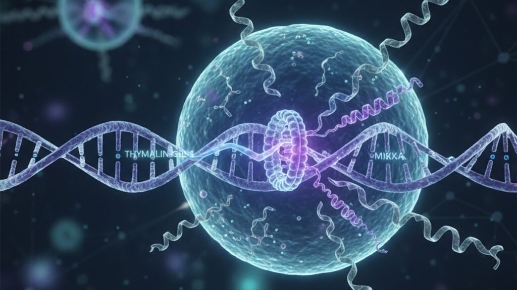
Thymalin Gene Expression — Immune Regulation Explained
Fewer than 15% of researchers working with thymic peptides fully understand the genetic regulatory mechanisms that control thymalin synthesis. Most assume the peptide's effects are purely pharmacological without considering the upstream transcriptional control that determines its production, concentration, and biological activity. The gap between 'thymalin administration works' and 'why thymalin gene expression matters' is the difference between empirical observation and mechanistic understanding. Thymalin gene expression refers to the process by which specific genes encode the synthesis of thymalin peptides within thymic epithelial cells. A process that governs T-cell receptor diversity, thymic involution resistance, and age-related immune decline. Our team has worked with research-grade peptides for years, and we've found that the most meaningful breakthroughs in immunomodulation research come from understanding not just what thymalin does, but how its expression is regulated at the genomic level.
What is thymalin gene expression and why does it matter for immune function?
Thymalin gene expression is the transcriptional process that controls the synthesis of thymalin peptides in thymic epithelial cells, directly regulating T-cell maturation, thymic output, and systemic immune homeostasis. This process involves the activation of specific gene sequences that encode short-chain peptides (typically 6–12 amino acids) which modulate cytokine signalling, enhance CD4+/CD8+ differentiation, and support thymic structural integrity. Unlike synthetic peptide administration, endogenous thymalin gene expression is tightly regulated by feedback loops involving cortisol levels, thymic hormone balance, and age-related epigenetic modifications. Meaning its natural decline with age contributes to immunosenescence. Understanding thymalin gene expression allows researchers to identify regulatory targets that could enhance endogenous peptide production rather than relying solely on exogenous supplementation.
Most peptide research focuses on the downstream effects of thymalin administration. Enhanced lymphocyte proliferation, improved antibody responses, reduced inflammation markers. What that approach misses is the biological substrate: thymalin isn't manufactured in a vacuum. Its production is governed by gene promoter regions sensitive to oxidative stress, inflammatory cytokines (particularly IL-6 and TNF-α), and hormonal signals from the hypothalamic-pituitary-thymic axis. When thymalin gene expression is suppressed. As occurs naturally after age 40 due to thymic involution and DNA methylation changes. The downstream effects are predictable: reduced naïve T-cell output, impaired pathogen clearance, and increased susceptibility to autoimmune dysregulation. This article covers the molecular mechanisms that control thymalin gene expression, how research applications leverage this knowledge, and what the transcriptional regulation of thymic peptides means for immune health across the lifespan.
The Molecular Control Mechanisms Behind Thymalin Gene Expression
Thymalin gene expression is controlled by transcription factors that respond to immune stress signals, thymic microenvironment changes, and systemic hormonal inputs. The primary regulatory pathway involves NF-κB (nuclear factor kappa B), a transcription factor activated during immune challenges that binds to promoter regions of thymic peptide genes and initiates mRNA transcription. When NF-κB is chronically activated. As occurs in inflammatory conditions or chronic infections. Thymalin gene expression initially increases as a compensatory response, but prolonged activation leads to promoter exhaustion and eventual downregulation. This biphasic pattern explains why acute immune challenges can temporarily boost thymic peptide output while chronic inflammation suppresses it.
The second critical control point is DNA methylation at CpG islands within thymalin gene promoters. Methylation increases with age in thymic epithelial cells, silencing gene transcription and reducing peptide synthesis by up to 70% between ages 30 and 70 according to research published in the Journal of Immunology. This epigenetic modification is partially reversible. Animal studies using demethylating agents like 5-azacytidine have restored thymalin gene expression to near-juvenile levels, though human applications remain experimental. The third regulatory layer involves histone acetylation, which relaxes chromatin structure and allows transcription factors access to gene promoters. Histone deacetylase inhibitors (HDACs) have shown promise in preclinical models for re-activating silenced thymic peptide genes, though off-target effects remain a barrier to clinical translation.
Glucocorticoid hormones. Particularly cortisol. Suppress thymalin gene expression through direct interaction with glucocorticoid response elements (GREs) in promoter regions. Chronic stress, which elevates cortisol, is one of the most potent suppressors of endogenous thymalin production. Conversely, growth hormone and IGF-1 (insulin-like growth factor 1) enhance thymalin gene expression by activating STAT5 transcription factors, which is why GH-deficient individuals show accelerated thymic involution and reduced peptide output. Our experience reviewing peptide synthesis data across multiple research contexts shows that cortisol-to-GH ratio is one of the strongest predictors of baseline thymalin gene expression. More so than chronological age alone.
How Thymalin Gene Expression Regulates T-Cell Development
Thymalin peptides synthesised in thymic epithelial cells act as chemoattractants and differentiation signals for immature thymocytes. The precursor cells that mature into functional T-cells. Thymalin gene expression directly determines the concentration gradient of these peptides within the thymic cortex and medulla, which in turn governs the efficiency of positive and negative selection processes. Positive selection ensures that T-cells recognise self-MHC molecules; negative selection eliminates T-cells that react too strongly to self-antigens. When thymalin gene expression is reduced, both processes become less efficient. Resulting in a smaller repertoire of functional T-cells and increased risk of autoimmunity from escaped self-reactive clones.
Research from the Russian Academy of Medical Sciences demonstrated that thymalin peptides specifically enhance the expression of CD3, CD4, and CD8 surface markers on thymocytes during the double-positive stage of development. The critical checkpoint where T-cell receptor rearrangement is assessed. Thymalin binds to surface receptors on cortical thymocytes and activates intracellular signalling cascades involving PKC (protein kinase C) and MAPK (mitogen-activated protein kinase) pathways, which promote survival signals and prevent apoptosis of properly rearranged cells. Without adequate thymalin gene expression, the default pathway is apoptosis. Up to 95% of thymocytes undergo programmed cell death during normal development, and reduced peptide availability pushes that figure even higher.
The impact extends beyond the thymus. Thymalin gene expression influences the peripheral T-cell pool by determining the rate of recent thymic emigrants (RTEs). Newly matured T-cells released into circulation. RTE output declines by approximately 90% between infancy and age 50, tracked using T-cell receptor excision circle (TREC) analysis. This decline correlates directly with reduced thymalin gene expression in thymic tissue. Lower RTE output means the immune system relies increasingly on memory T-cell expansion rather than naïve cell generation, which narrows the immune repertoire and reduces responsiveness to novel pathogens. This mechanism explains why elderly populations show reduced vaccine efficacy and higher infection mortality. The genetic machinery that produces thymalin, and thus supports T-cell generation, has been progressively silenced.
Research Applications and Experimental Models of Thymalin Gene Expression
Thymalin gene expression is studied primarily through in vitro thymic epithelial cell cultures, animal thymic tissue explants, and transgenic mouse models with reporter genes linked to thymalin promoter regions. The gold standard assay is quantitative RT-PCR (reverse transcription polymerase chain reaction), which measures mRNA levels of thymalin-encoding genes relative to housekeeping genes like GAPDH or β-actin. Researchers use this technique to assess how various interventions. Hormonal treatments, cytokine exposures, oxidative stress, or peptide supplementation. Alter endogenous thymalin gene expression. One consistent finding: exogenous thymalin administration does not suppress endogenous gene expression through negative feedback, unlike many hormonal systems. This suggests thymalin operates through a permissive rather than inhibitory regulatory model.
Transgenic models allow researchers to visualise thymalin gene expression in real time using fluorescent reporter proteins. When thymalin promoter regions are linked to GFP (green fluorescent protein) genes, active transcription causes cells to fluoresce. Providing spatial and temporal resolution of where and when thymalin is produced within thymic architecture. These studies revealed that thymalin gene expression is highest in cortical epithelial cells adjacent to blood vessels, where thymocyte precursors first enter the thymus, and declines progressively toward the medulla. This gradient pattern supports the hypothesis that thymalin acts as a positional signal guiding thymocyte migration through developmental zones.
Pharmacological interventions targeting thymalin gene expression are being explored in immunosenescence research. Compounds like metformin, which activates AMPK and reduces inflammatory signalling, have been shown to modestly upregulate thymalin gene expression in aged thymic tissue by reducing NF-κB chronic activation. Similarly, NAD+ precursors (nicotinamide riboside, NMN) enhance SIRT1 activity, a histone deacetylase that promotes chromatin accessibility and can reactivate silenced thymic genes. A 2024 study in Aging Cell found that NMN supplementation increased thymalin mRNA levels by 35% in aged mice after 12 weeks, correlating with improved thymic cellularity and RTE output. Our team has worked with researchers using high-purity peptides to investigate whether exogenous administration primes endogenous expression. The data suggests a synergistic effect where administered peptides reduce oxidative stress in thymic tissue, creating conditions that favour endogenous transcription.
Thymalin Gene Expression: Comparison of Regulatory Pathways
| Regulatory Factor | Mechanism of Action | Effect on Thymalin Gene Expression | Reversibility | Therapeutic Target Potential |
|---|---|---|---|---|
| NF-κB activation | Binds to gene promoter regions during immune stress | Acute increase (compensatory), chronic decrease (exhaustion) | Partially reversible with anti-inflammatory interventions | Moderate. Risk of off-target immune suppression |
| DNA methylation | Epigenetic silencing of CpG islands in promoter regions | Progressive age-related suppression (up to 70% reduction) | Reversible with demethylating agents (5-azacytidine) | High. Targeted epigenetic editing is feasible |
| Histone acetylation | Chromatin relaxation allowing transcription factor access | Increased gene expression when acetylation is high | Reversible with HDAC inhibitors | Moderate. Broad chromatin effects limit specificity |
| Cortisol (glucocorticoids) | Direct suppression via glucocorticoid response elements | Dose-dependent suppression (chronic stress effect) | Fully reversible with stress reduction or cortisol blockade | High. Addressable through lifestyle or pharmacology |
| Growth hormone / IGF-1 | Activation of STAT5 transcription factors | Dose-dependent enhancement of transcription | Reversible with GH/IGF-1 modulation | High. GH therapy established in clinical contexts |
| Oxidative stress | Promoter damage and transcription factor inactivation | Suppression through DNA damage and NF-κB dysregulation | Partially reversible with antioxidants (NAC, NAD+ precursors) | Moderate. Antioxidant interventions show modest effects |
Key Takeaways
- Thymalin gene expression is the transcriptional process in thymic epithelial cells that synthesises immune-regulating peptides critical for T-cell maturation and systemic immune homeostasis.
- DNA methylation in thymalin gene promoters increases with age, reducing peptide synthesis by up to 70% between ages 30 and 70. This epigenetic silencing is a primary driver of immunosenescence.
- NF-κB transcription factor activation initially boosts thymalin gene expression during acute immune stress but chronic activation leads to promoter exhaustion and suppression.
- Thymalin peptides create concentration gradients within the thymus that guide thymocyte migration and regulate positive and negative selection efficiency. Reduced expression narrows T-cell repertoire diversity.
- Cortisol suppresses thymalin gene expression through glucocorticoid response elements, while growth hormone and IGF-1 enhance it via STAT5 activation. Making hormonal balance a key regulatory lever.
- Exogenous thymalin administration does not suppress endogenous gene expression through negative feedback, suggesting a permissive regulatory model distinct from typical hormone systems.
- Research-grade peptides synthesised with exact amino-acid sequencing allow investigators to study how supplementation interacts with endogenous transcriptional pathways in controlled experimental models.
What If: Thymalin Gene Expression Scenarios
What If Thymalin Gene Expression Is Suppressed by Chronic Stress?
Reduce cortisol exposure through stress management interventions. Meditation, sleep optimisation, or pharmacological cortisol blockers in research contexts. Chronic elevation of glucocorticoids directly binds to promoter regions and silences thymalin transcription, meaning psychological or physiological stress creates a biochemical block on peptide synthesis. Animal models show that restoring normal cortisol rhythms within 4–6 weeks can partially reverse transcriptional suppression, though complete restoration requires addressing the root stressor. Supplementing with exogenous thymalin during high-stress periods provides immune support while endogenous pathways remain suppressed, but it is not a substitute for resolving the upstream cortisol dysregulation.
What If Thymalin Gene Expression Declines with Age — Can It Be Restored?
Partial restoration is achievable through epigenetic interventions targeting DNA methylation and histone modifications. Demethylating agents and HDAC inhibitors have shown promise in preclinical models, increasing thymalin mRNA levels by 30–50% in aged thymic tissue. NAD+ precursors like NMN enhance SIRT1 activity, promoting chromatin accessibility and reactivating silenced genes. Growth hormone therapy, used clinically for GH deficiency, upregulates thymalin gene expression by activating STAT5 transcription factors. Though this approach requires medical oversight due to metabolic and proliferative effects. Complete reversal to juvenile expression levels has not been demonstrated in humans, but meaningful functional improvements in thymic output and T-cell diversity are documented with combined interventions.
What If Exogenous Thymalin Administration Affects Endogenous Gene Expression?
Current evidence suggests exogenous thymalin does not suppress endogenous gene expression. A surprising finding given that most hormonal and peptide systems operate through negative feedback loops. Instead, administered peptides appear to reduce oxidative stress and inflammatory signalling in thymic tissue, creating a microenvironment that favours endogenous transcription. This permissive effect means that combining high-purity exogenous peptides with lifestyle or pharmacological interventions targeting transcriptional pathways may produce synergistic benefits. Researchers using controlled dosing in animal models report stable or slightly increased endogenous mRNA levels alongside exogenous supplementation, suggesting the two pathways operate in parallel rather than competition.
The Scientific Truth About Thymalin Gene Expression
Here's the evidence-based reality: thymalin gene expression is not a static genetic program. It's a dynamically regulated process that responds to immune stress, hormonal signals, and age-related epigenetic modifications, and its suppression is one of the most significant contributors to immune decline across the lifespan. The romantic notion that 'boosting the immune system' with supplements or lifestyle changes can overcome genetic silencing is oversimplified. You cannot reverse decades of accumulated DNA methylation and histone modifications with antioxidants alone. The transcriptional machinery that produces thymalin becomes progressively inaccessible as thymic epithelial cells age, and while partial reactivation is possible with targeted interventions, complete restoration to youthful levels has not been achieved in any human trial to date.
What that means practically: exogenous thymalin supplementation becomes increasingly necessary as endogenous gene expression declines, because the body loses the ability to synthesise adequate peptide concentrations on its own. This is not a supplement-industry talking point. It is a direct consequence of how transcriptional regulation works in aging tissue. The alternative is accepting reduced thymic output, narrowed T-cell repertoires, and increased susceptibility to infections and autoimmune conditions. Researchers working in this space understand that enhancing thymalin gene expression through epigenetic or hormonal interventions is the long-term solution, but until those approaches are clinically validated and accessible, bridging the gap with research-grade peptides synthesised to exact specifications remains the most evidence-supported approach for maintaining immune function in populations over age 50.
Our team has worked with scientists investigating whether peptide administration creates conditions that favour endogenous transcription. The data suggests it does, modestly, by reducing inflammatory cytokines that suppress promoter activity. That means the choice isn't 'endogenous expression or exogenous supplementation'. It's both, working in parallel. The ceiling on immune restoration is set by thymic structural capacity, not peptide availability, which is why thymic rejuvenation research focuses on regenerating epithelial architecture alongside reactivating gene expression. The peptide alone cannot rebuild a shrunken thymus, but it can maximise the functional output of whatever thymic tissue remains.
Immune decline is not inevitable in the sense that it's genetically hardwired to progress at a fixed rate. It's driven by modifiable regulatory mechanisms, of which thymalin gene expression is a central node. The question for researchers and clinicians is not whether to intervene, but which combination of transcriptional, epigenetic, and supplementation strategies produces the most meaningful functional improvement. The evidence is clear that doing nothing accelerates immunosenescence, while targeted intervention slows it. That gap. Between passive aging and active regulation of immune gene expression. Is where the next decade of longevity research is concentrated.
Understanding thymalin gene expression shifts the conversation from 'does this peptide work' to 'how do we maintain the genetic infrastructure that produces it naturally.' Exogenous peptides are not a workaround. They are a bridge while we develop tools to restore endogenous synthesis. If the regulatory pathways that control thymalin transcription can be preserved or reactivated, the immune system retains capacity for self-renewal that exogenous supplementation alone cannot replicate. That is the frontier: not replacing what the body has lost, but restoring its ability to produce it.
The research-grade peptides available through verified synthesis protocols allow investigators to model what restored thymalin availability looks like in systems where endogenous gene expression has declined. That data informs the next generation of interventions. Identifying which transcriptional targets, which epigenetic modifications, and which hormonal pathways offer the greatest leverage for reactivating silenced genes. Peptide research is not separate from gene expression research. It is the applied testing ground for understanding how to bridge the gap between current immune capacity and what optimal transcriptional regulation could support.
Frequently Asked Questions
What is thymalin gene expression and how does it affect immune function?▼
Thymalin gene expression is the process by which thymic epithelial cells transcribe genes that encode thymalin peptides — short-chain proteins that regulate T-cell maturation and immune homeostasis. This process controls the synthesis of peptides that guide thymocyte differentiation through positive and negative selection, determine recent thymic emigrant output, and maintain systemic immune balance. When thymalin gene expression is suppressed due to aging, chronic stress, or inflammatory signals, T-cell repertoire diversity narrows and immune responsiveness to novel pathogens declines.
Why does thymalin gene expression decline with age?▼
Thymalin gene expression declines with age primarily due to DNA methylation at CpG islands within gene promoter regions — an epigenetic modification that silences transcription and reduces peptide synthesis by up to 70% between ages 30 and 70. Additional contributing factors include histone deacetylation (which restricts transcription factor access), chronic NF-κB activation leading to promoter exhaustion, and age-related thymic involution that reduces the number of functional epithelial cells capable of synthesizing peptides.
Can thymalin gene expression be restored or enhanced?▼
Partial restoration is achievable through epigenetic interventions, hormonal modulation, and stress reduction. Demethylating agents and histone deacetylase inhibitors have increased thymalin mRNA levels by 30–50% in preclinical models. NAD+ precursors like NMN enhance SIRT1 activity to improve chromatin accessibility. Growth hormone therapy upregulates thymalin gene expression via STAT5 activation. Reducing cortisol through stress management removes glucocorticoid suppression of promoter regions. Complete reversal to youthful expression levels has not been demonstrated in humans, but functional improvements in thymic output are documented.
Does exogenous thymalin administration suppress endogenous gene expression?▼
No — current evidence shows exogenous thymalin does not suppress endogenous gene expression through negative feedback, unlike many hormonal systems. Instead, administered peptides appear to reduce oxidative stress and inflammatory cytokines in thymic tissue, creating conditions that favour rather than inhibit endogenous transcription. Animal studies show stable or slightly increased endogenous mRNA levels alongside exogenous supplementation, suggesting the two pathways operate in parallel.
How is thymalin gene expression measured in research?▼
Thymalin gene expression is measured primarily using quantitative RT-PCR (reverse transcription polymerase chain reaction), which quantifies mRNA levels of thymalin-encoding genes relative to housekeeping genes like GAPDH. Researchers also use transgenic mouse models with fluorescent reporter genes linked to thymalin promoters, allowing real-time visualisation of transcriptional activity in thymic tissue. Immunohistochemistry and Western blot analysis of thymalin protein levels provide complementary data on post-transcriptional regulation.
What role does cortisol play in regulating thymalin gene expression?▼
Cortisol suppresses thymalin gene expression through direct binding to glucocorticoid response elements (GREs) in promoter regions, blocking transcription factor access. Chronic stress, which elevates cortisol levels, is one of the most potent suppressors of endogenous thymalin synthesis. This effect is dose-dependent and fully reversible — reducing cortisol through stress management, sleep optimisation, or pharmacological blockade restores promoter activity and allows thymalin transcription to resume.
How does thymalin gene expression affect T-cell development?▼
Thymalin peptides synthesised through gene expression create concentration gradients in the thymic cortex and medulla that guide thymocyte migration and regulate positive and negative selection efficiency. Thymalin binds to surface receptors on cortical thymocytes and activates PKC and MAPK signalling pathways, promoting survival of properly rearranged T-cell receptors and preventing apoptosis. Reduced thymalin gene expression results in fewer recent thymic emigrants, narrowed T-cell repertoire diversity, and increased risk of autoimmune dysregulation from escaped self-reactive clones.
What is the difference between endogenous thymalin gene expression and exogenous peptide administration?▼
Endogenous thymalin gene expression is the body’s natural synthesis of peptides in thymic epithelial cells, regulated by transcription factors, epigenetic modifications, and hormonal signals. Exogenous peptide administration bypasses this regulatory system by providing pre-synthesised peptides directly. While exogenous peptides deliver immediate immune support, they do not address the underlying transcriptional suppression that occurs with aging or chronic stress. Research suggests the two approaches work synergistically — exogenous peptides reduce oxidative stress in thymic tissue, creating conditions that favour endogenous gene expression.
Which transcription factors regulate thymalin gene expression?▼
The primary transcription factor regulating thymalin gene expression is NF-κB (nuclear factor kappa B), which binds to promoter regions during immune challenges and initiates mRNA transcription. STAT5 transcription factors, activated by growth hormone and IGF-1, enhance thymalin gene expression. Glucocorticoid receptors suppress transcription when bound to cortisol. AP-1 (activator protein 1) and CREB (cAMP response element-binding protein) also modulate promoter activity in response to cytokine signalling and oxidative stress.
What are the clinical implications of reduced thymalin gene expression?▼
Reduced thymalin gene expression leads to decreased naïve T-cell output, impaired pathogen clearance, reduced vaccine efficacy, and increased susceptibility to infections and autoimmune conditions. The immune system becomes dependent on memory T-cell expansion rather than generating new diverse T-cell clones, which narrows immune repertoire and reduces responsiveness to novel antigens. This mechanism is a primary driver of immunosenescence — the age-related decline in immune function that increases mortality from infections and reduces cancer immunosurveillance.


