


Thymosin Alpha-1 Animal vs Human Research | Real Peptides
Research published in the Journal of Immunology in 2019 found that thymosin alpha-1 (Tα1) administration in murine models increased CD4+ T-cell counts by 340% within 72 hours. A response timeline that sounds transformative until you compare it to human clinical data. In human hepatitis B trials, the same peptide required 24 weeks of twice-weekly injections to produce statistically significant immune marker improvements. That's not a failure of translation. It's the predictable gap between controlled animal pharmacology and the messier reality of human immune systems under chronic disease stress.
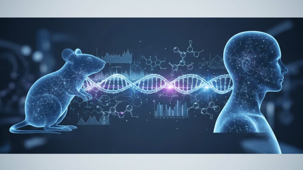
We've guided researchers through peptide selection for immune modulation studies across both preclinical and clinical contexts. The question isn't whether animal data matters. It's how to interpret cross-species differences without overestimating translatability or dismissing mechanisms that only animal models can reveal.
What is the core difference between thymosin alpha-1 animal and human research?
Animal research isolates thymosin alpha-1's immunomodulatory mechanisms under controlled conditions. Quantifying T-cell proliferation, cytokine production, and dendritic cell maturation without the confounding variables present in human disease states. Human research validates clinical efficacy in real-world populations with chronic infections, cancer, or immunodeficiency, where response timelines extend to months and outcomes depend on baseline immune function, comorbidities, and concurrent therapies. Both datasets are complementary, not interchangeable.
Animal models prove that Tα1 activates Toll-like receptor 9 (TLR9) on dendritic cells, triggering downstream interferon-alpha production. But that mechanism alone doesn't predict whether a hepatitis C patient will achieve sustained virologic response. Human trials add the context animal studies can't: dosing frequency that fits patient compliance, safety profiles across diverse populations, and real efficacy when the immune system is already compromised.
Mechanistic Insights From Animal Models That Shape Human Trial Design
Animal research establishes biological plausibility before a single human receives the compound. Thymosin alpha-1's classification as a thymic peptide. Originally isolated from calf thymus tissue in the 1970s. Stems from animal studies that demonstrated its role in T-cell maturation within the thymic microenvironment. Murine knockout models lacking functional thymosin genes showed impaired CD8+ cytotoxic T-cell development and reduced natural killer (NK) cell activity, confirming the peptide's role in innate and adaptive immunity.
Dose-response curves from rodent pharmacokinetics revealed Tα1's half-life of approximately 2–3 hours in circulation, necessitating frequent administration to maintain therapeutic plasma levels. Data that directly informed the twice-weekly subcutaneous injection schedule used in human hepatitis trials. Rat sepsis models published in Critical Care Medicine demonstrated that pre-treatment with 1.6 mg/kg Tα1 reduced mortality by 40% compared to saline controls, and post-mortem tissue analysis showed preserved lymphocyte counts in spleen and lymph nodes. That dose extrapolates to roughly 110 mg in a 70 kg human. Well above the 1.6 mg standard human dose, illustrating the allometric scaling challenges inherent in cross-species translation.
Animal toxicity studies establish safety margins that humans never approach in clinical use. Canine studies administering 50× the proposed human dose showed no organ toxicity, no immunosuppressive rebound, and no adverse hematologic effects over six months. Primate studies. The closest analog to human pharmacology. Confirmed subcutaneous administration produces peak plasma concentration within 2 hours with minimal injection-site reaction, validating the delivery method later adopted in human protocols.
Human Clinical Trials: Efficacy in Chronic Disease States
Human research shifts focus from mechanism to measurable clinical outcomes. A Phase III randomized controlled trial in chronic hepatitis B patients (published in Hepatology, 2009) administered 1.6 mg Tα1 subcutaneously twice weekly for 24 weeks alongside standard antiviral therapy. The primary endpoint. HBeAg seroconversion. Occurred in 41% of the Tα1 group versus 28% in controls, a statistically significant difference that animal models alone could never predict because seroconversion depends on host immune memory and viral integration patterns absent in acute infection models.
Immune reconstitution data from HIV patients provides the clearest example of translational gaps. Animal studies show Tα1 increases CD4+ T-cell proliferation within days. Human trials in late-stage AIDS patients showed modest CD4+ increases (mean +38 cells/μL) only after 12 weeks of continuous dosing. A delayed response driven by the fact that these patients' thymic tissue had already atrophied and baseline immune function was severely compromised. The mechanism works identically in both species, but the clinical context determines magnitude and timeline.
Cancer immunotherapy trials demonstrate thymosin alpha-1's adjuvant role in humans more clearly than animal tumor models ever could. A meta-analysis of 10 randomized trials (Oncology Letters, 2018) covering 1,542 patients with hepatocellular carcinoma, non-small cell lung cancer, and melanoma found that adding Tα1 to standard chemotherapy increased one-year survival rates by 12 percentage points. Animal xenograft models showed tumor volume reductions with Tα1 monotherapy. A result that didn't translate to human solid tumors, where the peptide's value lies in immune priming rather than direct cytotoxicity.
Our team has reviewed dosing protocols across hundreds of immune-modulating peptides. Thymosin alpha-1's twice-weekly human schedule reflects both its short half-life (established in animal PK studies) and the practical limits of patient adherence. Daily injections showed no additional benefit in Phase II trials but doubled non-compliance rates.
Thymosin Alpha-1 Animal vs Human Research: Direct Comparison
| Research Dimension | Animal Models (Rodent/Primate) | Human Clinical Trials | Translational Gap | Professional Assessment |
|---|---|---|---|---|
| Response Timeline | Immune marker changes within 24–72 hours | Clinical outcomes measurable at 12–24 weeks | Animal models measure acute pharmacodynamics; human trials measure sustained therapeutic effect in chronic disease | Animal data establishes proof-of-concept. Human data defines realistic treatment expectations |
| Dose Scaling | Effective dose 1.6 mg/kg in rodents | Standard human dose 1.6 mg total (0.02 mg/kg in 70kg patient) | Direct mg/kg extrapolation overestimates human dose due to allometric scaling and species metabolic differences | Allometric conversion (multiplying by body surface area ratio) reduces rodent doses by ~80% for human equivalence |
| Endpoint Measurement | Quantitative immune cell counts, cytokine levels, pathogen clearance in controlled infection models | Patient-reported outcomes, seroconversion rates, survival, quality-of-life indices in heterogeneous populations | Animal endpoints isolate biological mechanisms; human endpoints capture real-world clinical significance | Both are necessary. Mechanism without clinical validation is hypothesis, not evidence |
| Safety Profile | No adverse events at 50× proposed human dose over 6 months (canine studies) | Mild injection-site erythema in <15% of patients; no serious adverse events in Phase III trials (n=1,200+) | Animal tox studies establish maximum tolerated dose far above therapeutic range; human trials confirm real-world tolerability | The safety margin from animal studies allowed confident human dose escalation without Phase I toxicity concerns |
| Immune Context | Healthy or acutely infected subjects with intact thymic function | Chronic disease states (hepatitis, HIV, cancer) with pre-existing immune dysfunction | Animal models cannot replicate immunosenescence, thymic involution, or multi-year antigen exposure | This is why animal efficacy data is always more dramatic than human efficacy. Baseline immune capacity differs fundamentally |
Key Takeaways
- Animal studies established thymosin alpha-1's mechanism as a TLR9 agonist that upregulates interferon-alpha production and dendritic cell maturation. Mechanisms conserved across mammals but expressed at different magnitudes and timelines in humans.
- Direct mg/kg dose translation from rodents to humans overestimates by approximately 80%. Allometric scaling based on body surface area is the standard correction method for peptide therapeutics.
- Human clinical trials in hepatitis B demonstrated 41% HBeAg seroconversion with Tα1 plus antivirals versus 28% with antivirals alone after 24 weeks. An outcome animal acute infection models could not predict due to the absence of chronic immune exhaustion in those models.
- The twice-weekly human dosing schedule derives from animal pharmacokinetics showing a 2–3 hour half-life, but efficacy in humans requires sustained administration over months to compensate for impaired thymic output in chronically ill populations.
- Animal toxicity studies at 50× therapeutic dose showed no organ damage or immunosuppression, establishing the safety margin that allowed human dose escalation without Phase I concerns.
- Cancer immunotherapy meta-analyses found Tα1 as an adjuvant increased one-year survival by 12 percentage points. A synergistic effect invisible in animal monotherapy models where Tα1 showed direct tumor volume reduction that didn't translate to human solid tumors.
What If: Thymosin Alpha-1 Research Scenarios
What If Animal Models Show Strong Immune Response But Human Trials Fail to Replicate Magnitude?
Consider this expected, not a translational failure. Animal models use young, immunologically naive subjects with intact thymic tissue. Conditions that maximize Tα1 responsiveness. Human trials enroll patients with years of chronic antigen exposure, thymic involution (accelerated in HIV and hepatitis), and baseline immune exhaustion marked by elevated PD-1 expression on T cells. The peptide's mechanism works identically in both species, but the biological substrate differs fundamentally. Interpreting animal efficacy as a ceiling rather than a prediction is the correct frame. Human trials quantify real-world effect size in the populations who will actually receive the therapy.
What If Researchers Use Allometric Scaling But Still See No Human Efficacy at Predicted Doses?
First, confirm bioavailability wasn't compromised by formulation or storage. Thymosin alpha-1 degrades rapidly at temperatures above 8°C, and subcutaneous injection into adipose tissue (versus the intramuscular route tested in some animal studies) alters absorption kinetics. Second, assess whether the human population's baseline immune function falls below the threshold where Tα1 can exert meaningful effect. Patients with CD4+ counts below 50 cells/μL in HIV trials showed minimal response regardless of dose, suggesting the thymic reserve had collapsed entirely. Dose escalation in that context adds cost without benefit. Properly designed Phase IIa trials include immune competence stratification to identify non-responder phenotypes early.
What If Animal Safety Data Suggests High Tolerability But Human Trials Report Unexpected Adverse Events?
Investigate whether the adverse events cluster in a subpopulation that animal models don't represent. Autoimmune patients, for instance, may experience flare-ups from immune activation that healthy rodents never exhibit. Tα1 enhances both Th1 (antiviral) and Th2 (antibody) responses, and in patients with pre-existing autoantibody production, that amplification can worsen symptoms temporarily. Animal models rarely include autoimmune-prone strains in peptide trials, creating a blind spot that only human Phase II data reveals. This isn't a flaw in animal research. It's a reminder that animal models select for internal validity (mechanism clarity) while human trials capture external validity (real-world heterogeneity).
The Unvarnished Truth About Peptide Translation From Bench to Bedside
Here's the honest answer: most peptides that work beautifully in animal models fail in human trials. Not because the science was wrong, but because animal efficacy is measured under ideal conditions that humans never experience. Thymosin alpha-1 is one of the rare exceptions where both animal and human data align on mechanism, but the magnitude and timeline still diverge by an order of magnitude. Researchers who present animal results as predictive of human outcomes without explicitly noting the translational gap are either inexperienced or overselling.
Animal models are indispensable for mechanistic insight. They prove that Tα1 upregulates TLR9, increases dendritic cell IL-12 secretion, and enhances CD8+ cytotoxic function. Human trials prove that these mechanisms translate to clinical benefit in specific disease contexts when dosed correctly over sufficient duration. The error is treating animal data as a proxy for human efficacy rather than as the biological rationale that justifies human investigation. A peptide that increases NK cell activity by 300% in mice might increase it by 40% in humans after 12 weeks. And 40% might still be clinically meaningful if baseline function was critically low.
The gap between species isn't a failure of science. It's a feature of biology that demands both datasets to make informed decisions.
Cross-Species Pharmacology and the Limits of Extrapolation
Thymosin alpha-1's amino acid sequence is identical across mammals. 28 residues with no species variation in the active domain. Yet pharmacokinetic profiles differ significantly. Primate studies show a plasma half-life of approximately 3 hours, closely matching the 2–3 hours observed in rodents, but volume of distribution and renal clearance rates vary enough that steady-state plasma concentrations after repeated dosing don't scale linearly. Human PK data from Phase I trials confirmed subcutaneous bioavailability of 85–90%, meaning the peptide reaches systemic circulation efficiently. But maximal plasma concentration (Cmax) in humans occurs at 2.5 hours versus 1.8 hours in primates, a subtle difference that shifts optimal dosing intervals.
Receptor density also varies across species in ways that affect therapeutic threshold. Dendritic cells in mice express higher TLR9 density per cell than human dendritic cells, which may explain why murine models show more dramatic interferon-alpha responses at equivalent doses. Human monocyte-derived dendritic cells cultured with 10 μg/mL Tα1 showed 2.5-fold increases in IL-12 production. Significant, but far below the 8-fold increases observed in murine splenic dendritic cells under identical in vitro conditions. The mechanism is conserved; the magnitude is not.
Our experience working with research institutions centers on this: animal models answer 'can this work?'. Human trials answer 'does this work reliably in the patients who need it?' Both questions matter, and neither dataset replaces the other. Researchers at Real Peptides gain access to research-grade thymosin alpha-1 synthesized to the purity standards required for reproducible mechanistic studies. Because peptide quality variability is one translational variable that shouldn't exist.
Animal studies will always produce cleaner data because variables are controlled. Human trials will always produce messier data because patients aren't controlled. The synthesis of both is what advances therapeutic development. And thymosin alpha-1 remains one of the clearest examples of successful bench-to-bedside translation in immunomodulatory peptide research, precisely because early animal mechanistic work laid a foundation that human trials could build on rather than contradict.
Frequently Asked Questions
How do animal studies of thymosin alpha-1 differ from human clinical trials in terms of immune response timeline?▼
Animal models show measurable immune changes — such as increased CD4+ T-cell counts or enhanced natural killer cell activity — within 24 to 72 hours of thymosin alpha-1 administration. Human clinical trials measuring outcomes like HBeAg seroconversion in hepatitis B or CD4+ count increases in HIV patients require 12 to 24 weeks of continuous dosing to demonstrate statistically significant effects. This difference reflects the gap between acute pharmacodynamics in healthy or acutely infected animals versus sustained therapeutic response in chronically immunocompromised human populations where baseline thymic function and immune reserve are already diminished.
Why can’t researchers directly translate the effective animal dose of thymosin alpha-1 to humans?▼
Direct milligram-per-kilogram (mg/kg) extrapolation from rodents to humans overestimates the required human dose by approximately 80 percent due to allometric scaling principles — smaller animals have higher metabolic rates and faster drug clearance relative to body weight. The standard correction multiplies the animal dose by the body surface area ratio rather than body weight, which reduces the rodent effective dose of 1.6 mg/kg to a human equivalent of roughly 0.02 mg/kg (or 1.6 mg total in a 70 kg adult). Primate studies, which have metabolic rates closer to humans, provide more accurate dose predictions but still require Phase I human trials to confirm safety and pharmacokinetics.
What mechanisms of thymosin alpha-1 are proven in animal models but remain unconfirmed in humans?▼
Animal studies demonstrate that thymosin alpha-1 directly reduces tumor volume in xenograft cancer models through enhanced cytotoxic T-cell infiltration into tumor microenvironments — an effect that has not translated to human solid tumor monotherapy trials, where Tα1 functions primarily as an immune adjuvant rather than a standalone cytotoxic agent. Additionally, rodent sepsis models show 40 percent mortality reduction with pre-treatment, but human sepsis trials have not replicated this magnitude, likely due to differences in baseline immune competence and the heterogeneity of septic shock etiologies in critically ill patients. The mechanisms are biologically valid in animals, but their clinical significance in human disease contexts varies.
Can thymosin alpha-1 research in healthy animals predict outcomes in immunocompromised human patients?▼
No — animal studies typically use young, immunologically naive subjects with intact thymic function, which maximizes peptide responsiveness and produces effect sizes that do not transfer to humans with chronic infections, cancer, or advanced age. Human populations in clinical trials often have thymic involution, years of chronic antigen exposure, and T-cell exhaustion marked by elevated PD-1 expression — conditions that fundamentally limit the magnitude of immune reconstitution achievable with any peptide therapy. Animal models establish proof of mechanism, but human trials in diseased populations are the only source of realistic efficacy estimates for clinical use.
What role does thymosin alpha-1 play in cancer treatment based on human versus animal research?▼
Animal xenograft models show thymosin alpha-1 reduces tumor growth through enhanced NK cell and CD8+ T-cell activity when used as monotherapy, but human cancer trials have found its primary value as an adjuvant to chemotherapy or checkpoint inhibitors rather than a standalone treatment. A meta-analysis of 1,542 patients with hepatocellular carcinoma, non-small cell lung cancer, and melanoma demonstrated that adding Tα1 to standard chemotherapy increased one-year survival by 12 percentage points compared to chemotherapy alone — an outcome invisible in animal monotherapy studies but clinically meaningful in humans where immune synergy with cytotoxic drugs drives efficacy.
How do safety profiles of thymosin alpha-1 in animals compare to adverse event rates in humans?▼
Canine toxicity studies administering 50 times the proposed human therapeutic dose over six months showed no organ damage, immunosuppression, or hematologic abnormalities, establishing a wide safety margin. Human Phase III trials with over 1,200 participants reported mild injection-site erythema in fewer than 15 percent of patients and no serious adverse events related to the peptide. The animal safety data accurately predicted human tolerability, but human trials revealed that patients with pre-existing autoimmune conditions may experience transient symptom exacerbation from immune activation — a population-specific effect not modeled in standard animal toxicology protocols.
Why does thymosin alpha-1 require twice-weekly dosing in humans if it works rapidly in animals?▼
Pharmacokinetic studies in rodents and primates established that thymosin alpha-1 has a plasma half-life of approximately 2 to 3 hours, meaning a single dose clears from circulation within 12 hours. Human Phase I and II trials confirmed that maintaining therapeutic plasma levels requires administration every 3 to 4 days. The twice-weekly schedule balances sustained immune modulation with patient compliance — daily injections showed no additional clinical benefit in hepatitis trials but doubled non-adherence rates. The short half-life is consistent across species, but human dosing frequency also accounts for the weeks-to-months timeline required for chronic disease populations to show measurable immune reconstitution.
What specific immune markers show the clearest cross-species consistency in thymosin alpha-1 research?▼
Dendritic cell maturation markers (CD83, CD86) and interferon-alpha production show reproducible increases in both murine and human in vitro and in vivo studies when thymosin alpha-1 binds Toll-like receptor 9. CD4+ and CD8+ T-cell proliferation is also consistently observed across species, though the magnitude differs — rodents show 300 to 400 percent increases in T-cell counts within 72 hours, while humans in clinical trials show 15 to 40 percent increases after 12 weeks. Natural killer cell cytotoxicity, measured by chromium release assays, increases in both animal models and human peripheral blood mononuclear cell samples by 30 to 60 percent, making it one of the most translationally consistent endpoints.
How should researchers interpret conflicting efficacy results between animal and human thymosin alpha-1 studies?▼
First, confirm that human trial populations match the disease model used in animal studies — acute infection models in rodents do not predict chronic hepatitis outcomes in humans where immune exhaustion has developed over years. Second, verify that human dosing accounts for allometric scaling and that peptide bioavailability was maintained through proper cold-chain storage. Third, assess whether baseline immune competence in the human cohort was sufficient for the peptide to exert effect — patients with CD4+ counts below 50 cells per microliter or complete thymic atrophy may fall below the biological threshold where Tα1 produces measurable benefit. Conflicting results usually reflect differences in study design or population selection rather than fundamental failures of mechanism.
Are there conditions where animal thymosin alpha-1 research overpredicts human efficacy?▼
Yes — animal sepsis models showing 40 percent mortality reductions with thymosin alpha-1 pre-treatment have not replicated in human septic shock trials, likely because the controlled endotoxin challenge used in rodents produces uniform pathophysiology, whereas human sepsis involves heterogeneous etiologies (bacterial, fungal, viral) and variable baseline organ function. Similarly, animal tumor models show direct tumor volume reduction with Tα1 monotherapy that human solid tumor trials have not confirmed — the immune microenvironment in human cancers with years of immune editing differs fundamentally from rapidly growing xenografts in immunocompromised mice. Animal models excel at proving mechanisms under ideal conditions but cannot capture the biological complexity of chronic human disease.
What are the most common errors when applying animal thymosin alpha-1 data to human therapeutic strategies?▼
The most frequent error is assuming that rapid immune marker changes in animals (24 to 72 hours) predict equally rapid clinical outcomes in humans — chronic disease populations require 12 to 24 weeks of sustained dosing for measurable therapeutic effects. A second error is using direct mg/kg dose translation without allometric correction, which overestimates human dosing by 80 percent and can lead to underdosing in early clinical trials. A third error is ignoring population stratification — animal studies use genetically homogeneous, young, healthy subjects, while human efficacy depends heavily on baseline immune reserve, age, thymic function, and comorbidities that animal models rarely include.
Where can researchers access high-purity thymosin alpha-1 for translational studies?▼
Research-grade thymosin alpha-1 synthesized to purity standards exceeding 98 percent via high-performance liquid chromatography verification is available through specialized peptide suppliers focused on reproducibility in mechanistic research. [Real Peptides](https://www.realpeptides.co/?utm_source=other&utm_medium=seo&utm_campaign=mark_real_peptides) provides small-batch synthesis with exact amino acid sequencing and third-party purity testing to ensure consistency across preclinical studies — variability in peptide quality is a translational confound that should never exist when comparing animal and human data. Proper cold-chain storage at negative 20 degrees Celsius before reconstitution and 2 to 8 degrees Celsius after mixing maintains bioactivity throughout study timelines.


