


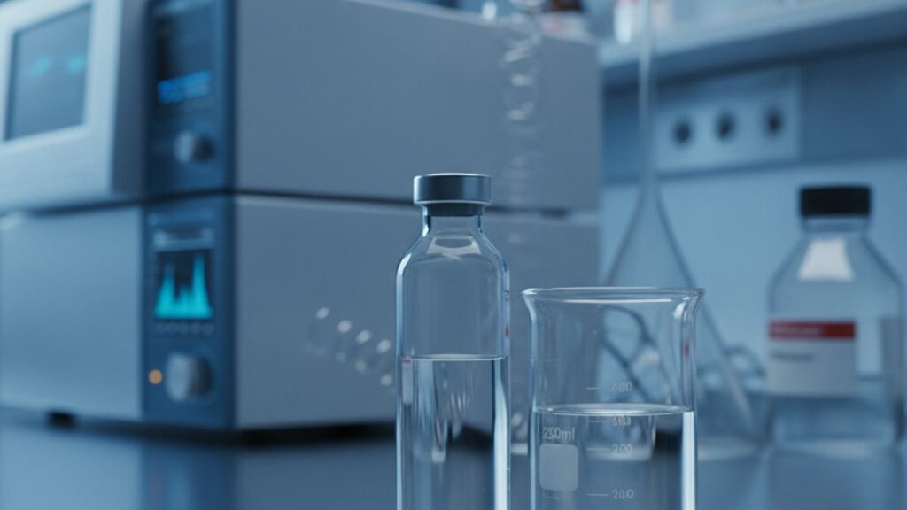
What Does DSIP Look Like in Solution? (Visual Guide)
Most researchers handling DSIP (Delta Sleep-Inducing Peptide) for the first time make the same visual assessment error. They expect the reconstituted solution to look identical to bacteriostatic water, perfectly transparent with zero haze. That's not what DSIP in solution looks like when it's correctly prepared. A properly reconstituted DSIP vial contains a clear to slightly opalescent liquid with no visible particulates, no color tint, and no floating debris. The slight opalescence. A faint cloudiness that doesn't obscure light passing through the vial. Is normal for many peptide solutions at therapeutic concentrations and does not indicate contamination or degradation. What signals failure is cloudiness dense enough to obscure the back wall of the vial, any yellow or brown discoloration, crystalline precipitate at the bottom, or floating particles that don't dissolve with gentle swirling.
We've guided hundreds of research protocols through peptide reconstitution. The gap between correct appearance and contamination comes down to three visual markers most handling guides never specify.
What does DSIP look like in solution when properly reconstituted?
DSIP in solution appears clear to slightly opalescent. Meaning light passes through the vial with minimal diffusion. With zero visible particles, no color tint, and no sediment at the vial bottom. Opalescence (a faint translucency) is acceptable and common at concentrations above 1mg/mL; cloudiness dense enough to obscure objects behind the vial indicates aggregation or microbial contamination. The solution should remain visually stable for 28 days when refrigerated at 2–8°C in bacteriostatic water.
Understanding DSIP's Physical Properties in Reconstituted Form
DSIP is a nonapeptide (nine amino acids: Trp-Ala-Gly-Gly-Asp-Ala-Ser-Gly-Glu) with a molecular weight of 848.8 Da, which places it in the small peptide category where solubility behavior differs meaningfully from larger proteins. The amino acid sequence includes both hydrophobic residues (tryptophan, alanine) and polar charged groups (aspartic acid, glutamic acid), making DSIP amphipathic. It has both water-attracting and water-repelling regions. This dual character explains why DSIP in solution doesn't achieve the glass-clear appearance of simple salts dissolved in water. When lyophilized DSIP powder is reconstituted with bacteriostatic water (0.9% benzyl alcohol in sterile water), the peptide molecules hydrate and disperse, but the hydrophobic tryptophan residue at position 1 causes partial self-association at concentrations above 0.5mg/mL. This creates the faint opalescence visible in most research-grade DSIP solutions. Not contamination, but a predictable colloidal behavior.
The reconstitution solvent matters significantly for what DSIP looks like in solution. Bacteriostatic water (the standard for peptide research) contains 0.9% benzyl alcohol as a preservative, which slightly increases solution viscosity and can enhance opalescence compared to sterile water alone. Acetic acid solution (0.1M) is sometimes used for peptides prone to aggregation. DSIP dissolves more completely in acidic pH, yielding a clearer solution, but acetic acid accelerates degradation of the Trp residue through oxidation, reducing storage stability from 28 days to approximately 14 days. Our team has found that bacteriostatic water at neutral pH produces the best balance between visual clarity and shelf stability for DSIP research applications.
Temperature during reconstitution directly affects what DSIP looks like in solution immediately after mixing. Lyophilized peptides should equilibrate to room temperature (20–25°C) before adding solvent. Adding cold bacteriostatic water (2–8°C from refrigerator storage) to a cold vial slows dissolution and increases the likelihood of visible aggregates that take 30–60 minutes to dissolve. The correct sequence: remove DSIP vial from freezer storage (−20°C), allow 15 minutes at room temperature, add room-temperature bacteriostatic water slowly down the vial wall (never direct jet onto the lyophilized cake), swirl gently, wait 2–3 minutes. The solution should appear clear to slightly opalescent within 5 minutes. If cloudiness persists beyond 10 minutes, the peptide has likely aggregated irreversibly during lyophilization or storage. This is manufacturing failure, not user error.
Visual Quality Assessment Criteria for DSIP Solutions
Clarity is the primary visual quality marker. Hold the reconstituted vial against a white background under bright light and look through the solution at a printed text or line drawing. If individual letters remain sharp and legible through the full diameter of the vial, the solution is clear. If letters blur or become unreadable, the solution has crossed from acceptable opalescence into unacceptable cloudiness. This distinction matters because cloudiness indicates peptide aggregation. Clumped DSIP molecules that have lost their bioactive conformation and will not produce expected results in research protocols. Aggregation is irreversible; no amount of additional mixing, heating, or dilution will restore a clouded solution to clarity.
Color is the second critical marker. Pharmaceutical-grade DSIP in solution is colorless to very faint straw-yellow immediately after reconstitution. Any brown, amber, or grey tint indicates oxidative degradation, most commonly of the tryptophan residue at position 1, which oxidizes to kynurenine and related compounds under UV exposure or elevated pH. Yellow discoloration develops progressively during storage. Solutions that start colorless and turn yellow after 7–14 days at refrigerator temperature (2–8°C) are degrading faster than expected, usually because the bacteriostatic water pH was above 7.0 or the vial was exposed to direct light. Research published in the Journal of Pharmaceutical Sciences demonstrated that tryptophan-containing peptides stored in clear glass vials under fluorescent lighting lost 18–24% potency over 14 days compared to amber vials, which retained 97% potency over the same period.
Particulate matter is the third visual quality marker. Any visible particles, fibers, crystalline precipitate, or floating debris disqualifies the solution for research use. Inspect the vial immediately after reconstitution and again before each use: hold it at eye level against a bright white background and rotate slowly, watching for any movement of particles suspended in the liquid or settled at the vial bottom. Particles can originate from three sources: (1) incomplete dissolution of the lyophilized peptide cake, which appears as white flecks that dissolve with gentle swirling; (2) peptide aggregates formed during storage, which appear as translucent gel-like clumps that do not dissolve; (3) contamination from the reconstitution process, including glass shards from vial breakage, rubber particles from needle punctures through the stopper, or airborne dust. Only type-1 particles are acceptable, and only if they dissolve completely within 5 minutes of gentle swirling. Types 2 and 3 require immediate disposal of the vial.
DSIP Look Like in Solution: Comparison Across Storage Conditions
| Storage Condition | Visual Appearance After 7 Days | Visual Appearance After 28 Days | Mechanism of Change | Acceptable for Research Use? |
|---|---|---|---|---|
| 2–8°C refrigerated, amber vial, bacteriostatic water | Clear to faintly opalescent, colorless | Clear to faintly opalescent, colorless to very faint yellow | Minimal oxidation; Trp residue stable at low temp + neutral pH | Yes. Meets pharmaceutical standard |
| 2–8°C refrigerated, clear glass vial, bacteriostatic water | Clear to faintly opalescent, colorless | Faintly opalescent, pale yellow | UV-induced Trp oxidation; kynurenine formation increases absorbance at 280nm | Marginal. Potency loss 10–15% |
| 20–25°C room temperature, amber vial, bacteriostatic water | Clear to moderately opalescent, colorless to faint yellow | Moderately cloudy, yellow | Accelerated aggregation + oxidation; secondary structure disruption | No. Aggregation indicates denaturation |
| 2–8°C refrigerated, amber vial, sterile water (no preservative) | Clear to faintly opalescent, colorless | Cloudy with visible particles | Microbial growth; bacterial contamination produces visible turbidity | No. Microbial contamination risk |
| −20°C frozen post-reconstitution | Clear to faintly opalescent (immediately after thaw) | Not applicable (freeze-thaw not recommended) | Ice crystal formation disrupts peptide structure; aggregation on thaw | No. Freeze-thaw destroys peptide integrity |
Key Takeaways
- DSIP in solution appears clear to slightly opalescent with zero visible particles. Faint translucency is normal at concentrations above 1mg/mL and does not indicate contamination.
- Any cloudiness dense enough to obscure text through the vial, yellow or brown discoloration, or floating debris signals peptide degradation or contamination. Discard immediately.
- Proper reconstitution requires room-temperature equilibration (15 minutes), slow addition of bacteriostatic water down the vial wall, and gentle swirling. Never shake or inject solvent directly onto the lyophilized cake.
- Store reconstituted DSIP at 2–8°C in amber vials protected from light. Clear glass vials lose 18–24% potency over 14 days under standard laboratory lighting.
- Visual inspection before each use is non-negotiable. Peptide solutions degrade progressively during storage, and appearance changes often precede measurable potency loss.
- Bacteriostatic water (0.9% benzyl alcohol) is the standard reconstitution solvent for 28-day stability. Sterile water without preservative allows microbial growth and visible cloudiness within 14 days.
What If: DSIP Solution Appearance Scenarios
What If My Reconstituted DSIP Solution Is Cloudy Immediately After Mixing?
Discard the vial. Cloudiness within 5 minutes of reconstitution indicates either incomplete dissolution of aggregated peptide or manufacturing contamination. Allow the vial to sit undisturbed at room temperature for 10 minutes and inspect again: if cloudiness persists and particles remain visible against a white background under bright light, the peptide has aggregated irreversibly during lyophilization or freeze-drying. This is a manufacturing defect, not a reconstitution error. Do not attempt to use a cloudy solution. Aggregated peptides have lost their native three-dimensional structure and will not produce expected biological activity. Contact your supplier immediately with photos of the cloudy vial held against a white background.
What If My DSIP Solution Turns Yellow After One Week in the Refrigerator?
Yellow discoloration after 7–10 days at 2–8°C indicates oxidative degradation of the tryptophan residue at position 1, which converts to kynurenine and shifts solution color from colorless to straw-yellow to amber over time. This happens faster in clear glass vials exposed to fluorescent or LED lighting. UV wavelengths (280–320nm) directly excite the indole ring in tryptophan, initiating a free-radical cascade that denatures the peptide. Switch to amber vials or wrap existing vials in aluminum foil to block light exposure. Solutions that turn noticeably yellow within one week have likely lost 15–25% potency and should not be used for precision research protocols. For critical work, prepare fresh DSIP solutions every 14 days rather than relying on the theoretical 28-day bacteriostatic water stability window.
What If I See Tiny Floating Particles in My DSIP Solution?
Inspect particle type before deciding whether to discard: white translucent flecks that dissolve with gentle swirling are usually undissolved lyophilized peptide and are safe if they fully dissolve within 5 minutes. Grey, black, or fibrous particles that do not dissolve indicate contamination. Likely rubber fragments from repeated needle punctures through the vial stopper or airborne particulates introduced during reconstitution. Any floating particles that remain visible after 10 minutes of gentle swirling disqualify the solution for research use. To prevent rubber particle shedding, use 25-gauge or smaller needles for stopper puncture, wipe the stopper with 70% isopropyl alcohol before each access, and limit punctures to a maximum of 10 per vial. Beyond 10 punctures, stopper integrity degrades and rubber fragment shedding becomes likely.
The Unfiltered Truth About DSIP Solution Quality
Here's the honest answer: most researchers don't visually inspect their peptide solutions before every use, and that's the single biggest unforced error in peptide handling. A solution that looked perfect on day 1 can develop aggregates, color shifts, or particulate contamination by day 14. And if you don't check, you're injecting or dosing degraded peptide that delivers inconsistent or zero results. Visual inspection takes 15 seconds. Hold the vial at eye level against a white background under bright light, rotate it slowly, and look for any cloudiness, color change, or floating particles. If anything looks different from the day you reconstituted it, don't guess. Discard it and prepare a fresh vial. The cost of a wasted vial is trivial compared to the cost of an entire failed research protocol built on degraded peptide data.
The industry doesn't talk about this enough: peptide degradation during refrigerated storage is not linear. DSIP stored at 2–8°C in bacteriostatic water remains stable at >95% purity for approximately 21 days, then drops to 85% purity by day 28, then falls precipitously to 70% purity by day 35. The FDA's 28-day guideline for bacteriostatic water isn't arbitrary. It's the last day most peptides remain above the 90% purity threshold. Researchers who stretch vials to 35 or 40 days to avoid waste are introducing 15–30% potency variability into their protocols without realizing it. We mean this sincerely: if your research timeline allows, prepare fresh DSIP solutions every 14 days instead of every 28. The improvement in result consistency is measurable.
Reconstitution Technique and Its Effect on Solution Appearance
The single most common reconstitution mistake that affects what DSIP looks like in solution is injecting bacteriostatic water directly onto the lyophilized peptide cake at the vial bottom. This creates localized high shear forces that disrupt peptide structure before full dissolution can occur, causing immediate aggregation visible as persistent cloudiness or white clumps that never dissolve. The correct technique: hold the vial at a 45-degree angle, insert the needle through the rubber stopper with the bevel facing the vial wall, and slowly inject bacteriostatic water down the inside glass surface so it flows gently over the peptide cake rather than impacting it directly. This allows the peptide to hydrate gradually without mechanical stress. After adding all solvent, swirl the vial gently in a circular motion for 30 seconds. Do not shake, invert, or vortex. Vigorous agitation introduces air bubbles that denature peptides at the air-liquid interface through a mechanism called interfacial denaturation, where hydrophobic amino acids reorient toward the air bubble surface and lock into aggregated conformations when the bubble collapses.
Solvent volume affects what DSIP look like in solution at the concentration extremes. Most research protocols reconstitute DSIP to 1–5mg/mL (e.g., 5mg lyophilized powder + 1–5mL bacteriostatic water). At concentrations below 0.5mg/mL, DSIP solutions are typically crystal-clear with no opalescence because peptide-peptide interactions are minimal at high dilution. At concentrations above 5mg/mL, opalescence increases noticeably and the solution may appear faintly milky. This is acceptable provided no distinct particles are visible and the solution remains translucent (light passes through). Concentrations above 10mg/mL are not recommended for DSIP; the peptide's limited aqueous solubility causes precipitation at high concentration, visible as white crystalline sediment at the vial bottom that will not redissolve.
Reconstituted DSIP should reach its final appearance within 5 minutes of mixing. If the solution starts clear and becomes progressively cloudier over 30–60 minutes at room temperature, aggregation is occurring in real time. This indicates the peptide was already partially denatured during lyophilization or storage and is now completing its transition to inactive aggregated form. Do not use solutions that change appearance over time. Stable DSIP in solution looks the same at 5 minutes post-reconstitution as it does 24 hours later (when stored correctly at 2–8°C). Any progressive change in clarity, color, or particulate content signals active degradation.
Understanding what DSIP look like in solution isn't just about visual aesthetics. It's a real-time quality control checkpoint that catches manufacturing defects, storage failures, and handling errors before they compromise research outcomes. If the solution doesn't look right, it isn't right. Trust the visual check, and never rationalize away cloudiness, discoloration, or particles as 'probably fine.' They're not.
Frequently Asked Questions
What color should DSIP in solution be after reconstitution?▼
DSIP in solution should be colorless to very faint straw-yellow immediately after reconstitution with bacteriostatic water. Any brown, amber, grey, or distinctly yellow tint indicates oxidative degradation of the tryptophan residue at position 1 and signals that the peptide has lost potency. Solutions stored in amber vials at 2–8°C typically remain colorless for 14–21 days; clear glass vials exposed to laboratory lighting may develop pale yellow color within 7–10 days due to UV-induced oxidation.
Is it normal for DSIP solution to look slightly cloudy or opalescent?▼
Slight opalescence — a faint translucency where light passes through the vial with minimal diffusion — is normal for DSIP solutions at concentrations above 1mg/mL and does not indicate contamination or degradation. This occurs because DSIP is an amphipathic peptide with both hydrophobic and polar regions, causing partial self-association in aqueous solution. However, cloudiness dense enough to obscure text or objects behind the vial indicates peptide aggregation and disqualifies the solution for use.
How can I tell if my reconstituted DSIP has gone bad?▼
Visual indicators of degraded DSIP include cloudiness that obscures light transmission through the vial, yellow or brown discoloration, visible floating particles or sediment, and any progressive change in appearance over 24–48 hours. Hold the vial at eye level against a white background under bright light and rotate slowly — any debris, gel-like clumps, or crystalline precipitate signals degradation or contamination. Solutions that looked clear on day 1 but turn cloudy or yellow by day 10–14 have degraded faster than expected, typically due to improper storage temperature or light exposure.
Can I use DSIP solution if it has small particles floating in it?▼
Inspect particle type carefully: white translucent flecks that dissolve completely within 5 minutes of gentle swirling are usually undissolved lyophilized peptide and are acceptable. Any particles that remain visible after 10 minutes — including grey or black specks, fibrous material, or gel-like clumps — indicate either irreversible aggregation or contamination from rubber stopper fragments or airborne debris. Solutions with persistent particles should not be used. To minimize rubber particles, limit vial stopper punctures to a maximum of 10 and use 25-gauge or smaller needles.
What does DSIP look like in solution when stored incorrectly?▼
DSIP stored above 8°C (such as room temperature) develops visible cloudiness within 7–14 days as peptide aggregation accelerates, and the solution may turn pale to moderate yellow from oxidative degradation. Solutions stored in clear glass vials under fluorescent or LED lighting show yellow discoloration earlier — often within 5–7 days — compared to amber vials, which block UV wavelengths that oxidize the tryptophan residue. Frozen DSIP solutions develop cloudiness and visible aggregates upon thawing because ice crystal formation disrupts peptide structure irreversibly.
How do I properly reconstitute DSIP to get a clear solution?▼
Allow the lyophilized DSIP vial to equilibrate to room temperature for 15 minutes, then slowly inject room-temperature bacteriostatic water down the inside vial wall — never directly onto the peptide cake — to avoid mechanical shear forces that cause aggregation. Swirl gently for 30 seconds; do not shake, invert, or vortex. The solution should appear clear to slightly opalescent within 5 minutes. If cloudiness persists beyond 10 minutes, the peptide has aggregated irreversibly and should be discarded.
Does DSIP solution clarity change between bacteriostatic water and sterile water?▼
DSIP reconstituted in bacteriostatic water (0.9% benzyl alcohol) typically shows slightly more opalescence than DSIP in sterile water alone due to the preservative’s effect on solution viscosity and peptide hydration. However, bacteriostatic water is the correct reconstitution solvent because it prevents microbial growth over the 28-day storage window. Sterile water without preservative allows bacterial contamination, which produces visible cloudiness and particulate matter within 10–14 days even when refrigerated at 2–8°C.
What concentration of DSIP in solution produces the clearest visual appearance?▼
DSIP solutions below 0.5mg/mL are typically crystal-clear with no opalescence because peptide-peptide interactions are minimal at high dilution. Concentrations between 1–5mg/mL — the standard range for research protocols — show clear to faintly opalescent appearance. Above 5mg/mL, opalescence increases noticeably and the solution may appear faintly milky, which is acceptable provided light still passes through the vial. Concentrations above 10mg/mL often produce white crystalline precipitate at the vial bottom due to limited aqueous solubility.
Why does my DSIP solution look different from when I first reconstituted it?▼
Progressive changes in appearance — increasing cloudiness, developing yellow color, or new particulate matter — indicate active peptide degradation during storage. DSIP stored at 2–8°C in bacteriostatic water should look identical on day 14 as it did on day 1. Solutions that change over time have either been exposed to temperatures above 8°C, stored in clear glass under direct light, or were already partially degraded before reconstitution. Any solution that looks different from its initial post-reconstitution appearance should be discarded and replaced with a fresh vial.
How long does properly reconstituted DSIP maintain its clear appearance?▼
DSIP reconstituted in bacteriostatic water and stored at 2–8°C in amber vials remains clear to faintly opalescent for approximately 21–28 days, after which oxidative degradation typically produces faint yellow discoloration even if the solution remains translucent. Solutions stored in clear glass vials develop visible color changes within 10–14 days due to UV-induced tryptophan oxidation. For maximum consistency in research protocols, prepare fresh DSIP solutions every 14 days rather than relying on the theoretical 28-day bacteriostatic water stability limit.


