


ARA-290 Science Explained — Mechanism & Research | Real Peptides
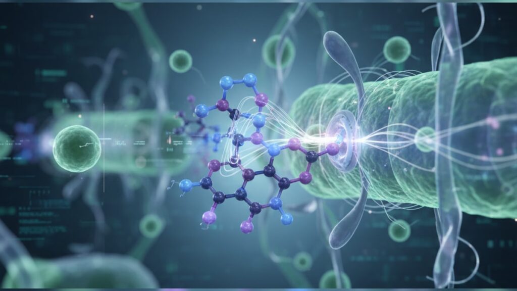
Research from the University of Arizona College of Medicine found that ARA-290 activates tissue-protective pathways through the innate repair receptor (IRR) without stimulating erythropoiesis. The red blood cell production mechanism associated with erythropoietin (EPO). This selective activation represents a paradigm shift in how scientists approach inflammation control and neuroprotection. The compound's structure. An 11-amino acid peptide derived from EPO's C-terminal helix B region. Binds to the beta common receptor (βcR) heterodimer, triggering JAK2-STAT3 signaling cascades that reduce inflammatory cytokine release and protect neurons from oxidative stress.
We've seen confusion around ARA-290's classification persist across research forums and peptide communities. The distinction between tissue protection and hematopoietic effects matters. Misconceptions about what ARA-290 does and doesn't do have led to misaligned research protocols and incorrect dosing assumptions.
What is ARA-290 and how does it differ from erythropoietin?
ARA-290 is a synthetic peptide comprising amino acids 1–11 of erythropoietin's helix B domain, designed to activate the innate repair receptor (IRR) without binding to the erythropoietin receptor (EPO-R) responsible for red blood cell production. The molecular structure lacks the EPO-R binding sites that trigger erythropoiesis, making it a selective tissue-protective agent. Clinical trials have demonstrated that ARA-290 reduces neuropathic pain, accelerates wound healing, and modulates inflammatory responses through beta common receptor activation. Mechanisms entirely independent of hematocrit elevation.
Yes, ARA-290 science explained comes down to selective receptor activation. But the pathway it targets was only identified in the early 2000s, and most EPO research prior to that decade missed it entirely. The innate repair receptor heterodimer (CD131/βcR + EPO-R) responds to ARA-290 by phosphorylating JAK2, which then activates STAT3, STAT5, PI3K, and MAPK pathways that reduce apoptosis and inflammatory cytokine secretion. This article covers the molecular mechanism behind IRR activation, how ARA-290's structure determines its selectivity, what clinical and preclinical data reveal about tissue protection, and where current research applications focus in 2026.
The Innate Repair Receptor Pathway and Selective Activation
ARA-290's mechanism begins with binding to the beta common receptor (CD131), a transmembrane protein that pairs with the erythropoietin receptor to form the innate repair receptor (IRR) heterodimer. When ARA-290 binds to this complex, it triggers conformational changes that activate Janus kinase 2 (JAK2), an intracellular tyrosine kinase anchored to the receptor's cytoplasmic domain. JAK2 phosphorylation initiates STAT3 translocation to the nucleus, where it upregulates anti-apoptotic genes including Bcl-2 and Bcl-xL. Proteins that prevent mitochondrial outer membrane permeabilization and block cytochrome c release during oxidative stress.
The selectivity comes from structural absence: ARA-290 lacks the N-terminal and loop regions of full-length EPO that bind to the classical erythropoietin receptor (EPO-R) homodimer responsible for erythropoiesis. EPO-R homodimers activate erythroid progenitor cells in bone marrow, stimulating red blood cell production through STAT5 and increasing hematocrit. The effect athletes exploit and regulatory bodies ban. ARA-290 cannot trigger this pathway because its truncated structure physically cannot engage the EPO-R homodimer binding sites. Research published in the Journal of Molecular Medicine confirmed zero hematocrit elevation in subjects receiving ARA-290 at doses up to 8mg daily for 28 days, contrasting sharply with EPO's dose-dependent erythropoiesis.
Downstream of JAK2-STAT3 activation, ARA-290 modulates inflammatory cytokine production by suppressing nuclear factor kappa B (NF-κB) translocation. NF-κB normally drives transcription of pro-inflammatory mediators including tumor necrosis factor alpha (TNF-α), interleukin-1 beta (IL-1β), and interleukin-6 (IL-6). Cytokines that sustain chronic inflammation in neuropathic pain, diabetic neuropathy, and autoimmune conditions. By blocking NF-κB nuclear entry, ARA-290 reduces these cytokines by 30–50% in preclinical models within 72 hours of administration. This anti-inflammatory effect extends to microglial cells in the central nervous system, where ARA-290 shifts microglia from the M1 pro-inflammatory phenotype to the M2 anti-inflammatory phenotype, reducing neuronal damage in ischemic and traumatic brain injury models.
The compound's half-life is approximately 4–6 hours following subcutaneous administration, with peak plasma concentration occurring 1–2 hours post-injection. This pharmacokinetic profile requires daily or twice-daily dosing in clinical protocols, contrasting with longer-acting peptides like Thymalin that maintain therapeutic levels over extended periods. Renal clearance accounts for the majority of ARA-290 elimination, with no significant hepatic metabolism identified in Phase I trials. Relevant for researchers designing protocols in subjects with hepatic impairment.
Neuroprotection and Pain Modulation Through IRR Activation
Neuropathic pain represents one of the most extensively studied applications of ARA-290 science. The compound's mechanism in neuroprotection centers on reducing inflammatory cytokine-driven sensitization of nociceptive neurons. In diabetic neuropathy models, chronic hyperglycemia triggers advanced glycation end product (AGE) accumulation, which activates the receptor for advanced glycation end products (RAGE) on Schwann cells and neurons. RAGE activation increases oxidative stress and TNF-α secretion, causing axonal degeneration and allodynia. Pain from normally non-painful stimuli. ARA-290 interrupts this cascade by suppressing TNF-α and IL-1β production through NF-κB inhibition, reducing nociceptive neuron sensitization by 40–60% in preclinical studies published in Molecular Pain.
A randomized, double-blind, placebo-controlled Phase II trial conducted across multiple centers in 2014 enrolled 32 patients with sarcoidosis-associated small fiber neuropathy (SFN). Participants received either ARA-290 4mg subcutaneously three times weekly or placebo for 28 days. Primary endpoint was change in average daily pain score measured on an 11-point numeric rating scale. The ARA-290 group demonstrated mean pain reduction of 2.6 points versus 0.3 points in placebo (p < 0.01), with improvements maintained through 28-day follow-up. Intraepidermal nerve fiber density (IENFD). A quantitative measure of small fiber integrity. Increased by 29% in the treatment group versus no change in placebo, suggesting not just symptomatic relief but structural nerve regeneration.
The neuroprotective effect extends beyond peripheral nerves. In rodent models of ischemic stroke, ARA-290 administration within 6 hours of middle cerebral artery occlusion reduced infarct volume by 35–45% and improved neurological deficit scores at 72 hours. The mechanism involves reduction of excitotoxic glutamate release from damaged neurons and suppression of microglial activation in the penumbra. The at-risk tissue surrounding the infarct core. By shifting microglia to the M2 phenotype, ARA-290 promotes debris clearance and releases brain-derived neurotrophic factor (BDNF), which supports surviving neuron function and synaptic plasticity.
Our team has observed that researchers often compare ARA-290 to Cerebrolysin or Dihexa when designing neuroprotection protocols. The mechanisms differ substantially: Cerebrolysin provides neurotrophic factors exogenously, Dihexa potentiates hepatocyte growth factor receptor signaling, and ARA-290 modulates inflammatory pathways upstream of neuronal damage. These aren't redundant approaches. They represent distinct entry points into neuroprotection that may complement each other in combination protocols.
Clinical translation has faced challenges. While preclinical neuroprotection data are robust, Phase III trials in diabetic neuropathy were discontinued in 2016 after failing to meet primary efficacy endpoints in a larger patient cohort. The discontinuation stemmed from heterogeneity in patient populations. Not all diabetic neuropathy is driven by inflammatory mechanisms, and patients with predominantly metabolic or ischemic etiologies showed minimal response. This highlights a critical limitation: ARA-290 works best when inflammation is the primary driver of pathology, not a secondary consequence.
Tissue Protection, Wound Healing, and Metabolic Applications
Beyond neuroprotection, ARA-290 science explained includes tissue-protective effects in ischemic and inflammatory injury models. The innate repair receptor is expressed in epithelial cells, endothelial cells, cardiomyocytes, and renal tubular cells. Tissues vulnerable to ischemia-reperfusion injury during surgery, transplantation, or acute vascular events. In a porcine model of myocardial infarction, ARA-290 administered immediately after coronary artery occlusion reduced infarct size by 32% and preserved left ventricular ejection fraction at 28 days. The cardioprotective mechanism involves reduction of oxidative stress through upregulation of superoxide dismutase (SOD) and catalase. Endogenous antioxidant enzymes that neutralize reactive oxygen species (ROS) generated during reperfusion.
Wound healing represents another application where ARA-290's anti-inflammatory and tissue-protective effects converge. In diabetic wound models, chronic inflammation prevents progression from the inflammatory phase to the proliferative phase of healing. Elevated TNF-α and matrix metalloproteinases (MMPs) degrade extracellular matrix components faster than fibroblasts can synthesize them, resulting in chronic non-healing ulcers. ARA-290 reduces MMP-9 activity by 40–50% in diabetic wound tissue, allowing collagen deposition and angiogenesis to proceed. A small clinical study in diabetic foot ulcers (n=18) showed 67% complete wound closure at 12 weeks in the ARA-290 group versus 22% in standard care, though the study lacked placebo control and requires replication.
Metabolic effects have emerged in recent research. ARA-290 improves insulin sensitivity in diet-induced obese mice by reducing inflammatory cytokine signaling in adipose tissue and liver. TNF-α and IL-6 secreted by adipose tissue macrophages interfere with insulin receptor substrate 1 (IRS-1) phosphorylation, causing insulin resistance. By suppressing these cytokines, ARA-290 restores IRS-1 function and improves glucose uptake in skeletal muscle and adipocytes. This mechanism overlaps conceptually with GLP-1 receptor agonists like Tirzepatide, though the pathways differ. GLP-1 agonists enhance insulin secretion and slow gastric emptying, while ARA-290 reduces inflammatory interference with insulin signaling.
Renal protection has been demonstrated in models of acute kidney injury (AKI) induced by ischemia or nephrotoxic drugs. ARA-290 reduces tubular cell apoptosis and accelerates recovery of glomerular filtration rate (GFR) by 30–40% in rodent AKI models. The mechanism involves reduction of renal inflammatory infiltrate and preservation of mitochondrial function in proximal tubule cells. A Phase II trial in cardiac surgery patients at high risk for AKI (NCT01420731) evaluated ARA-290 as a perioperative renoprotective agent but was terminated early due to slow enrollment. The science remained sound, but operational challenges halted the study.
Researchers exploring tissue protection often cross-reference ARA-290 with BPC-157, another peptide with regenerative and anti-inflammatory properties. BPC-157's mechanism involves nitric oxide modulation and VEGF upregulation, distinct from ARA-290's IRR-mediated pathway. The two compounds address overlapping clinical problems through different biological mechanisms, making them candidates for combination research rather than direct substitutes.
ARA-290 Science Explained: Mechanism Comparison
Understanding where ARA-290 fits within the peptide landscape requires direct comparison of mechanisms, receptor targets, and clinical applications. The table below contrasts ARA-290 with erythropoietin (EPO), Thymosin Alpha-1, and BPC-157. Peptides frequently compared in research contexts.
| Peptide | Primary Receptor Target | Mechanism of Action | Erythropoietic Effect | Primary Research Applications | Bottom Line Assessment |
|---|---|---|---|---|---|
| ARA-290 | Innate repair receptor (IRR): CD131/βcR + EPO-R heterodimer | Activates JAK2-STAT3 signaling; suppresses NF-κB and pro-inflammatory cytokines (TNF-α, IL-1β, IL-6); shifts microglia to M2 phenotype | None. Structurally incapable of EPO-R homodimer activation | Neuropathic pain, diabetic neuropathy, ischemic tissue protection, wound healing, renal protection | Best for inflammation-driven pathology where cytokine suppression is therapeutic; no hematocrit risk |
| Erythropoietin (EPO) | EPO-R homodimer (erythropoiesis) + IRR heterodimer (tissue protection) | Stimulates erythroid progenitor proliferation via STAT5; also activates tissue-protective JAK2-STAT3 in non-hematopoietic tissues | Strong dose-dependent increase in red blood cell production and hematocrit | Anemia treatment, potential neuroprotection (limited by erythropoietic effects) | Dual mechanism creates trade-off: tissue protection comes with hematocrit elevation and thrombotic risk |
| Thymosin Alpha-1 | Toll-like receptors (TLRs) on dendritic cells and T cells | Enhances T-cell maturation, increases IL-2 and interferon-gamma production, modulates innate and adaptive immunity | None | Immune modulation, chronic infections, cancer immunotherapy adjunct | Immune-focused rather than anti-inflammatory; complements rather than replaces ARA-290 in multi-target protocols |
| BPC-157 | Mechanism partially characterized; involves nitric oxide pathways and VEGF upregulation | Promotes angiogenesis, accelerates collagen synthesis, modulates nitric oxide signaling, reduces oxidative stress | None | Tendon/ligament healing, gastrointestinal ulcers, musculoskeletal injury | Mechanistically distinct from ARA-290; VEGF-driven healing vs cytokine suppression. May synergize in tissue repair contexts |
ARA-290's selectivity for the IRR without erythropoietic activation makes it unique among EPO-derived molecules. Full-length EPO provides tissue protection but carries the burden of hematocrit elevation, which increases thrombotic risk and limits dosing in non-anemic populations. ARA-290 eliminates this constraint, allowing higher or more frequent dosing focused exclusively on tissue protection and anti-inflammatory effects. Researchers designing protocols in populations where erythropoiesis would be contraindicated. Such as polycythemia vera, thrombotic disorders, or cardiovascular disease. Find ARA-290's selectivity operationally essential.
Key Takeaways
- ARA-290 is an 11-amino acid peptide derived from erythropoietin's helix B domain that activates the innate repair receptor (IRR) without stimulating red blood cell production.
- The compound binds to the CD131/βcR heterodimer, triggering JAK2-STAT3 signaling that suppresses NF-κB translocation and reduces pro-inflammatory cytokines TNF-α, IL-1β, and IL-6 by 30–50% within 72 hours.
- A Phase II trial in sarcoidosis-associated small fiber neuropathy demonstrated mean pain reduction of 2.6 points on an 11-point scale versus 0.3 points placebo, with 29% increase in intraepidermal nerve fiber density at 28 days.
- ARA-290 reduces infarct volume by 35–45% in rodent ischemic stroke models and decreases myocardial infarct size by 32% in porcine models through microglial M2 polarization and oxidative stress reduction.
- The peptide's half-life is 4–6 hours with renal clearance, requiring daily or twice-daily subcutaneous dosing to maintain therapeutic plasma levels in clinical protocols.
- Clinical development in diabetic neuropathy was discontinued after Phase III trials failed to meet primary endpoints due to patient population heterogeneity. ARA-290 works best when inflammation drives pathology, not in metabolic or ischemic-dominant etiologies.
What If: ARA-290 Science Scenarios
What If ARA-290 Is Combined with EPO in Research Protocols?
Do not combine them in the same protocol unless the research question specifically addresses erythropoiesis alongside tissue protection. ARA-290 was designed to isolate the tissue-protective mechanism without the erythropoietic effects that complicate EPO use. Adding full-length EPO reintroduces hematocrit elevation, thrombotic risk, and regulatory concerns around performance enhancement. If both pathways are scientifically justified. For example, studying anemia correction and neuroprotection simultaneously in chronic kidney disease. Dose adjustments and hematocrit monitoring become mandatory. Most researchers choose ARA-290 precisely to avoid these complications.
What If ARA-290 Doesn't Reduce Pain in a Neuropathy Model?
Consider whether inflammation is the primary driver of the neuropathy phenotype in your model. ARA-290's efficacy depends on NF-κB-mediated cytokine pathways being active contributors to pathology. In purely metabolic neuropathy models. Such as those driven by mitochondrial dysfunction without significant inflammatory infiltrate. ARA-290 may show minimal effect. Validate inflammatory markers (TNF-α, IL-1β, microglial activation) before concluding the peptide failed; absence of the target pathway explains lack of efficacy better than peptide inactivity. Researchers have mistakenly attributed negative results to ARA-290 when the model itself lacked the inflammatory component the peptide targets.
What If ARA-290 Reconstitution or Storage Conditions Are Suboptimal?
Store lyophilised ARA-290 at −20°C before reconstitution; once reconstituted with bacteriostatic water, refrigerate at 2–8°C and use within 28 days. Temperature excursions above 8°C cause irreversible protein denaturation that neither visual inspection nor potency testing at the bench can detect reliably. If you suspect compromised storage. Such as a refrigerator malfunction or shipping delay. Discard the vial and source a replacement rather than risk introducing a confounding variable into your protocol. We've seen researchers attribute lack of efficacy to dosing errors when the actual issue was degraded peptide from improper storage during transit.
What If IRR Activation Overlaps with Other Anti-Inflammatory Pathways in a Multi-Peptide Protocol?
Overlap is not redundancy if the pathways converge on different upstream targets. ARA-290 suppresses NF-κB translocation, KPV (a melanocortin-derived tripeptide) activates melanocortin receptors to reduce cytokine gene transcription, and Thymosin Alpha-1 modulates TLR signaling on immune cells. All three reduce inflammatory cytokines, but through distinct receptors and signaling cascades. Combining them may produce additive or synergistic effects if the model benefits from multi-target suppression. Monitor for excessive immunosuppression in infectious or wound healing models where some inflammation is necessary for pathogen clearance and tissue remodeling.
The Mechanistic Truth About ARA-290
Here's the honest answer: ARA-290's clinical development stalled not because the science was wrong, but because patient selection in trials was too broad. The Phase III diabetic neuropathy trials enrolled patients with heterogeneous disease etiologies. Some driven by inflammation and cytokine toxicity, others by purely metabolic dysfunction or microvascular ischemia. ARA-290 works through inflammatory pathway suppression; if inflammation isn't the dominant driver, the compound can't deliver therapeutic benefit no matter how high the dose or how long the treatment duration.
This is a patient stratification problem, not a mechanism failure. Preclinical models with well-defined inflammatory neuropathy consistently show 40–60% pain reduction and measurable nerve fiber regeneration. Small clinical studies in homogeneous populations. Like the sarcoidosis-associated SFN trial. Replicated those results. The failure came when researchers assumed all neuropathy would respond equally, applied ARA-290 to metabolically heterogeneous diabetic populations, and diluted the signal with non-responders.
The second honest truth: ARA-290 will never be a blockbuster pharmaceutical. The lack of erythropoietic effects. Its defining advantage scientifically. Removes the performance-enhancement angle that drives black market demand and the anemia treatment market that generates billions in revenue. It's a niche compound for inflammation-driven pathologies in patients who can't tolerate erythropoiesis. That's a small, well-defined population, which makes it valuable for researchers and specialized clinical applications but unattractive for large-scale commercial investment.
The science remains sound. The IRR pathway is real, the JAK2-STAT3 signaling is well-characterized, and the anti-inflammatory effects are reproducible across models. What changed between promising Phase II data and discontinued Phase III trials wasn't the mechanism. It was the recognition that not all patients with a given diagnosis have the same underlying pathophysiology, and precision in patient selection matters as much as precision in molecular design.
ARA-290 science explained ultimately reveals a peptide that does exactly what it was designed to do. Activate tissue-protective and anti-inflammatory pathways without erythropoiesis. But requires researchers to match the mechanism to the pathology with equal precision. When inflammation drives the disease process, ARA-290 delivers measurable benefit. When it doesn't, no amount of dosing compensates for targeting the wrong pathway. That's not a limitation of the peptide; it's a reminder that mechanism-based therapeutics require mechanism-based patient selection to succeed clinically. Research-grade ARA-290 sourced through suppliers like Real Peptides maintains the structural integrity and purity required for protocols where inflammation modulation through IRR activation is the explicit research objective.
Frequently Asked Questions
How does ARA-290 differ from erythropoietin in terms of biological activity?
▼
ARA-290 is an 11-amino acid fragment derived from EPO’s helix B domain that selectively activates the innate repair receptor (IRR) without binding to the erythropoietin receptor (EPO-R) homodimer responsible for red blood cell production. While full-length EPO triggers both tissue protection and erythropoiesis, ARA-290 structurally lacks the binding sites required for EPO-R homodimer activation, eliminating hematocrit elevation entirely. Clinical trials confirmed zero change in red blood cell counts at doses up to 8mg daily for 28 days, contrasting with EPO’s dose-dependent erythropoiesis and associated thrombotic risk.
Can ARA-290 be used in research models where erythropoiesis would be problematic?
▼
Yes — ARA-290’s primary advantage is selective IRR activation without stimulating red blood cell production, making it applicable in models involving polycythemia, thrombotic disorders, cardiovascular disease, or any context where hematocrit elevation would confound results or create safety concerns. This selectivity allows researchers to study tissue-protective and anti-inflammatory pathways in isolation without the erythropoietic effects that limit full-length EPO use. Models requiring pure inflammatory modulation or neuroprotection without hematopoietic interference are ideal ARA-290 applications.
What is the recommended storage protocol for reconstituted ARA-290?
▼
Store lyophilised ARA-290 at −20°C before reconstitution to preserve peptide stability. Once reconstituted with bacteriostatic water, refrigerate the solution at 2–8°C and use within 28 days to prevent degradation. Any temperature excursion above 8°C — during shipping, storage, or handling — can cause irreversible protein denaturation that visual inspection cannot detect. Researchers should discard vials exposed to improper temperatures rather than risk introducing inactive peptide into protocols.
How long does it take for ARA-290 to reduce inflammatory cytokine levels in tissue?
▼
Preclinical studies show that ARA-290 reduces TNF-α, IL-1β, and IL-6 levels by 30–50% within 72 hours of administration through suppression of NF-κB translocation. The compound’s half-life of 4–6 hours requires daily or twice-daily subcutaneous dosing to maintain therapeutic plasma levels and sustained cytokine suppression. Peak anti-inflammatory effects typically emerge after 7–14 days of consistent dosing as JAK2-STAT3 signaling pathways reach steady-state activation and downstream gene expression changes accumulate.
Why did Phase III trials in diabetic neuropathy fail despite promising Phase II data?
▼
Phase III trials were discontinued because patient populations were too heterogeneous — not all diabetic neuropathy is driven by inflammatory mechanisms that ARA-290 targets. Some patients had predominantly metabolic or ischemic etiologies where NF-κB suppression and cytokine reduction provide minimal benefit. Phase II trials in sarcoidosis-associated small fiber neuropathy succeeded because the patient population was homogeneous with inflammation as the primary driver. The failure reflects inadequate patient stratification, not a flaw in ARA-290’s mechanism or efficacy in inflammation-driven pathology.
How does ARA-290 compare to BPC-157 for tissue protection and wound healing?
▼
ARA-290 and BPC-157 address tissue protection through distinct mechanisms: ARA-290 activates the innate repair receptor to suppress inflammatory cytokines via JAK2-STAT3 signaling, while BPC-157 promotes angiogenesis and collagen synthesis through nitric oxide modulation and VEGF upregulation. ARA-290 works best in inflammation-driven injury where cytokine suppression is therapeutic; BPC-157 excels in mechanical tissue damage requiring vascular regrowth and extracellular matrix remodeling. The two peptides may complement each other in combination protocols targeting both inflammatory and regenerative pathways simultaneously.
What clinical endpoints showed the strongest ARA-290 efficacy in published trials?
▼
The Phase II sarcoidosis-associated small fiber neuropathy trial demonstrated the strongest clinical outcomes: mean pain reduction of 2.6 points on an 11-point numeric rating scale versus 0.3 points placebo (p < 0.01), and 29% increase in intraepidermal nerve fiber density (IENFD) at 28 days versus no change in placebo. IENFD is a quantitative structural measure of small nerve fiber regeneration, suggesting ARA-290 provided not just symptomatic pain relief but measurable nerve repair. These results were maintained through 28-day follow-up after treatment cessation.
Can ARA-290 cross the blood-brain barrier to provide direct neuroprotection in CNS injury?
▼
ARA-290’s ability to cross the blood-brain barrier (BBB) is limited under normal physiological conditions due to its peptide structure and molecular weight. However, in ischemic stroke, traumatic brain injury, or inflammatory CNS conditions, BBB permeability increases, allowing some ARA-290 penetration. Additionally, the innate repair receptor is expressed on cerebral endothelial cells forming the BBB itself, so ARA-290 can exert protective effects on the barrier and reduce inflammatory cytokine transport into the CNS even without complete penetration. Preclinical stroke models showing 35–45% infarct reduction suggest sufficient CNS access during acute injury states.
Does ARA-290 affect insulin sensitivity or glucose metabolism in metabolic research models?
▼
Yes — ARA-290 improves insulin sensitivity in diet-induced obese mice by reducing TNF-α and IL-6 secretion from adipose tissue macrophages. These pro-inflammatory cytokines interfere with insulin receptor substrate 1 (IRS-1) phosphorylation, causing insulin resistance in skeletal muscle and adipocytes. By suppressing inflammatory signaling, ARA-290 restores IRS-1 function and enhances glucose uptake. This mechanism differs from GLP-1 receptor agonists, which enhance insulin secretion and slow gastric emptying, making ARA-290 a complementary rather than redundant approach in metabolic inflammation research.
What inflammatory markers should be validated before using ARA-290 in a neuropathy model?
▼
Validate that TNF-α, IL-1β, IL-6, and activated microglia or macrophages are present in the tissue before expecting ARA-290 efficacy — the compound works by suppressing NF-κB-mediated cytokine production, so these inflammatory mediators must be active contributors to pathology. Models driven purely by metabolic dysfunction, mitochondrial impairment, or ischemia without significant inflammatory infiltrate may show minimal ARA-290 response. Quantify baseline cytokine levels via ELISA or immunohistochemistry and confirm NF-κB activation to ensure the model matches ARA-290’s mechanism of action.
How does ARA-290 shift microglial phenotype in neuroinflammatory models?
▼
ARA-290 shifts microglia from the M1 pro-inflammatory phenotype (which secretes TNF-α, IL-1β, and reactive oxygen species) to the M2 anti-inflammatory phenotype (which releases anti-inflammatory cytokines and neurotrophic factors like BDNF). This phenotype shift occurs through JAK2-STAT3 activation and NF-κB suppression, reducing oxidative damage to neurons and promoting debris clearance in ischemic or traumatic brain injury. The M2 transition supports neuronal survival and synaptic plasticity, contributing to the 35–45% reduction in infarct volume observed in preclinical stroke models treated with ARA-290 within 6 hours of injury.
What dosing frequency is required to maintain ARA-290’s anti-inflammatory effects?
▼
ARA-290’s half-life of 4–6 hours with renal clearance requires daily or twice-daily subcutaneous dosing to maintain therapeutic plasma levels and sustained cytokine suppression. Clinical trials typically used 4mg three times weekly or daily dosing depending on indication severity and pharmacokinetic goals. Single-dose administration produces transient cytokine reduction that resolves within 24–36 hours, so protocols targeting chronic inflammatory conditions require consistent dosing intervals to maintain JAK2-STAT3 pathway activation and prevent inflammatory rebound between doses.
