
Research notes
Blog
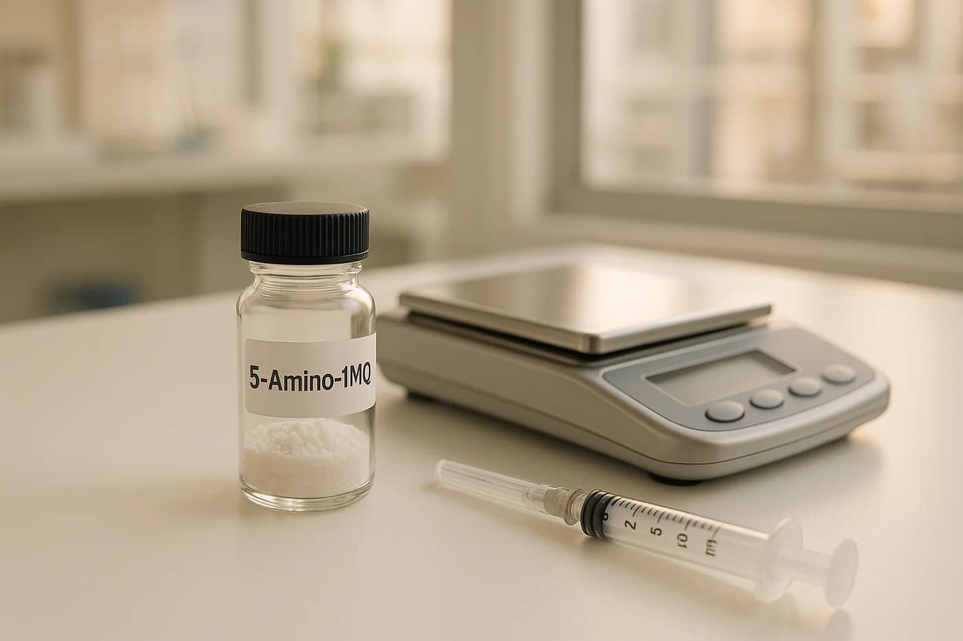
How to take 5 Amino 1MQ
Storage, stability and correct laboratory handling of 5 Amino 1MQ for research use only. Not for human or veterinary use.
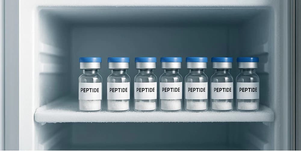
How to Store Research Peptides: Temperature Guide
How to Store Research Peptides: Temperature Guide Storing research peptides correctly is critical to maintaining their stability and ensuring reliable research results. Here’s what you need to know: Lyophilized Peptides: Store at room temperature for short-term use, refrigerate at 39°F for medium-term, and freeze at -4°F or -112°F for long-term preservation. Protect them from moisture and light. Reconstituted Peptides: Keep refrigerated at 36–46°F for up to 30 days. For longer storage, freeze in single-use aliquots at -4°F or -112°F to prevent degradation from freeze-thaw cycles. Key Factors to Avoid: Temperature fluctuations, moisture, light exposure, and repeated freeze-thaw cycles can degrade peptides. Proper storage practices save time, resources, and ensure consistent research outcomes. Best Way To Store Your Peptides: Maximum Potency (Lyophilized & Reconstitute) Ipamorelin BPC-157 Peptide Stability and Degradation Factors To ensure high-quality research outcomes, it’s crucial to understand what causes peptides to degrade. Environmental and chemical factors play a significant role in breaking down peptides, making proper storage practices a cornerstone of reliable experimentation. Factors That Affect Stability Temperature is one of the biggest culprits behind peptide degradation. Even minor temperature increases can accelerate harmful reactions, which is why consistent cold storage is essential to preserve peptide structure and functionality. Light exposure, especially to UV light, can damage peptides by breaking down specific amino acid residues. Aromatic amino acids like phenylalanine and tryptophan are particularly vulnerable to photochemical damage. Shielding peptides from both natural sunlight and artificial light sources can prevent this type of degradation. Moisture poses another serious threat. Humidity exposure can lead to hydrolysis, particularly in lyophilized peptides. Water molecules break peptide bonds, causing fragmentation and reducing biological activity. Even small amounts of moisture from the air can initiate this process. Oxygen exposure speeds up oxidation, especially in peptides containing cysteine, methionine, or tryptophan residues. Oxidation becomes even more problematic with repeated freeze-thaw cycles and in high-pH environments. Reducing air exposure when handling peptides can help mitigate these effects. pH levels also play a key role in peptide stability. Peptides exposed to pH levels above 8 are at greater risk of degradation. The surrounding chemical environment significantly influences how quickly peptides break down. These factors collectively contribute to specific degradation pathways, each of which can impact peptide integrity. Common Degradation Pathways Oxidation is a frequent issue, particularly for peptides with cysteine and methionine residues. High pH levels can accelerate this process, altering amino acid structures and diminishing peptide effectiveness. Hydrolysis occurs when water molecules break peptide bonds, causing the chain to fragment. Larger peptides are often more susceptible to this process, and asparagine residues may undergo dehydration, leading to chain cleavage. Deamidation affects sequences containing asparagine-glycine or glutamine-glycine combinations. This process alters the charge and structure of the peptide, which can compromise its biological activity and research applications. Aggregation happens when peptide molecules clump together, forming larger complexes that may precipitate out of solution. Peptides with extensive β-sheet structures are particularly prone to aggregation under heat or improper storage conditions. Disulfide exchange can occur in peptides with cysteine residues, leading to incorrect bond formations and structural alterations. Temperature fluctuations and improper pH levels can worsen this issue. Impact on Research Outcomes Peptide degradation has a direct and often detrimental impact on research reliability and reproducibility. When peptides lose their structural integrity, experimental results can become inconsistent, wasting valuable time and resources. Poor thermostability has already caused significant resource losses in peptide-based research. Alarmingly, only 11% of peptides advance from phase I clinical trials to drug approval. Even small deviations in storage temperature can trigger degradation, leading researchers to unknowingly use compromised materials. This not only delays progress but also drains funding that could be directed toward more productive endeavors. By maintaining optimal storage conditions, researchers can ensure peptide quality remains consistent, allowing them to focus on achieving meaningful scientific breakthroughs without being sidetracked by storage-related problems. Storage Conditions for Lyophilized Peptides Lyophilized peptides are known for their stability compared to liquid forms, making them a reliable choice for various research purposes. Maintaining proper storage conditions is critical to preserving their integrity and ensuring consistent experimental results. Short-Term Storage Guidelines For short-term storage, lyophilized peptides can typically remain stable at room temperature for several weeks. This stability is due to the freeze-drying process, which removes moisture that could otherwise lead to degradation. If you need to store peptides for up to three months, refrigeration at 2–8°C (36–46°F) is recommended. This temperature range keeps the peptides stable while ensuring they remain easily accessible. Always keep peptides in their original, sealed containers to protect them from moisture exposure. For longer storage durations, consider the methods outlined below. Long-Term Storage Methods When storing lyophilized peptides for extended periods, freezing is essential. For storage lasting several months to five years, keep the peptides as powders at –20°C (–4°F) in a desiccator to maintain a dry environment. For particularly sensitive peptides, ultra-low temperatures of –80°C (–112°F) provide optimal conditions to prevent degradation. To further protect peptides, combine freezing with desiccant use to avoid hydrolytic damage. Additionally, store peptides in UV-resistant containers or in dark environments to prevent light-induced degradation. Preventing Contamination Preventing contamination is a crucial step in handling lyophilized peptides. Before opening a vial, allow it to equilibrate to room temperature in a desiccator. This prevents condensation and moisture uptake that could compromise the peptide. “To prevent uptake of moisture from the air on the cold surface of the peptide or on the inside of its container, allow the peptide to come to room temperature before opening.” – peptidesciences.com When working with peptides, handle them quickly and reseal the containers immediately to limit air exposure. For additional protection, reseal vials under a dry, inert gas like nitrogen or argon. This step is especially important for peptides containing sensitive amino acids such as cysteine, methionine, or tryptophan. To minimize contamination risks further, consider aliquoting peptides into smaller portions. This reduces the need for repeated freeze-thaw cycles, which can degrade the product over time. sbb-itb-9529e2d Storage Guidelines for Reconstituted Peptides When it comes
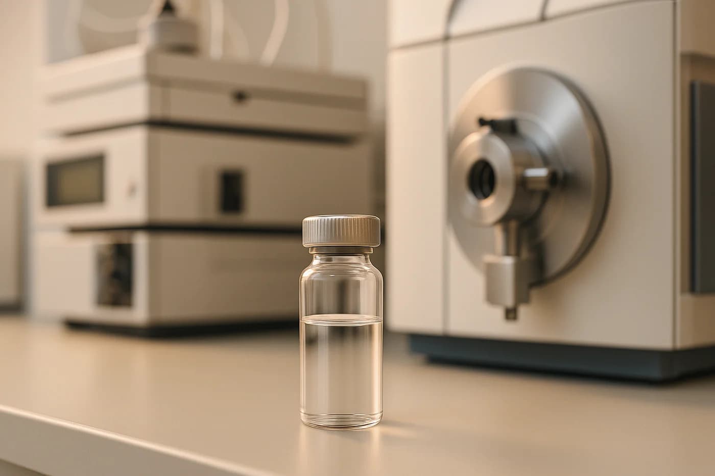
5 Key Factors in Peptide Purity Testing
5 Key Factors in Peptide Purity Testing When it comes to peptide purity testing, precision is everything. Impurities, even in small amounts, can skew research results, waste resources, and compromise safety in clinical applications. Here’s what you need to know: Peptide Purity Levels: Clinical-grade peptides require ≥98% purity, while early-stage research may allow >90%. Testing Methods: High-Performance Liquid Chromatography (HPLC), Mass Spectrometry (MS), and Endotoxin Screening are the main techniques. HPLC: The gold standard for purity analysis, separating peptides from contaminants based on chemical properties. MS: Confirms molecular identity and detects impurities that HPLC might miss. Endotoxin Screening: Ensures peptides are free from bacterial toxins, critical for biological applications. Impurity Identification: Detects synthesis errors, degradation products, and contaminants like truncated sequences or oxidized residues. Quick Tip: For accurate results, combine HPLC with MS and follow strict quality control practices. This approach ensures compliance with regulatory standards and reliable data for research or therapeutic use. how to understand peptide detection reports ChinaPeptides(HPLC /ESI-MS COA test Peptidie synethesis) High-Performance Liquid Chromatography (HPLC) Analysis HPLC is a powerful method for analyzing peptide purity, offering precise separation of target peptides from contaminants. By leveraging molecular interactions with solvent phases, HPLC delivers the detailed insights researchers need for accurate assessments. The process involves pushing a liquid sample through a column filled with small particles. As the sample moves, molecules separate based on their chemical properties, creating distinct patterns that can be analyzed. Reversed-phase HPLC (RP-HPLC) is the go-to choice for peptide analysis due to its reliability and efficiency. It uses columns packed with hydrophobic materials, often C18 phases, to separate peptides based on their hydrophobicity and polar interactions. More hydrophobic molecules take longer to pass through, creating clear separation patterns for analysis. HPLC Analysis Steps Achieving reliable results with HPLC requires careful preparation and execution. Sample preparation: Properly dissolve peptide samples and filter them through a 0.22 μm membrane to eliminate particulates that could clog the system or distort results. Mobile phase preparation: Use a solvent mix, typically acetonitrile with formic or trifluoroacetic acid, to maintain pH and ionization for consistent separation. Column selection: Choose a column based on the peptide’s properties. While C18 columns are widely used, factors like molecular weight and polarity may call for alternatives like XBridge Peptide BEH Columns, which handle a broader pH range and minimize secondary interactions. Gradient elution method: Gradually increase the proportion of organic solvent during the run to separate complex mixtures into individual components. Detection: Use UV detection at 214 nm and 280 nm, as peptide bonds absorb strongly in the far UV range. Many labs also use supplementary detectors like photodiode array (PDA) or mass spectrometry for comprehensive data. For instance, workflows often include solubilizing and filtering samples, optimizing columns (e.g., switching between C18 and C8 phases), fine-tuning gradients based on peptide characteristics, and employing multi-wavelength detection to calculate purity with precision. Temperature control is another key factor. Raising the column temperature can improve hydrophobic peptide solubility, sharpen peaks, and enhance separation accuracy. After each analysis, cleaning the column and flow path is essential to prevent cross-contamination and maintain consistent performance. Reading Chromatograms Interpreting HPLC chromatograms accurately is vital for determining peptide purity. Each peak represents one or more separated molecules, with the peak area corresponding to the concentration of each component. To calculate purity, use the formula: Percent purity = (Area of main peak / Total peak area) × 100. This provides the percentage of the desired peptide relative to all detected components. The retention time of each peak identifies compounds by comparison with known standards. The target peptide typically produces the largest peak, while impurities appear as smaller peaks at different retention times. Broad or asymmetrical peaks may indicate impurities or analytical issues, making it essential to achieve clear separation of the main peak from others. For example, a biotech company working on a 22-mer therapeutic peptide targeting a GPCR receptor initially found 88.1% purity with 11.9% impurities. Further analysis identified contaminants like a truncated variant and oxidized methionine. By re-purifying with a steeper gradient, reduced temperature, and switching from a C18 to a Phenyl-hexyl column for better resolution of aromatic regions, they improved purity to 98.5%. This optimization saved about $12,000 by avoiding a full re-synthesis and enabled a successful submission to Health Canada without data-related issues. Diode array detectors (DAD) can provide further validation by analyzing the UV spectrum across peaks. Consistent spectra usually indicate pure compounds, while variations suggest multiple components. Replicate injections and internal standards help ensure repeatability and correct for injection or detector inconsistencies, which is especially important for regulatory and quality control purposes. Finally, measuring resolution between peaks confirms adequate separation. Poor resolution can lead to overlapping peaks, skewing purity calculations and potentially jeopardizing research accuracy or compliance with regulatory standards. Mass Spectrometry (MS) for Verification Mass spectrometry takes peptide purity testing to the next level by identifying compounds based on their exact molecular weight and exposing impurities that HPLC alone might miss. It’s particularly effective at detecting post-translational modifications, sequence variants, and trace-level impurities – issues that often escape other testing methods. This precision is especially important for synthetic peptides, where impurities can closely resemble the target peptide, making them challenging to differentiate with traditional techniques. The process works by ionizing peptide molecules and measuring their mass-to-charge ratios, creating a unique mass spectrum. This spectrum acts like a molecular fingerprint, confirming the identity of the peptide and highlighting any contaminants. Let’s explore the two main MS techniques used for this purpose. Mass Spectrometry Methods When it comes to peptide purity testing, two key MS approaches stand out: LC-MS and MALDI-TOF. LC-MS (Liquid Chromatography-Mass Spectrometry): This method pairs liquid chromatography’s separation capabilities with the identification power of mass spectrometry. It delivers both qualitative and quantitative insights in one experiment, capable of detecting impurities at concentrations as low as 0.1% of the active pharmaceutical ingredient. MALDI-TOF (Matrix-Assisted Laser Desorption/Ionization Time-of-Flight): Known for its ability to analyze intact masses, this technique is widely used for testing the identity
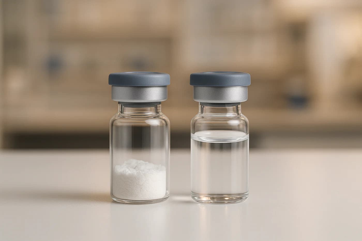
Lyophilized vs Liquid Peptides: Lab Comparison
Lyophilized vs Liquid Peptides: Lab Comparison When deciding between lyophilized and liquid peptides for your lab work, the right choice depends on your specific needs. Here’s a quick breakdown: Lyophilized Peptides: These are freeze-dried into a stable powder, offering long-term shelf life (up to years) when stored at -20°C to -80°C. They are ideal for long-term projects, international shipping without cold chain logistics, and experiments requiring stability. However, they require reconstitution and careful handling to avoid moisture absorption. Liquid Peptides: These are pre-dissolved and ready to use, saving time in immediate applications. They work well for short-term experiments but have limited stability (1–2 weeks at 4°C, up to 1 year at -80°C) and require stringent storage and handling to prevent contamination and degradation. Quick Comparison: Aspect Lyophilized Peptides Liquid Peptides Shelf Life Years (-20°C to -80°C) Weeks to months (-20°C to -80°C) Storage Room temperature shipping possible Requires constant cold chain logistics Preparation Needs reconstitution Ready to use Contamination Risk Low High Best For Long-term storage, stability Immediate use, time-sensitive experiments Lyophilized peptides are the go-to for durability and cost efficiency, while liquid peptides are better for convenience and quick turnaround experiments. Choose based on your lab’s workflow and storage capabilities. 1. Lyophilized Peptides Stability and Shelf Life Lyophilized peptides are an excellent choice for long-term storage. The freeze-drying process removes about 95% of the water content from the peptides, creating a dry powder that is far more resistant to degradation compared to liquid formulations. These peptides can remain stable for several years when stored at -20°C (-4°F), whereas liquid peptides typically last only up to 30 days at 4°C (39°F). Additionally, the reduced moisture content in lyophilized peptides significantly lowers the risk of bacterial degradation, a common issue in liquid forms. Storage and Handling When it comes to storage, lyophilized peptides offer greater flexibility than their liquid counterparts. For long-term preservation – spanning months to years – storing them in a freezer at -80°C (-112°F) is ideal. For shorter periods, refrigeration at 4°C (39°F) is sufficient. Before opening a vial, it’s important to let it reach room temperature to prevent moisture condensation. Once opened, weigh out the required amount quickly and reseal the vial immediately, as peptides can rapidly absorb moisture from the air. Certain peptides require extra precautions due to their amino acid composition. For example, peptides containing cysteine, methionine, or tryptophan should be stored in an oxygen-free environment and resealed under dry, inert gas like nitrogen or argon to prevent oxidation. Meanwhile, peptides with amino acids like aspartic acid, glutamic acid, lysine, arginine, or histidine benefit from storage in a desiccator to limit moisture exposure. To avoid structural degradation from repeated freeze-thaw cycles, aliquoting peptides into smaller, separate vials is highly recommended. These careful storage practices ensure that lyophilized peptides retain their functionality for a wide range of laboratory uses. Application Suitability Lyophilized peptides are particularly well-suited for scenarios requiring stability, ease of transportation, and long-term storage. Their dry, lightweight form makes them easier and more cost-effective to ship internationally. Unlike liquid formulations, which necessitate cold chain shipping and constant temperature monitoring, lyophilized peptides can often be transported at room temperature using standard shipping methods. This makes them a practical choice for researchers and labs across the globe. It’s worth noting that some peptides, especially those with short sequences, are highly hygroscopic. After lyophilization, they may appear as a gel or may be nearly invisible in the vial. This is not a sign of degradation but rather a result of their natural tendency to absorb moisture from the environment. Proper handling and storage can help maintain their integrity and performance. 2. Liquid Peptides Stability and Shelf Life Liquid peptides break down more quickly compared to their lyophilized counterparts due to factors like solvent type, pH levels, and the specific amino acids in their composition. Typically, their shelf life is 1–2 weeks at +4°C (39°F), 3–4 months at -20°C (-4°F), and up to 1 year at -80°C (-112°F). This is significantly shorter than lyophilized peptides, which can remain stable for several years when stored properly. Certain amino acids, such as Asn, Gln, Cys, Met, Trp, or Tyr, make peptides more prone to degradation. Storage and Handling Proper storage and handling are crucial to maintaining the integrity of liquid peptides. Since they are highly susceptible to bacterial contamination, storing them in solution is not ideal. If storage in solution is unavoidable, use sterile buffers with a pH between 5 and 6, or freeze solutions with a pH above 8 immediately. To prevent degradation, aliquot the peptide solution to avoid repeated freeze-thaw cycles, and store it at temperatures of -20°C (-4°F) or colder. Filtering the solution through a 0.2 µm filter can also reduce the risk of bacterial contamination. For peptides vulnerable to oxidation – especially those containing Trp, Met, or Cys – use oxygen-free water or buffers with reducing agents to limit damage. These stringent handling requirements highlight the challenges of liquid peptides compared to the more stable lyophilized versions. Application Suitability Liquid peptides are particularly useful for experiments requiring immediate application. Their ready-to-use nature eliminates the need for reconstitution, which is especially important in experiments involving peptide hormones that circulate in extremely low concentrations (0.1–100 pM). Even small losses during reconstitution could significantly affect experimental results. However, peptides containing Cys, Met, Trp, Asp, Gln, or N-terminal Glu are especially unstable in solution. Their use is practical only if they are applied immediately or stored under highly controlled conditions. While liquid formulations can simplify workflows by cutting down preparation time and reducing handling errors, these advantages must be carefully weighed against their shorter shelf life and more demanding storage requirements. Lyophilization of Peptides and Oligonucleotides sbb-itb-9529e2d Advantages and Disadvantages Choosing between lyophilized and liquid peptides depends on the specific needs of a laboratory. Both formats come with their own set of benefits and challenges that influence how experiments are conducted and their overall outcomes. Aspect Lyophilized Peptides Liquid Peptides Stability Stable for months or even years when stored

Top 7 Cognitive Research Peptides in 2025
Top 7 Cognitive Research Peptides in 2025 Peptides are transforming cognitive research by targeting specific neural processes like memory, learning, and neuroprotection. In 2025, seven peptides stand out for their potential in laboratory studies: Semax: Modulates neurotransmitters and boosts BDNF for memory and attention studies. Selank: Enhances GABA activity, reduces anxiety, and supports memory. Cerebrolysin: Promotes neuroprotection, neurogenesis, and recovery in stroke and brain injury models. Pinealon: Influences gene expression, aiding memory and aging-related research. NAD+: Supports mitochondrial function and reduces neuroinflammation for aging and neurodegeneration studies. Dihexa: Stimulates synapse formation and improves neural connectivity. P21: Aids neurogenesis and synaptic function, focusing on Alzheimer’s and cognitive decline. These peptides offer diverse mechanisms for studying brain health, from reducing inflammation to enhancing synaptic plasticity. Each compound is strictly for research use, adhering to regulatory standards. Quick Comparison Peptide Primary Function Key Application Unique Strength Semax Boosts BDNF, modulates neurotransmitters Memory and attention research Multi-targeted brain support Selank Enhances GABA and serotonin activity Anxiety and memory studies Combines calming and cognitive benefits Cerebrolysin Neuroprotection and regeneration Stroke and brain injury recovery Dual protection and repair mechanisms Pinealon Directly influences gene expression Memory and aging-related studies DNA-level impact on brain function NAD+ Supports mitochondrial health Aging and neurodegeneration research Addresses cellular energy and inflammation Dihexa Stimulates synaptic growth Alzheimer’s and neural repair studies High efficacy at low doses P21 Enhances neurogenesis Alzheimer’s and cognitive aging Stable, small molecule crossing brain barrier These compounds represent the forefront of cognitive science, providing researchers with tools to investigate brain function and potential therapies for neurodegenerative diseases. 47 – Peptides for Cognitive Function Selection Criteria The selection process for these seven cognitive research peptides adheres to strict standards to ensure both scientific reliability and laboratory applicability. Each peptide was carefully evaluated based on criteria that align with the highest benchmarks in cognitive neuroscience research. The first and most critical criterion is a peer-reviewed research foundation. A thorough literature review identified 54 relevant studies. This ensures that only peptides backed by substantial scientific evidence are included. Another key factor is laboratory relevance, focusing on validated research models. Each peptide demonstrates measurable outcomes in cognitive assessments, such as memory formation, learning processes, and neuroprotective mechanisms. The National Institutes of Health highlights the importance of this approach: “Translating these discoveries to the clinic will depend on the quality of the target validation process plus confidence in a causative link between the modulation of a target(s) and its functional physiological consequence(s) associated with treating or preventing a disease state.” Brain protection effects were also a major consideration. Each peptide exhibits neuroprotective properties through mechanisms like reducing inflammation, combating oxidative stress, and regulating neurotransmitters. For example, elevated homocysteine levels are linked to 12–31% of Alzheimer’s cases, underscoring the importance of compounds capable of addressing multiple pathways involved in cognitive decline. The selection process further requires convergent evidence from diverse research methods. The NIH emphasizes this approach: “Comprehensive target validation is a low-throughput process that involves more than a single method or assay. It depends on convergent evidence collected from a variety of studies.” Each peptide included in the selection has demonstrated effectiveness in both in vitro (test tube or cell culture) and in vivo (animal or human) studies, providing robust validation. To complement this scientific rigor, stringent manufacturing and quality control standards are applied. Real Peptides ensures ISO-certified manufacturing processes and ≥99% purity, verified through independent analytical testing. Purity is essential to guarantee the identity, activity, and efficacy of peptides used in research. Given that peptide- and protein-based drugs account for roughly 25% of the global pharmaceutical market, maintaining these standards is vital. “ISO certification ensures your partner follows internationally recognized standards, which translates into reliable, safe, and compliant products every time.” Additionally, comprehensive documentation and supply chain traceability add another layer of transparency, meeting stringent regulatory requirements. This meticulous approach helps identify and resolve potential issues early, ensuring consistent and reproducible results in laboratory studies. 1. Semax Semax is a synthetic analog derived from a fragment of the adrenocorticotropic hormone (ACTH). It has gained attention in cognitive research for its potential to influence learning and memory processes. Below, we’ll break down its mechanism of action, laboratory uses, notable research findings, and its role in cognitive studies. Mechanism of Action Semax works by interacting with several key systems in the brain. It modulates the hippocampal BDNF/TrkB pathway, activates serotonergic and dopaminergic systems, and engages melanocortin receptors. Additionally, it inhibits enkephalinase enzymes, which play a role in neuropeptide regulation. In laboratory studies, Semax has been shown to boost BDNF synthesis in astrocytes taken from the basal forebrain of rats, supporting the cellular processes that underlie learning and memory. Laboratory Applications Researchers use Semax in experiments focusing on memory enhancement, neuroprotection against oxidative stress, and the study of receptor-mediated cognitive pathways. Its role in studying melanocortin receptors (specifically MC4R and MC5R) helps scientists explore how these receptors contribute to cognitive improvements. Semax is also used to investigate the brain’s serotonin system, the regulation of BDNF expression in the hippocampus, and its effects on immune response and vascular health during ischemic conditions. Key Research Findings Research highlights several promising outcomes with Semax. For instance, a single 50 μg/kg dose has been shown to increase BDNF protein levels and TrkB phosphorylation in the hippocampus of rats. A 1996 trial demonstrated that Semax improved attention and memory in healthy individuals experiencing fatigue. More recently, a 2022 study by Sciacca and colleagues found that Semax can inhibit amyloid-beta fibril formation and reduce the toxicity of oligomers when used at a 1:5 molar ratio with Aβ. Comparative Advantages Semax has proven to be a valuable tool in cognitive research due to its multi-targeted approach to enhancing brain function. It acts quickly and has minimal reported side effects, making it particularly appealing for experimental use. Its effectiveness has been demonstrated in various settings, including in vitro experiments, animal studies, and human trials, offering researchers a versatile option for studying cognitive mechanisms and potential therapeutic applications. 2. Selank Selank is
Peptide Degradation: Prevention Methods
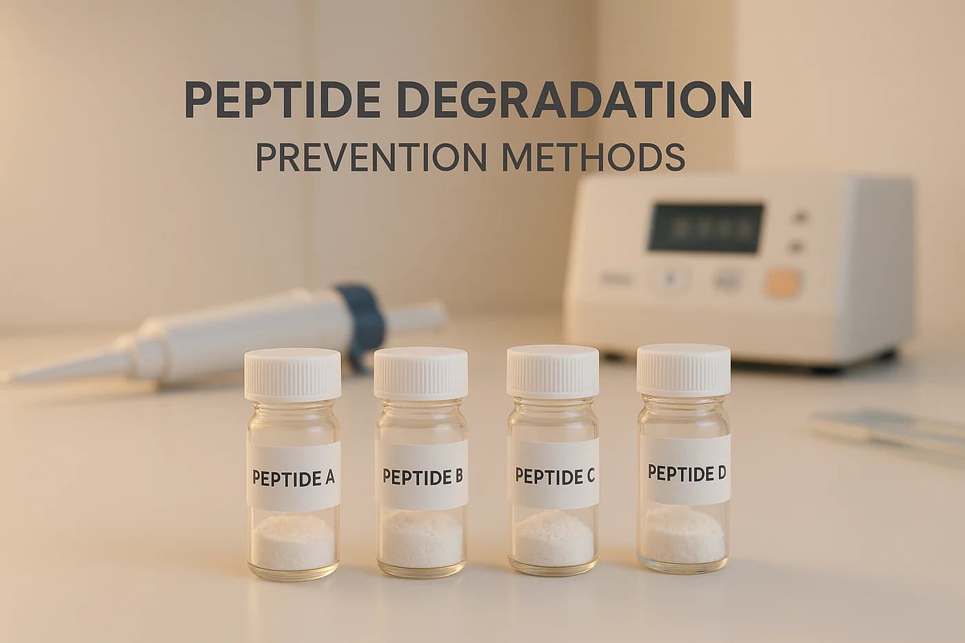
Peptide Degradation: Prevention Methods Peptides are delicate molecules prone to degradation, which can ruin experiments and waste resources. To maintain their stability, you need to address factors like storage, handling, and chemical vulnerabilities. Here’s a quick summary of the key strategies: Storage: Freeze peptides at -20°C or -80°C for long-term preservation. Lyophilization (freeze-drying) is ideal for extending shelf life. Handling: Avoid repeated freeze-thaw cycles by using single-use aliquots. Prevent moisture and oxygen exposure to limit hydrolysis and oxidation. Chemical Modifications: Protect peptides with terminal modifications or non-natural amino acids to resist enzymatic breakdown. Environmental Control: Keep peptides away from light, heat, and fluctuating temperatures to prevent structural changes. Additives: Use antioxidants and pH buffers to stabilize oxidation-prone residues and prevent chemical changes. Best Way To Store Your Peptides: Maximum Potency (Lyophilized & Reconstitute) Ipamorelin BPC-157 Best Storage Conditions for Peptides Storing peptides correctly is essential to maintain their stability and effectiveness over time. By carefully controlling factors like temperature, moisture, and oxygen, you can preserve peptide integrity for years instead of just days. Correct Storage Temperatures Temperature is the most critical factor when it comes to preserving peptides. For short-term storage, keep them at 4°C (39°F). For long-term storage, aim for −20°C (−4°F) or lower. The best option for minimizing degradation – down to as little as 0.001% – is −80°C (−112°F). If you’re planning to store peptides for extended periods, strict freezing protocols are a must. Be cautious about temperature fluctuations, as they can destabilize peptides. Avoid frost-free freezers entirely, as their defrost cycles lead to temperature swings that speed up degradation. Heat and sunlight are also major threats, so always store peptides in cold, dark conditions to maintain their quality. Before opening a peptide container, let it sit at room temperature first. This simple step prevents condensation, which could lead to water uptake and trigger hydrolysis reactions. How Lyophilization Helps Lyophilization, or freeze-drying, is a game-changer for peptide storage. By removing water, lyophilization transforms peptides into a stable, dry powder that lasts much longer than liquid forms. In fact, lyophilized peptides can remain stable at room temperature for several weeks. For long-term storage, keep them in tightly sealed containers at temperatures below -15°C (5°F), with -50°C (-58°F) or lower being ideal. When handling lyophilized peptides, work quickly. Weigh out the required amount and reseal the container immediately to limit exposure to moisture. Even brief contact with humid air can undo the benefits of lyophilization. This method pairs well with cold storage by eliminating water – a key factor in peptide degradation. Protecting from Moisture and Oxygen Moisture and oxygen are the biggest threats to peptide stability. Peptides should never be stored in solution for long periods, even in sterile, oxygen-free environments, as this can lead to gradual chemical degradation. Some amino acids, like tryptophan, methionine, and cysteine, make peptides especially prone to oxidation. For these sensitive peptides, use oxygen-free water or buffers and consider adding reducing agents like 1,4-dithio-dl-threitol (DTT). Cysteine-containing peptides need extra care – dissolve them in degassed acidic buffers to prevent oxidation, as thiol groups rapidly form disulfides at pH levels above 7. To protect peptides, store them in tightly sealed containers with desiccants to absorb any residual moisture. Before opening, let the container reach room temperature in a desiccator to avoid condensation. If you need to store peptide solutions, divide them into small aliquots and freeze them at temperatures below -15°C (5°F). This reduces the risk of repeated freeze-thaw cycles and exposure to air. Safe Handling and Mixing Methods Once you’ve established proper storage, careful handling and mixing are the next steps to ensure your peptides remain stable and effective. How you manage these processes can make or break your experiments, so attention to detail is key. Selecting Proper Solvents Choosing the right solvent is critical for dissolving peptides without causing damage. Start by considering the peptide’s amino acid composition to determine its solubility. For acidic peptides, try dissolving them in a 0.1M ammonium bicarbonate solution, then dilute with water to reach a neutral pH. For basic peptides, begin with 25% acetic acid and follow with water dilution. Neutral peptides may require organic solvents like acetonitrile (ACN), dimethylsulfoxide (DMSO), or dimethylformamide (DMF). Be cautious, though – peptides containing cysteine or methionine can degrade in DMSO, and DMF may release dimethylamine over time, which could react with protecting groups and introduce impurities. Make sure the peptide completely dissolves in the initial solvent before moving to buffered solutions. If you encounter slow dissolution, brief sonication might help, or you can re-lyophilize the peptide and try a different solvent. In many cases, sterile distilled water or a diluted acetic acid solution (0.1%) works well, but always test solubility on a small sample first. Reducing Light and Heat Exposure Both light and heat can quietly degrade peptides, so it’s essential to shield them from these elements. Store lyophilized peptides in cool, dim environments, away from direct sunlight or heat sources. During reconstitution, avoid unnecessary warming and use gentle sonication to limit thermal stress. For peptides that are particularly sensitive to light, handle them in low-light conditions and keep exposure to heat and light at a minimum throughout the process. Using Small Portions to Avoid Freeze-Thaw Damage Repeated freeze-thaw cycles can quickly compromise peptide stability. To avoid this, divide your peptides into smaller, single-use aliquots based on your experimental needs. This approach reduces temperature fluctuations and limits exposure to air. When working with peptide solutions, store aliquots in tightly sealed vials at –20°C (–4°F). Thaw them at room temperature, use them promptly, and reseal the vials immediately. For added protection, you might consider using cryoprotectants. Back in 1949, Christopher Polge found that adding glycerol to freezing solutions significantly improved cell survival. For peptides in solution, use sterile buffers with a pH between 5 and 6, aliquot the solution right away, and freeze. If the pH is above 8, always freeze the solution when it’s not in use. sbb-itb-9529e2d Chemical Changes and Protective Agents When storage and handling methods
Complete Guide to Peptide Synthesis Methods
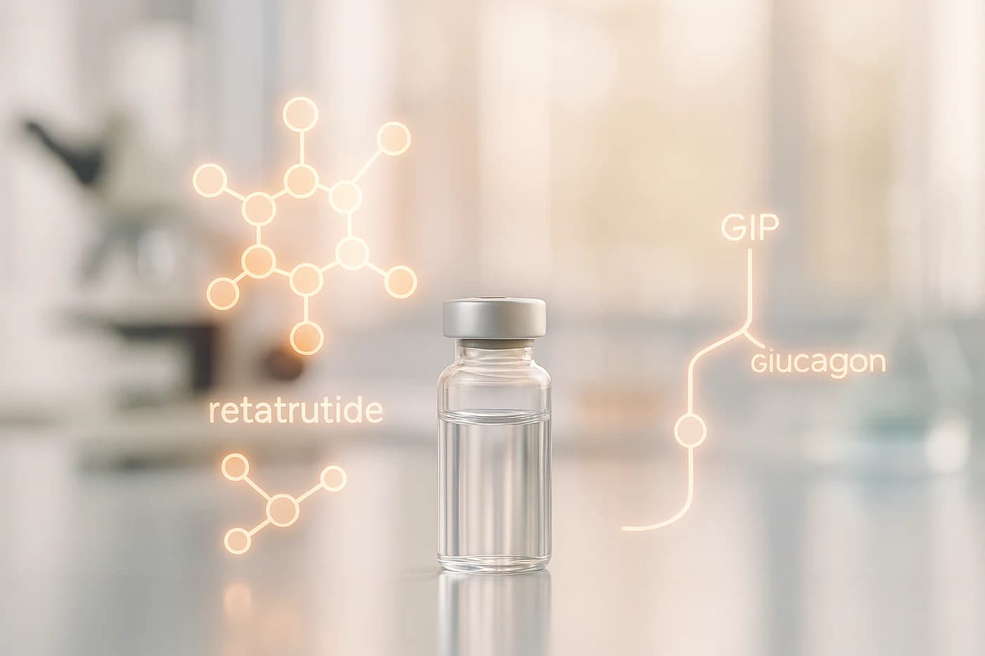
Complete Guide to Peptide Synthesis Methods Peptide synthesis is a critical process in biomedical research and drug development, offering precise control over biological pathways. With over 60 peptide-based drugs approved in the last 20 years and a market projected to reach $68.83 billion by 2028, synthetic peptides are transforming fields like diabetes treatment, oncology, and regenerative medicine. Key methods include: Solid-Phase Peptide Synthesis (SPPS): Efficient for long peptides and high-throughput applications but requires precise conditions. Liquid-Phase Peptide Synthesis (LPPS): Ideal for short peptides and hydrophobic sequences but involves extensive purification. Hybrid Methods: Combine SPPS and LPPS for complex peptides or large-scale production. Advancements in automation, reagents, and quality control are improving synthesis efficiency and reliability. Whether you’re using SPPS for small-scale research or LPPS for industrial production, optimizing each step ensures high-purity peptides for research and clinical applications. Solid Phase Peptide Synthesis Hands On Procedures from swelling resin to HPLC & MS analysis Main Peptide Synthesis Methods Peptide synthesis relies on three main approaches: solid-phase, liquid-phase, and hybrid methods. The choice between these depends on factors like peptide length, complexity, and budget. Below, we’ll explore the unique processes, advantages, and limitations of each method. Solid-Phase Peptide Synthesis (SPPS) Solid-phase peptide synthesis is the go-to method in research labs worldwide, thanks to its efficiency and versatility. Developed by Robert Bruce Merrifield, who earned the Nobel Prize in Chemistry in 1984 for this groundbreaking innovation, SPPS works by anchoring the peptide to an insoluble resin and extending it step by step. This eliminates the need for intermediate purification, as by-products are easily washed away while the peptide remains bound to the resin. SPPS is widely used in drug development, vaccine research, biomaterials, and general laboratory studies. Its high throughput and scalability make it particularly useful for creating combinatorial peptide libraries and conducting high-throughput screening. The process is often automated, further boosting efficiency. The Fmoc/tBu protection strategy, which simplifies modifications and cleavage, has become the standard for SPPS. By 1994, 98% of laboratories were using Fmoc chemistry. However, success in SPPS requires precise reaction conditions, including proper mixing to prevent resin bead clumping. Liquid-Phase Peptide Synthesis (LPPS) Liquid-phase peptide synthesis (LPPS) involves linking amino acids in solution using dehydration condensation and coupling agents. This method is particularly effective for creating short peptides or fragments and is often used for large-scale production of small peptides. LPPS is especially useful for synthesizing hydrophobic peptides that tend to aggregate on resin during SPPS. It also offers better solubility control for complex sequences. However, LPPS requires extensive purification steps, including extraction and chromatography, after each coupling reaction. While it can be more expensive due to higher solvent and reagent usage, LPPS remains a valuable tool for specific applications where SPPS might fall short. Feature SPPS LPPS Best for Long peptides, high throughput Short peptides, SAR studies Purification Minimal, resin-supported Extensive solvent work Time Efficiency Fast Slower Automation Highly automatable Manual or semi-automated Scalability Medium to large scale Small to medium scale Advanced and Hybrid Methods Hybrid methods combine elements of SPPS and LPPS to overcome the limitations of each. These approaches are particularly useful for synthesizing complex peptides with intricate structures or modifications that single methods struggle to achieve. For example, a hybrid technique might start with SPPS or LPPS to build a peptide backbone, followed by incorporating advanced modifications or scaling up production using recombinant systems. A notable application of hybrid methods is the production of Enfuvirtide (T20), an HIV membrane fusion inhibitor. This 36-amino acid peptide was commercially manufactured at a scale of several tons per year using a combination of solid-phase and solution-phase chemistry. Hybrid methods are especially valuable for large peptides, where the failure rate for chemical synthesis alone can reach 20% for sequences over 30 amino acids. Ongoing advancements continue to refine hybrid techniques. Innovations such as Molecular Hiving™, which uses soluble anchors, and multi-column solvent gradient purification (MCSGP) for continuous purification, are pushing the boundaries of peptide synthesis. Additionally, advanced analytical tools like mass spectrometry and NMR spectroscopy play a critical role in ensuring the quality and consistency of synthesized peptides. When deciding between synthetic and recombinant methods, researchers should weigh factors like peptide complexity, required production volume, cost, and timelines. This careful evaluation helps determine the best approach for specific research or industrial needs. Tools and Strategies for Peptide Synthesis Creating peptides successfully demands precise equipment, high-quality reagents, and problem-solving techniques tailored to each step of the process. Required Equipment and Reagents Modern peptide synthesis relies heavily on automated peptide synthesizers, which streamline production and ensure consistency. These synthesizers come in various types, such as batch systems for large-scale production, continuous flow models for improved efficiency, and library synthesizers capable of generating multiple peptides simultaneously. AAPPTec offers synthesizers for every scale, from small research quantities to industrial production reaching multiple kilograms. “Automated solid phase peptide synthesizers from AAPPTec are versatile, easy to use instruments that produce high-quality peptides economically.” – AAPPTec For manual synthesis, it’s essential to treat glassware with a silylating agent to avoid resin adhesion. Use orbital, wrist-action, or overhead stirrers, as magnetic stirrers can damage resin beads. Fmoc-protected amino acids are the go-to building blocks for their mild deprotection and compatibility with automation. For sequences prone to aggregation, Boc-protected amino acids may be more effective. Coupling reagents play a critical role in ensuring efficient amino acid incorporation. Here’s a quick comparison: Coupling Reagent Solubility (M) Stability in DMF Reactivity Key Features HATU 0.45 Excellent Highest Ideal for hindered couplings but costly HBTU 0.5 Excellent Good Reliable for routine synthesis; may cause guanidinylation COMU 1.5 Low Highest Great for tough couplings; safer than explosive reagents PyBOP >1.5 Moderate Good Effective in situ activation; avoids guanidinylation Reagents like HATU and PyAOP are particularly efficient due to the unique properties of HOAt, while Oxyma-based reagents (e.g., COMU) outperform traditional HOBt-based options. Solvents are another key factor, making up 80–90% of the nonaqueous mass used in synthesis. While DMF remains a staple, 2-MeTHF offers better performance in coupling steps. Solvents
HPLC vs MS: Peptide Analysis Comparison
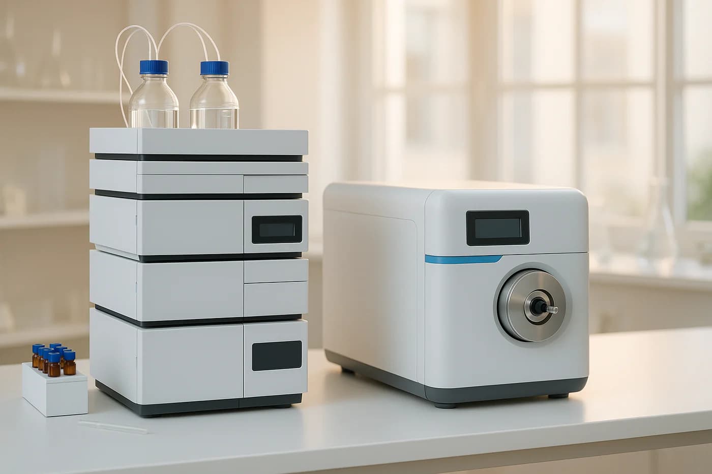
HPLC vs MS: Peptide Analysis Comparison Looking to analyze peptides? You’ll likely rely on two primary methods: High-Performance Liquid Chromatography (HPLC) and Mass Spectrometry (MS). Each has its strengths and limitations, and the best choice depends on your lab’s goals, budget, and analytical needs. Key Takeaways: HPLC is ideal for separating and quantifying peptides, offering high resolution and reliability for purity checks. It’s widely used for routine quality control and can detect impurities below 0.1%. MS excels in identifying molecular weight, structure, and sequence. It’s highly sensitive (down to femtomolar levels) and is unmatched for detecting post-translational modifications or analyzing complex mixtures. Cost Considerations: HPLC systems are more affordable upfront ($10,000–$40,000), while MS systems can range from $40,000 to over $500,000. Maintenance costs also differ significantly. Combined Use: Many labs pair HPLC with MS (LC-MS) for a powerful combination of separation and detailed molecular analysis. Quick Comparison: Factor HPLC MS Cost $10,000–$40,000 $40,000–$500,000+ Annual Maintenance $5,000–$15,000 $10,000–$20,000+ Purity Verification Excellent High specificity with mass-based data Structural Analysis Limited Detailed molecular and sequence insights Sensitivity Good Extremely sensitive (femtomol–zeptomol) Training Moderate Requires specialized training Choosing the Right Method: HPLC: Best for routine purity analysis and cost-effective separation. MS: Ideal for detailed structural data, sequence analysis, and detecting trace impurities. Both (LC-MS): Use together for a complete peptide analysis toolkit, combining HPLC’s separation with MS’s detailed identification. For labs in the U.S., aligning with regulatory standards like GLP/GMP and FDA requirements is critical. Whether you’re verifying purity or diving into molecular details, understanding the strengths of each method will help you make the right choice. Step-by-step LC-MS Peptide Mapping High-Performance Liquid Chromatography (HPLC) Explained HPLC is a well-established method used in U.S. laboratories to separate, quantify, and identify peptides. Let’s break down how this technique works and why it’s so widely used. How HPLC Works At its core, HPLC separates compounds by pushing a mobile phase through a specialized column under high pressure. This process relies on the physical and chemical properties of molecules to distinguish them. The most common method for peptide analysis is Reverse-Phase HPLC (RP-HPLC). This technique separates compounds based on their hydrophobicity. In this setup, a polar mobile phase – typically a mix of water and acetonitrile – interacts with a non-polar, surface-modified silica column. Polar molecules pass through first, while less polar ones take longer due to stronger interactions with the non-polar stationary phase. To ensure clear, sharp peaks, trifluoroacetic acid (TFA) is often added to maintain a low pH. The separated peptides are then detected using UV absorption at 214–215 nm. Depending on the analysis, the system can run in either an isocratic or gradient mode. Other HPLC approaches include size-exclusion chromatography (SEC) for separating molecules by size and ion-exchange chromatography (IEX) for distinguishing based on charge. Despite these options, RP-HPLC remains the go-to method due to its efficiency and reliability. This operational precision makes HPLC a cornerstone of peptide analysis, as we’ll explore further. HPLC Advantages for Peptide Analysis HPLC stands out for its ability to deliver high resolution and sensitivity, making it invaluable in fields like biomedicine and proteomics. For example, it can detect impurities at levels below 0.1% of the main compound. This level of precision ensures the purity needed for pharmaceutical, clinical, and research applications. It’s especially critical in quality control, where separating similar compounds can mean the difference between success and failure. RP-HPLC also excels in speed and efficiency, making it ideal for labs handling numerous samples. Standardized purification protocols simplify the process, reducing both analysis time and the need for constant adjustments. A practical example of its importance is in Alzheimer’s disease research, where analyzing the purity and concentration of beta-amyloid (Aβ) peptides is essential for tracking disease progression. “HPLC peptide purity analysis is a vital tool across numerous fields due to its efficiency and precision. From drug development and clinical diagnostics to the food industry and environmental science, HPLC ensures the purity and stability of peptide molecules.” – MtoZ Biolabs While RP-HPLC is the most popular, other modes like HILIC and CEX are also used. In certain cases, these methods can even outperform RP-HPLC for specific peptide mixtures. HPLC Drawbacks and Limitations Despite its strengths, HPLC does have its challenges. Unlike Mass Spectrometry, it provides limited structural information, which can be a disadvantage when detailed molecular characterization is required. Additionally, HPLC systems are expensive to purchase and maintain, and their operation requires skilled technicians. Sample preparation can be time-intensive, and separation times may stretch for complex mixtures. Another issue is sensitivity to technical variables. Small changes in mobile phase composition, column condition, or temperature can affect reproducibility. Regular maintenance is essential, and the costs can add up quickly. Handling peptides also requires care. Materials like polypropylene are preferred to minimize non-specific binding, and organic solvent concentrations should stay below 75% to prevent peptide precipitation. Adding modifiers can help improve solubility. Finally, while HPLC is generally fast, its combination with Mass Spectrometry (LC-MS) introduces additional hurdles. Proteomic studies, for example, must contend with the vast dynamic range of protein concentrations in samples like plasma. The most abundant proteins, such as serum albumin, dominate, while others, like cytokines, exist at much lower levels – spanning a range of over 10 orders of magnitude. These limitations highlight why many labs pair HPLC with other techniques. By combining HPLC’s reliable separation capabilities with complementary methods, researchers can achieve a more comprehensive understanding of peptide properties and behavior. Mass Spectrometry (MS) Explained Mass spectrometry (MS) takes a completely different approach to peptide analysis compared to HPLC. Instead of relying on separation techniques, MS identifies and quantifies peptides by measuring their mass-to-charge ratios. This method provides detailed insights into molecular weight and structure. How Mass Spectrometry Works Mass spectrometry works by analyzing the mass-to-charge ratio (m/z) of ions, making it possible to identify and measure molecules in complex mixtures. The process involves several steps: ionization, acceleration, deflection, detection, and data processing. A mass spectrometer typically includes three main components: an ion source, a mass
Optimizing Peptide Blends for Multiplex Assays
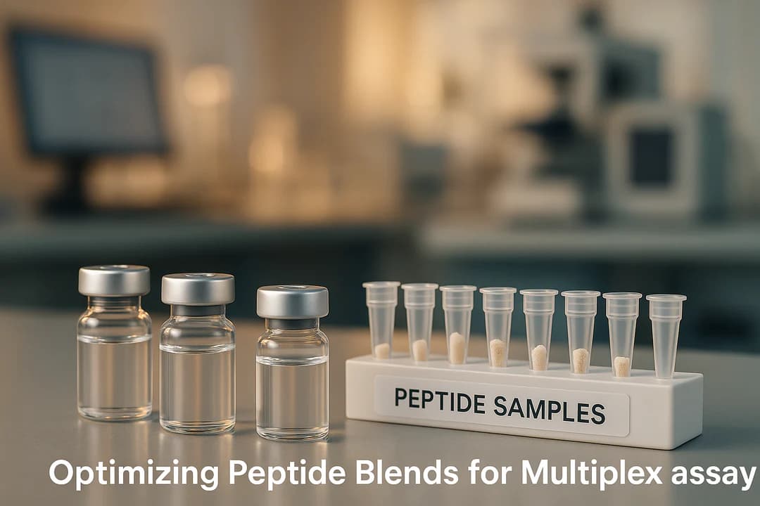
Optimizing Peptide Blends for Multiplex Assays Peptide blends enhance multiplex assays by improving sensitivity, reducing sample volume, and cutting costs. These assays measure multiple analytes simultaneously, enabling efficient biomarker research. Key steps include: Peptide Compatibility: Avoid cross-reactivity through systematic testing. Concentration Balance: Achieve precise sensitivity and specificity. Matrix Stability: Use stable isotope-labeled peptides and optimized storage to minimize degradation. Mass spectrometry and immunoassays are the primary platforms, each excelling in specific areas like sensitivity or multiplexing capacity. High-quality peptides (≥99% purity) are essential for reliable results. For streamlined workflows, pre-blended peptide stacks simplify preparation and ensure consistency. Bottom line: Careful optimization and quality peptides are critical for accurate, reproducible multiplex assays. Sample Prep and Handling Tips for MILLIPLEX® Multiplex Assays Design Considerations for Peptide Blends Developing effective peptide blends for multiplex assays demands attention to three key factors: peptide compatibility, concentration balance, and matrix stability. These elements are critical for ensuring the reliability and efficiency of the assay. Let’s dive into each of these considerations. Peptide Compatibility and Cross-Reactivity A major hurdle in multiplex assay design is avoiding interference between peptides. Cross-reactivity between detection antibodies and immobilized capture ligands can severely limit the number of proteins analyzed in a single assay. Antibodies that work well in monoplex assays may behave unpredictably in multiplex formats, leading to nonspecific binding, increased background noise, and reduced sensitivity. To address this, you can evaluate peptide compatibility through a three-step interference test: Step 1: Incubate individual proteins with a complete detection antibody cocktail to identify antibodies that bind to unintended targets. Step 2: Test a full protein mixture with individual detection antibodies to detect cross-reactive binding. Step 3: Run antibody cocktails with one antibody removed against complete protein mixtures to pinpoint specific cross-reactivity patterns. In immuno-SRM assays, mass spectrometry-based detection can help mitigate off-target antibody binding. Once compatibility is confirmed, the next step is to fine-tune concentration levels for optimal performance. Balancing Concentration for Sensitivity and Specificity Striking the right balance in concentration is essential. High affinity without specificity can lead to false positives, while low concentrations might miss targets present in smaller amounts. Achieving this balance often involves screening multiple candidates. For immunoassays, a coefficient of variation (CV) of 15% is generally acceptable. The FDA further recommends that measured values fall within 85–115% of the true value on average, and within 80–120% at concentration extremes. Selecting antibodies with both high affinity and specificity is critical. To monitor recovery, you can spike chemically labeled peptides or proteins into the assay and adjust concentrations as needed during multiple reaction monitoring (MRM). Additionally, multiplex immunoassays require thorough testing to ensure no cross-reactivity occurs between antibodies and other analytes in the mix. Proper concentration optimization ensures accurate and reproducible results. Matrix Effects and Stability Management Sample matrices, such as plasma or serum, can significantly impact peptide stability and assay performance. Human peptidase genes account for 3.1% of all encoded proteins, and most peptides have short half-lives ranging from 2 to 30 minutes. These factors make it essential to address matrix effects and stability challenges. To minimize nonspecific adsorption, pre-rinse pipette tips and use low-adsorption materials. Solubility can be improved by incorporating 5–60% organic solvents and adjusting the pH with 0.1–5% acid solutions. Using stable isotope-labeled (SIL) peptides as internal standards is another effective strategy. SIL peptides help normalize sample-specific ion suppression, ensuring accurate quantification. For peptide selection, aim for sequences with an SSRCalc score between 10 and 45, and avoid reactive residues like cysteine and methionine or motifs that inhibit trypsin cleavage, as these can lead to missed cleavages. Storage conditions also play a crucial role. Dry peptides (0.1–1 mg aliquots) should be stored under inert gas or in a desiccator at –20°C or lower for long-term use. When sourcing peptides, opt for purified ones with greater than 95% chemical purity and greater than 99% isotopic purity. Vendors like Real Peptides (https://realpeptides.co) provide high-quality peptides with HPLC-verified ≥99% purity and thorough endotoxin screening, ensuring consistency across batches. To maintain peptide integrity during preparation and analysis, reduce disulfide bonds and alkylate cysteine residues to prevent reformation. Use quenching reagents, such as aqueous acidic solutions or organic solvents, to halt enzymatic reactions. Additionally, controlling incubation and analysis temperatures is vital to avoid thermal degradation. Purified peptides, which undergo chromatographic refinement and amino acid analysis (AAA) for precise net content determination, are preferred over crude peptides, which may lack the purity required for accurate quantification. Step-by-Step Peptide Blend Optimization Workflow Fine-tuning peptide blends for multiplex assays demands a structured process, typically spanning about three months for MRM–MS assays. Initial Design and Testing Start by selecting peptides based on the target proteins and the specific requirements of your assay. Peptide hormones generally fall within a concentration range of 0.1 to 100 pM. Begin with small-scale compatibility tests to establish baseline metrics like signal intensity, retention time, and peak shape. These initial tests can help pinpoint potential issues before combining peptides into a more complex blend. Be mindful of nonspecific adsorption during this phase. Perform multiple transfer tests using containers and pipette tips to evaluate and reduce peptide adsorption. Adding surfactants early can help mitigate adsorption problems, while experimenting with different solvent compositions and pH levels can improve peptide solubility and overall assay performance. The insights gained here pave the way for the next step: incorporating robust internal standards. Adding Internal Standards and Controls Stable Isotope-Labeled (SIL) peptides are the go-to choice for internal standards in multiplex assays. For calibration, use recombinant protein standards alongside SIL peptides. While the AQUA approach with SIL peptides offers precision, it may underestimate protein concentrations when compared to recombinant standards. To maintain data integrity, incorporate multiple levels of quality control materials to monitor assay performance. This is especially crucial since protein extraction from tissue can account for up to 72% of quantification variability. Matrix effects can vary depending on the peptide, so it’s critical to evaluate each internal standard within the relevant matrix. A standard that works well in a simple buffer might behave differently in
What is retatrutide
What is Retatrutide Retatrutide (LY3437943) is a synthetic peptide designed for research purposes, targeting three hormone receptors: GLP-1, GIP, and glucagon. It binds most effectively to the GIP receptor, making it a tool for studying metabolic processes like weight regulation, glucose control, and liver fat reduction. Key features include: Triple-receptor activation: GLP-1, GIP, and glucagon receptors. Research findings: A 2024 study showed an 82.4% reduction in liver fat over 24 weeks. Delivery and half-life: Subcutaneous injection with a 6-day half-life, allowing weekly dosing. Applications: Used in obesity, diabetes, and regenerative medicine studies. Though promising in research, it is not approved for human use and is available only for laboratory studies. Proper handling and storage are essential for maintaining its integrity. How Retatrutide Works Molecular Structure and Receptor Targeting Retatrutide is a synthetic peptide made up of 39 amino acids, built on a GIP peptide backbone. To improve its stability, it includes three non-standard amino acids – two α-amino isobutyric acids and one α-methyl-L-leucine – placed at specific positions (residues 2, 20, and 13). Its structure forms a continuous helix, with the N-terminal segment interacting with receptor transmembrane domains and the C-terminal engaging extracellular regions of GLP-1, GIP, and glucagon receptors. Modifications in the N-terminal (residues 4–9) and mid-region (residues 10–21) allow it to effectively activate all three receptors. Interestingly, retatrutide shows about 8.9 times greater potency at the GIP receptor, while its potency at the glucagon and GLP-1 receptors is comparatively lower, with relative factors of 0.3 and 0.4, respectively. These unique molecular characteristics contribute to its strong pharmacokinetic performance. Pharmacokinetics and Dosing Retatrutide’s pharmacokinetics make it well-suited for extended research studies. It has a Tmax range of 8–72 hours and achieves 80% bioavailability when administered subcutaneously. Its systemic exposure increases proportionally with doses ranging from 0.25 mg to 15 mg. The peptide has a mean steady-state volume of distribution of approximately 10.3 liters, with an apparent clearance rate of 0.061 L/h, resulting in a half-life of about six days – ideal for once-weekly dosing. Retatrutide is primarily metabolized through peptide hydrolysis and beta-oxidation of its fatty acid component. Metabolites are excreted via urine and feces, while the intact peptide remains undetectable. Importantly, its pharmacokinetic profile remains consistent across both healthy individuals and those with metabolic conditions. Effects on Metabolic Pathways Retatrutide works by activating GLP-1 and GIP receptors, boosting glucose-dependent insulin secretion, and engaging the glucagon receptor to regulate hepatic glucose production. This multi-receptor approach helps maintain glucose balance while also influencing energy metabolism. It enhances nutrient uptake and energy expenditure, showing higher effectiveness at lower doses compared to single-receptor agonists. Beyond glucose regulation, retatrutide has been shown to improve lipid profiles by lowering triglycerides and increasing HDL cholesterol. It may also reduce insulin resistance linked to elevated non-esterified fatty acids, commonly seen in obesity. Additionally, its impact on appetite control makes it a valuable tool for studying feeding behavior and energy balance. Preclinical studies using obese C57/B16 male mice have demonstrated dose-dependent effects on gastric emptying and metabolic parameters under standardized conditions. For researchers exploring these pathways, retatrutide is available for purchase through Real Peptides. Research Applications Metabolic Research Studies Retatrutide plays a crucial role in research focused on obesity, type 2 diabetes, and metabolic disorders. As a triple receptor agonist targeting GLP-1, GIP, and glucagon receptors, it helps scientists explore how activating these pathways together influences weight regulation and glucose metabolism in experimental settings. Animal studies demonstrate that a 10 nmol/kg dose of retatrutide significantly reduces body weight within just 10 days, outperforming single-receptor agonists. These studies also report a noticeable reduction in food intake, highlighting its importance in investigating appetite regulation. In diabetic db/db mice, daily subcutaneous injections of retatrutide surpass the effects of liraglutide and tirzepatide by reducing not only food consumption and body weight but also markers of inflammation and fibrosis. Research involving female mice with NAFLD shows that retatrutide effectively normalizes liver fat levels and decreases steatosis markers in a dose-dependent manner. These findings open the door for exploring its broader potential in tissue repair and metabolic health. Regenerative Medicine Studies Early studies suggest that retatrutide may aid in tissue repair by activating GLP-1R, GIPR, and GCGR pathways simultaneously. For instance, research on metabolic dysfunction-associated steatohepatitis (MASH) has used retatrutide to better understand liver regeneration mechanisms. Additionally, its impact on inflammatory and fibrotic mediators has been leveraged to study tissue remodeling processes. Laboratory Model Compatibility Retatrutide has proven to be highly compatible with various laboratory models, making it a versatile tool for research. Mouse models such as C57BL/6, C57BL/KSJ, and db/db respond strongly to subcutaneous doses of approximately 10 nmol/kg. Studies reveal that retatrutide delays gastric emptying in a dose-dependent manner, making it useful for examining digestive physiology. In models of diabetic kidney disease, db/db mice consistently show improvements in both kidney protection and metabolic health during 10-week treatment protocols with daily injections. For researchers seeking reliable results, high-quality retatrutide is available through Real Peptides, ensuring consistency and reproducibility in experiments. Benefits and Limitations for Laboratory Use Benefits of Retatrutide Retatrutide offers valuable opportunities for laboratory research, thanks to its unique mechanism as a triple receptor agonist targeting GLP-1, GIP, and glucagon receptors. This allows scientists to explore complex metabolic processes that single-receptor agonists can’t fully address. One of its standout features is its high potency across all three receptors, with particularly strong binding to the GIP receptor. This makes it a powerful tool for studying metabolic interactions. The compound’s weekly dosing schedule is another advantage, as it supports long-term research while maintaining predictable, dose-proportional characteristics. This consistency is especially useful when examining dose-response relationships. A 2024 study by Ma et al. demonstrated the compound’s effectiveness. In experiments with db/db mice, a 10 nmol/kg daily dose over 10 weeks significantly reduced body weight and food intake compared to liraglutide and tirzepatide. Additionally, the study showed notable improvements in liver enzyme levels, cholesterol, and inflammatory markers, highlighting retatrutide’s potential in metabolic research. However, while these benefits are promising, there are certain challenges to
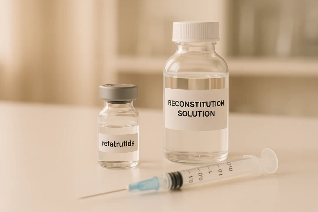
Is reconstitution solution good to reconstitute retatrutide?
Storage, stability and correct laboratory handling of reconstitution solution good to reconstitute retatrutide for research use only. Not for human or veterinary use.
Is Retatrutide Natural?
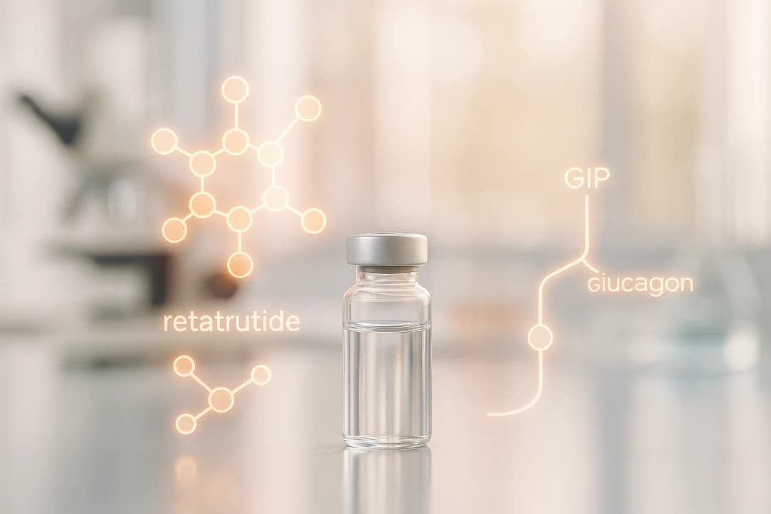
Is Retatrutide Natural? No, retatrutide is not natural. It is a fully synthetic peptide developed in laboratories. Retatrutide is a 39-amino acid compound designed to activate three specific hormone receptors: GLP-1, GIP, and glucagon receptors. Unlike natural peptides, which are produced by living organisms, retatrutide is chemically synthesized using advanced lab techniques like solid-phase peptide synthesis (SPPS). This synthetic design offers precise control over its structure, stability, and function, making it suitable for research on metabolic conditions like obesity and type 2 diabetes. Its laboratory origin ensures high purity, consistency, and tailored modifications that natural peptides cannot achieve. Retatrutide’s Chemical Structure and Classification Molecular Structure of Retatrutide Retatrutide is a synthetic peptide made up of 39 amino acids, designed using a GIP peptide backbone. Its amino acid sequence is YA¹QGTFTSDYSIL²LDKK⁴AQA¹AFIEYLLEGGPSSGAPPPS³, with the superscripted numbers denoting specific chemical modifications that distinguish it as synthetic. The peptide includes three non-standard amino acid residues that are central to its function: two α-amino isobutyric acids (Aib) and one α-methyl-L-leucine (αMeL), located at positions 2, 20, and 13, respectively. Each modification serves a purpose: Aib2 enhances resistance to dipeptidyl peptidase 4 (DPP4) cleavage, Aib20 boosts GIP receptor activity, and αMeL13 fine-tunes GLP-1 receptor activity. Structurally, retatrutide features a continuous helical design. This allows its N-terminal segment to penetrate the receptor’s transmembrane domain, while the C-terminal segment interacts with various receptor components, including the extracellular loops and the N-terminal α-helix of the extracellular domain for both GLP-1 and GIP receptors. This intricate architecture highlights its synthetic origin. Is Retatrutide Found in Nature? Retatrutide is not naturally occurring [13, 15]. It is entirely synthetic, created through laboratory processes [12, 15]. Unlike naturally secreted hormones like GLP-1, GIP, or glucagon, which each typically target one or two receptors, retatrutide is engineered to activate GLP-1, GIP, and glucagon receptors simultaneously. Its receptor activity also varies. Retatrutide is most potent at the human GIP receptor (EC50: 0.0643 nM), moderately potent at the GLP-1 receptor (EC50: 0.775 nM), and less potent at the glucagon receptor (EC50: 5.79 nM). Compared to natural hormones, it is about 8.9 times more potent at the GIP receptor but less potent at the GLP-1 and glucagon receptors by factors of 0.4 and 0.3, respectively. Why Scientists Create Synthetic Peptides Synthetic peptides like retatrutide are designed to address the limitations of natural compounds and achieve specific pharmacological goals. One major advantage is improved stability and extended duration. Retatrutide, for example, has a six-day half-life, enabling once-weekly dosing, whereas natural peptides typically have much shorter half-lives. Its ability to target multiple receptors simultaneously further underscores the benefits of synthetic design. Natural peptides rarely engage multiple receptor pathways with optimal potency. By engineering retatrutide to activate GLP-1, GIP, and glucagon receptors in a coordinated way, scientists can explore complex metabolic processes that are challenging to study with individual natural hormones. Additionally, synthetic methods allow for fine-tuning peptide properties. Retatrutide’s modified amino acids are a prime example of how receptor binding, stability, and activity can be optimized. The effectiveness of this approach has been demonstrated in clinical research. In June 2023, Eli Lilly and Company reported results from a Phase 2 trial showing that retatrutide led to a mean weight reduction of up to 17.5% at 24 weeks in adults with obesity or overweight. Participants receiving the highest dose (12 mg) achieved a 24.2% mean weight reduction after 48 weeks. For researchers looking for high-quality synthetic peptides, Real Peptides offers retatrutide and other compounds designed for research purposes. Their products undergo strict quality control to ensure consistent performance in laboratory experiments. Learn more about their offerings at www.realpeptides.co. Natural vs. Synthetic Peptides: Main Differences Defining Natural and Synthetic Peptides Natural peptides are short chains of amino acids naturally produced by living organisms. These peptides are found in plants, animals, and microorganisms, created through biological processes. To obtain them, scientists use isolation techniques to extract these compounds from their complex biological environments. Synthetic peptides, on the other hand, are made in laboratories using chemical synthesis methods. These lab-created peptides can either replicate natural peptides or feature entirely new sequences and structures, giving researchers full control over their design and composition. Below is a closer look at how these two types of peptides differ. Side-by-Side Comparison The choice between natural and synthetic peptides often depends on specific research goals. Key differences in their production methods, purity, and applications are outlined in the table below: Attribute Natural Peptides Synthetic Peptides Source Extracted from organisms Created via chemical synthesis Manufacturing Process Isolation from biological material Solid-phase peptide synthesis (SPPS) Purity Variable High (≥99%) Stability Lower Higher Research Applications Limited Broad and customizable Synthetic peptides stand out for their scalability, high purity, and ability to be tailored to specific research needs. They also provide faster production times compared to natural peptides. These advantages make them a preferred choice for studies requiring consistency and reliability. For researchers in need of high-quality synthetic peptides like retatrutide, Real Peptides offers compounds that meet stringent quality standards. Their products undergo thorough quality control to ensure dependable performance in laboratory experiments. Visit www.realpeptides.co to explore their range of peptide solutions tailored to research needs. How Retatrutide is Made in the Laboratory Solid-Phase Peptide Synthesis (SPPS) Method Retatrutide is synthesized using solid-phase peptide synthesis (SPPS), a method that constructs peptides step-by-step by adding protected amino acids to a growing chain anchored to a solid resin. The process begins with attaching the C-terminal amino acid to the resin. Then, the protective group on the amino acid’s N-terminal is removed, allowing the next N-protected amino acid to bind. This cycle of deprotection and coupling continues until the peptide sequence is fully assembled. For retatrutide, the SPPS process is fine-tuned with specific adjustments. These include selective removal of protective groups and the careful integration of peptide fragments to minimize errors like deletion or racemization while ensuring a high level of purity. Techniques like Fmoc (9-fluorenylmethyloxycarbonyl) and Boc (tert-butyloxycarbonyl) protection are commonly used, along with coupling reagents to activate the carboxyl group of each